
Pre-reconstruction calibration, data correction, and material decomposition method and apparatus for photon-counting spectrally-resolving x-ray detectors and x-ray imaging

a detector and x-ray imaging technology, applied in the field of pre-reconstruction calibration, data correction, and material decomposition methods and apparatuses for photon counting spectrally-resolving x-ray detectors and x-ray imaging, can solve the problems of loss of photon count and a distortion of photon energy, artifacts in the reconstructed image, and measurement of photon energy cannot be uniquely related to incident photon energy

Active Publication Date: 2016-10-06
TOSHIBA MEDICAL SYST CORP
View PDF8 Cites 14 Cited by
  • Summary
  • Abstract
  • Description
  • Claims
  • Application Information

AI Technical Summary

Benefits of technology

The patent text discusses the use of photon-counting detectors in computed tomography (CT) systems and the challenges associated with accurately detecting and measuring X-rays transmitted through the body. The technical effects of the patent include the development of methods for calibrating and correcting for detector artifacts, as well as the use of spectral CT to improve material differentiation and beam hardening correction. The patent also discusses the impact of pileup, ballistic deficit, polar effects, and characteristic X-ray escape on image quality and proposes solutions to improve image quality and accuracy in reconstructing the X-ray source.

Problems solved by technology

However, due to factors such as interaction depth and ballistic deficit, the measured photon energy cannot be related to incident photon energy uniquely.
Furthermore, at high flux, pulse-pileup may also cause a loss in photon count and a distortion in photon energy.
The distorted spectral signal can cause artifacts in the reconstructed images.
Furthermore, the dead time is not a constant for a given readout circuit due to the location of the pulse formation within the detector cell.
If the characteristic X-rays escape from the detector, the detector signal is incorrect and the loss of energy incurred manifests itself as errors in the output spectrum of the detectors.
Thus, the measured spectral signal can be distorted and may cause artifacts in the reconstructed image.
Uncorrected, each of the discussed measurement / detector artifacts distorts the detected spectrum relative to the incident spectrum ultimately degrading the quality of reconstructed images and the material decomposition derived from the data.

Method used


Image

  • Pre-reconstruction calibration, data correction, and material decomposition method and apparatus for photon-counting spectrally-resolving x-ray detectors and x-ray imaging
  • Pre-reconstruction calibration, data correction, and material decomposition method and apparatus for photon-counting spectrally-resolving x-ray detectors and x-ray imaging
  • Pre-reconstruction calibration, data correction, and material decomposition method and apparatus for photon-counting spectrally-resolving x-ray detectors and x-ray imaging

Examples


Embodiment Construction

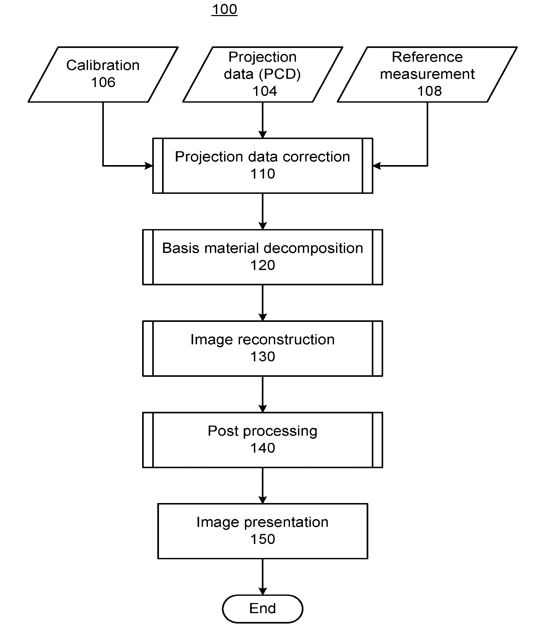
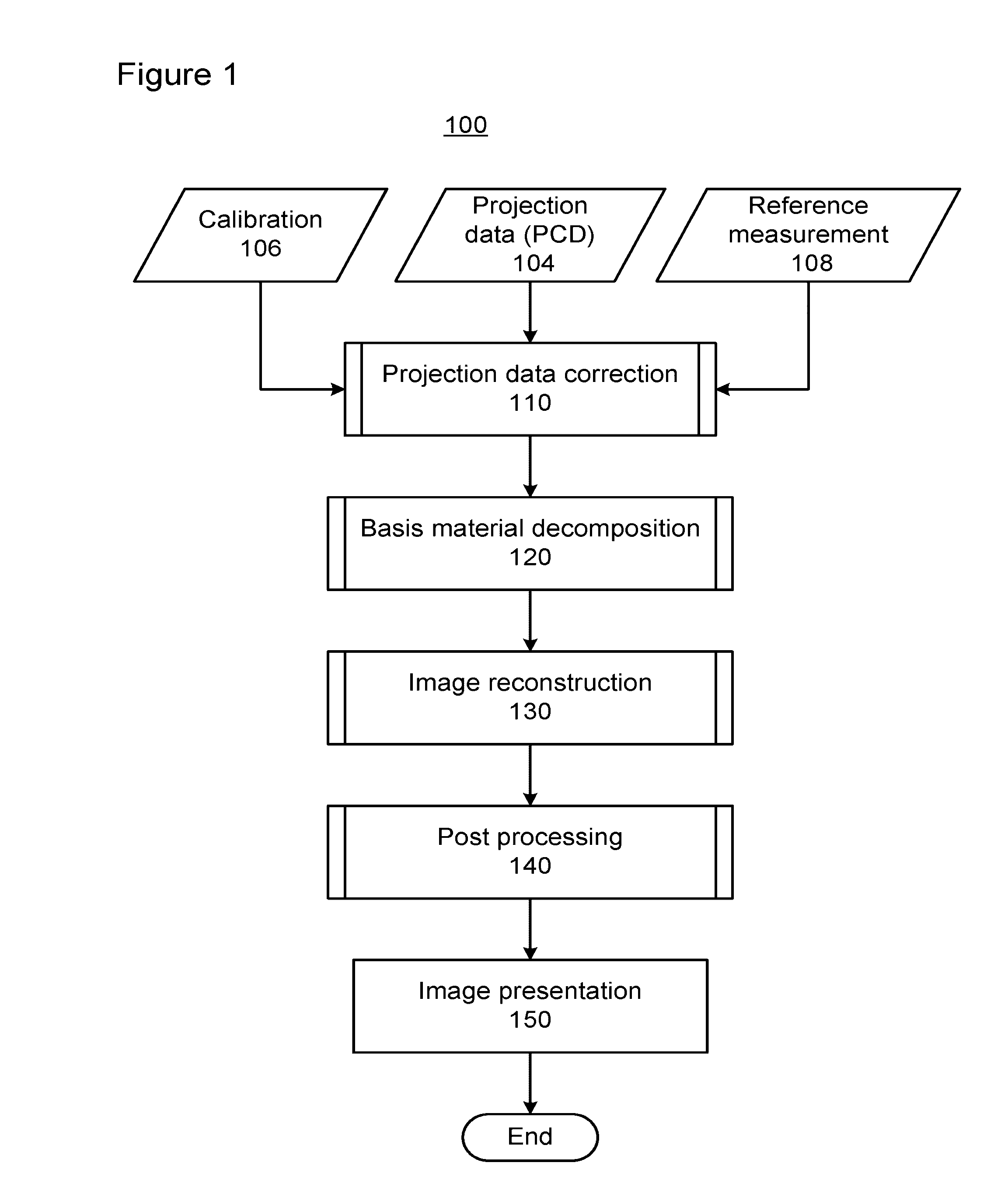
[0027]Referring now to the drawings, wherein like reference numerals designate identical or corresponding parts throughout the several views, FIG. 1 shows a flow diagram of a method for reconstructing an image of an object OBJ based on a series of projection measurements of the object OBJ performed at different projection directions (i.e., computed tomography (CT) using projective measurements). The data processing is a product of three inputs, including: calibration values 106, projection data 104, and reference measurement values 108. The projection data have multiple spectral components, making it compatible with material decomposition based on the different spectral absorption characteristics of high-Z and low-Z materials (i.e., greater contribution of photoelectric absorption for bone (high-Z) than for water (low-Z)). In addition to being applicable to CT applications as shown in FIG. 1, processes 110 and 120 are also applicable to non-CT applications involving projective measu...


PUM


Abstract

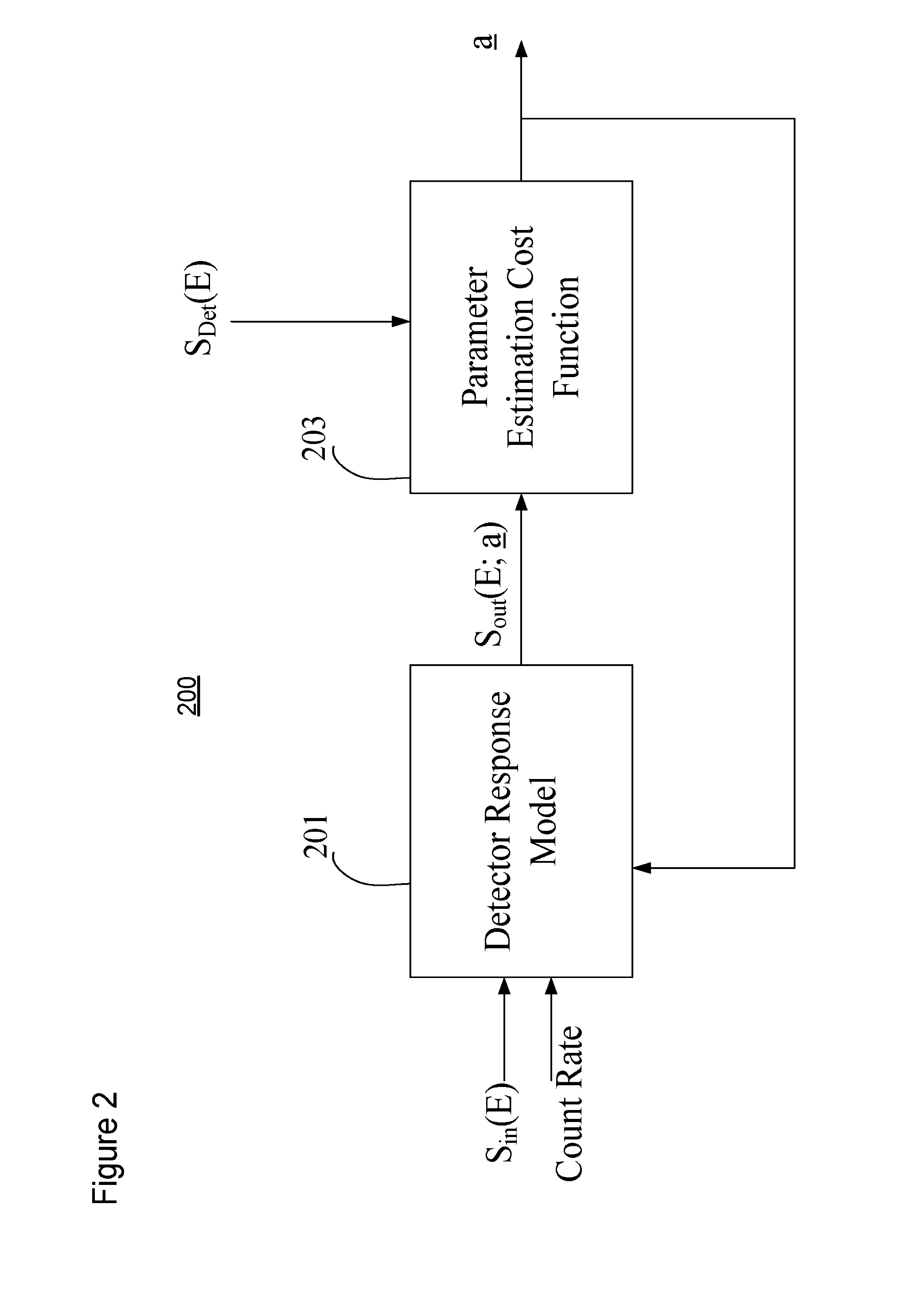
An apparatus and method of processing X-ray projection data obtained using photon-counting detectors and having multiple spectral components. The processing of the projection data includes correcting for nonlinear detector response, where the detector response model includes: pileup, ballistic deficit effects, polar effects, and characteristic X-ray escape. The processing of the projection data also includes a material decomposition mapping the projection data from spectral components into material components corresponding to high-Z and low-Z materials. The material decomposition includes a noise balancing process where the allocation of spectral components between a high-energy and a low-energy combination of spectral components is adjusted such that both high- and low-energy components have signal-to-noise ratios of similar magnitude. For computed tomography (CT) applications, material decomposition can be followed by image reconstruction and then image post-processing and presentation. For non-CT applications, material decomposition can be followed by image post-processing and presentation.

Description

BACKGROUND[0001]1. Field[0002]This disclosure relates to data processing of X-ray projection data obtained using spectrally-resolving photon-counting X-ray detectors for both computed tomography (CT) and non-CT applications, and more particularly relates to the data processing steps of calibration, correcting for detector artifacts, measurement artifacts, and performing material decomposition of X-ray projection data.[0003]2. Description of the Related Art[0004]Computed tomography (CT) systems and methods are widely used, particularly for medical imaging and diagnosis. CT systems generally create images of one or more sectional slices through a subject's body. A radiation source, such as an X-ray source, irradiates the body from one side. A collimator, generally adjacent to the X-ray source, limits the angular extent of the X-ray beam, so that radiation impinging on the body is substantially confined to a planar region defining a cross-sectional slice of the body. At least one detec...

Claims


Application Information

Patent Timeline
IPC IPC(8): A61B6/00G06T5/00G06T11/00
CPCA61B6/585A61B6/482G06T11/005G06T2207/10116G06T2211/424G06T2211/421G06T2207/10016G06T5/002A61B6/032A61B6/405A61B6/4241A61B6/4266A61B6/5205G01T1/17G01T1/2985G06T2211/408
Inventor ZOU, YUWANG, XIAOLANPETSCHKE, ADAMCAO, CHUNGUANGZHANG, YUEXING
Owner TOSHIBA MEDICAL SYST CORP
Learn More