
Lung, lobe, and fissure imaging systems and methods

a computed tomography and fissure imaging technology, applied in the field of computed tomography imaging, can solve the problems of difficult automated lobar segmentation, difficult lobe segmentation, and limited diagnostic imaging role in clinical practi

Active Publication Date: 2015-09-10
RGT UNIV OF CALIFORNIA
View PDF5 Cites 55 Cited by
  • Summary
  • Abstract
  • Description
  • Claims
  • Application Information

AI Technical Summary

Benefits of technology

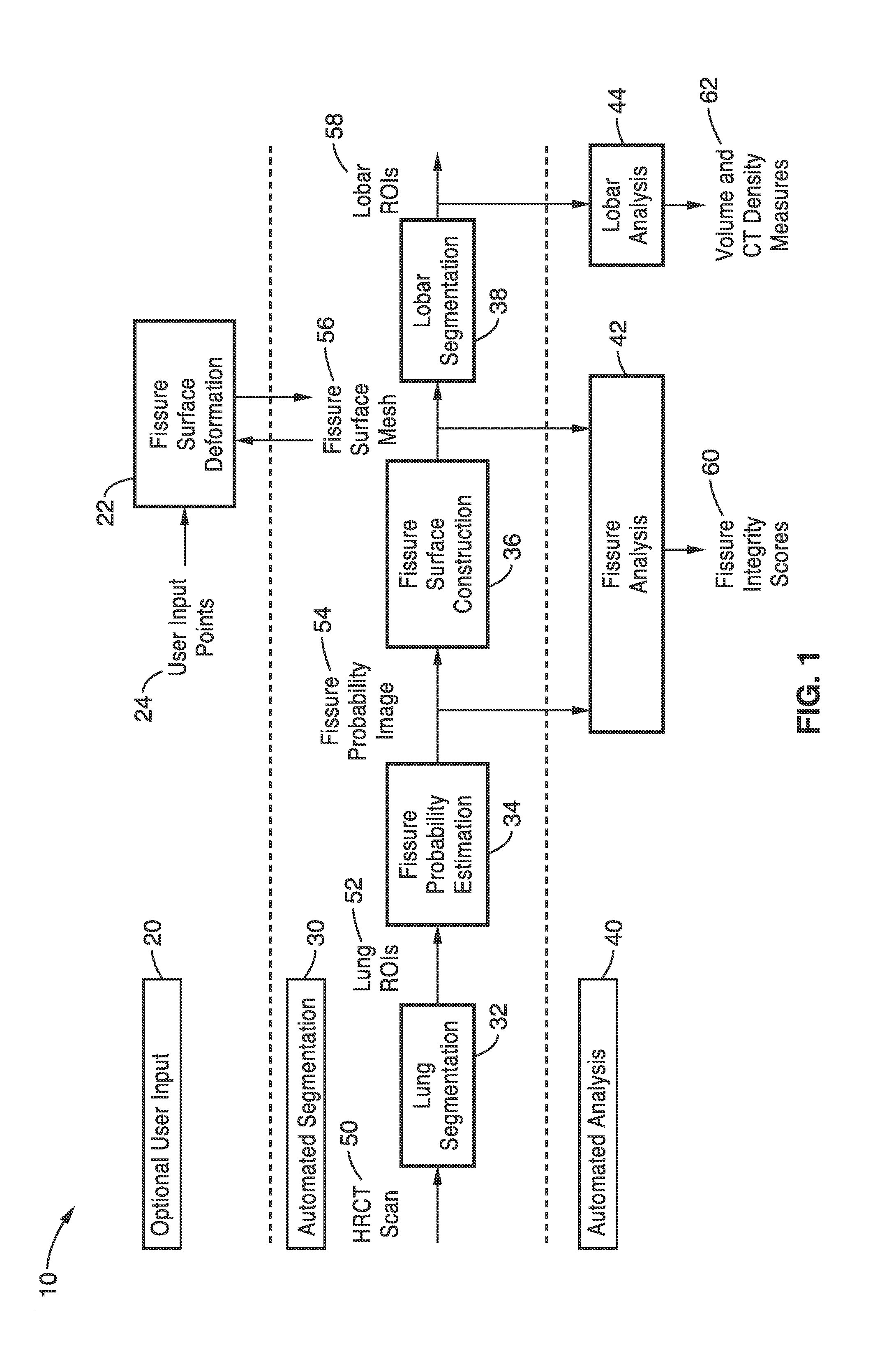
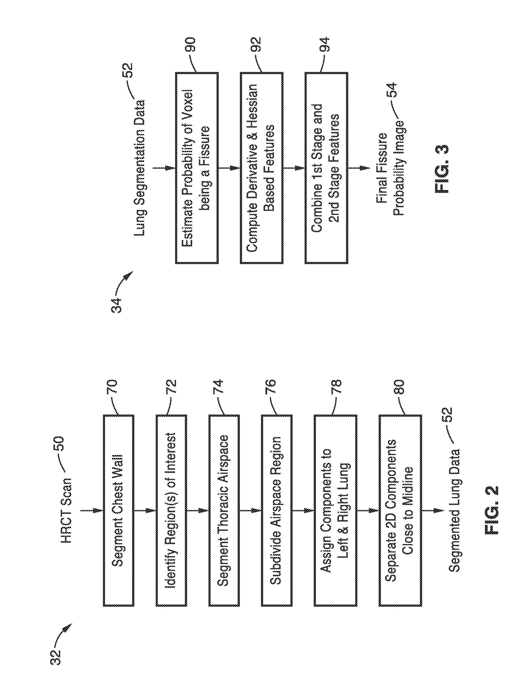
The present invention is about a system and method for analyzing lung images using automated and interactive techniques. The method involves segmenting the lung fields in CT images using a combination of thresholding, region-growing, and fuzzy logic. Additionally, a maximum cost path algorithm is used to segment the anterior and posterior junction lines of lung CT image data. The invention has technical effects in improving the accuracy and efficiency of lung image analysis, which can aid in the diagnosis and treatment of various lung-related diseases.

Problems solved by technology

However, the role of diagnostic imaging has generally been limited to visual inspection in clinical practice.
However, subdivision (segmentation) of the lungs into lobes is much more challenging because they are separated by only a thin layer of tissue (fissure).
Automated lobar segmentation is much more difficult and a small number of research groups are currently working on this problem.
Current approaches have not as yet yielded a robust automated lobar segmentation that is reliable enough to be used routinely in clinical practice.
These challenges arise because partial volume averaging of the thin fissural plane leads to very faint, incomplete edges that indicate the lobar boundary.
Also, in many patients the fissures are anatomically incomplete (i.e., they do not extend all the way across the lung parenchyma).
Anatomic and pathological variants in shape and location of fissures also create problems, particularly when disease such as emphysema is present, as this leads to severe deformities.
The minor fissure of the right lung is particularly difficult to identify because it tends to run parallel to the scan plane and is more variable in shape.

Method used


Image

  • Lung, lobe, and fissure imaging systems and methods
  • Lung, lobe, and fissure imaging systems and methods
  • Lung, lobe, and fissure imaging systems and methods

Examples


experiment 1

[0081]c. Experiment 1

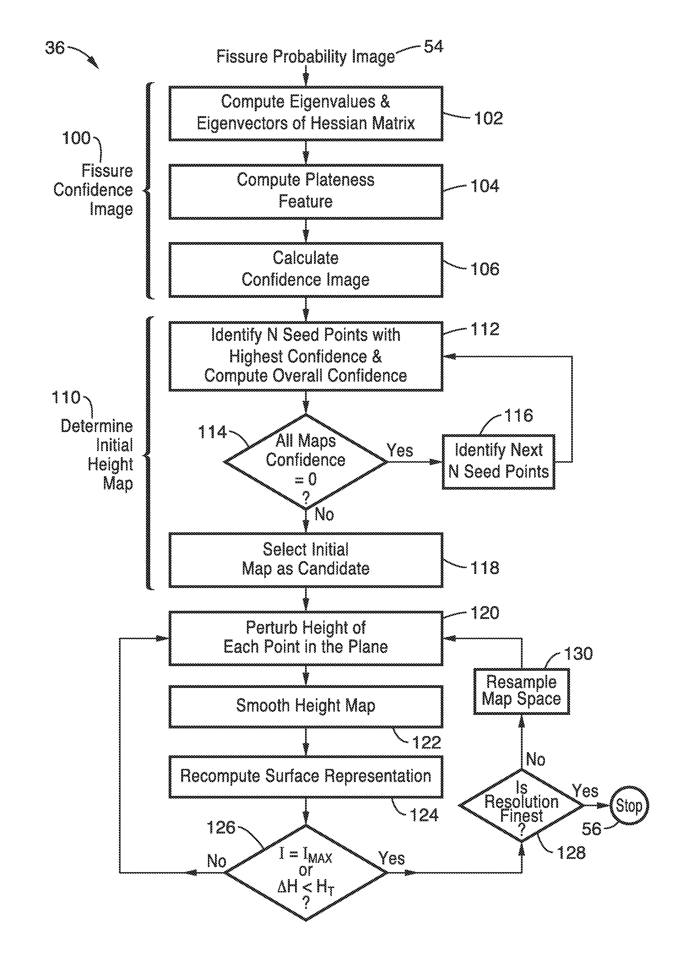
[0082]Training of the supervised fissure enhancement filter and tuning of the parameters for the method in sections A and B above was performed using a total of 18 chest CT scans obtained from a research database, with slice thickness and in-plane resolution ranging from 0.55 to 0.78 mm and from 0.5 to 1.25 mm respectively. The method was tested on chest CT scans from another research database, consisting of 41 scans from different patients with severe emphysema and forced expiratory volume in one second (FEV1)<45%. The slice thickness and in-plane resolution of the test set ranged from 0.5 to 1.0 mm and 0.54 to 0.83 mm respectively.

[0083]Approximate nearest neighbor searching was used to implement the KNN classifiers of the supervised fissure enhancement filter, where the number of nearest neighbor K was set to fifteen and an error bound ε of 0.5 was used. To reduce computational time, the multiple scales used for computing the confidence and performing the evo...

experiment 2

[0089]d. Experiment 2

[0090]Training of the supervised fissure enhancement filter and tuning of the parameters for the method described above in sections a and b was performed using a total of 18 chest CT scans obtained from a research database, with slice thickness and in-plane resolution ranging from 0.50 to 1.25 mm and 0.55 to 0.78 mm respectively. The method was tested on chest CT scans from another research database, consisting of 22 scans from different patients with either idiopathic pulmonary fibrosis or severe emphysema (forced expiratory volume in one second <45%). The slice thickness and in-plane resolution of the test set ranged from 0.6 to 3.0 mm and 0.59 to 0.80 mm respectively.

[0091]Accordingly, the systems and methods of the present invention provide a powerful automation combined with intuitive, human intervention and feedback to achieve a robust, widely-applicable system that can even handle the most diseased or abnormal images where fully automated segmentation is ...


PUM


Abstract

An automated or semi-automated system and methods are disclosed that provide a rapid and repeatable method for identifying lung, lobe, and fissure voxels in CT images, and allowing for quantitative lung assessment and fissure integrity analysis. An automated or semi-automated segmentation and editing system and methods are also disclosed for lung segmentation and identification of lung fissures.

Description

CROSS-REFERENCE TO RELATED APPLICATIONS[0001]This application is a 35 U.S.C. §111(a) continuation of PCT international application number PCT / US2013 / 057753 filed on Aug. 31, 2013, incorporated herein by reference in its entirety, which claims priority to, and the benefit of, U.S. provisional patent application Ser. No. 61 / 700,829 filed on Sep. 13, 2012, incorporated herein by reference in its entirety. Priority is claimed to each of the foregoing applications.[0002]The above-referenced PCT international application was published as PCT International Publication No. WO 2014 / 042902 on Mar. 20, 2014, which publication is incorporated herein by reference in its entirety.STATEMENT REGARDING FEDERALLY SPONSORED RESEARCH OR DEVELOPMENT[0003]Not ApplicableINCORPORATION-BY-REFERENCE OF COMPUTER PROGRAM APPENDIX[0004]Not ApplicableNOTICE OF MATERIAL SUBJECT TO COPYRIGHT PROTECTION[0005]A portion of the material in this patent document is subject to copyright protection under the copyright law...

Claims


Application Information

Patent Timeline
IPC IPC(8): G06T7/00G06T15/08G06T7/60
CPCG06T7/0012G06T7/0038G06T7/0081G06T7/0083G06T7/602G06T7/608G06T2207/20141G06T2207/20148G06T2207/10081G06T2207/20076G06T2207/20044G06T2207/20116G06T2207/20136G06T15/08G06T2200/04G06T2207/20101G06T2207/20156G06T2207/30061A61B6/032G06T7/11G06T7/12G06T7/187G06T7/136
Inventor BROWN, MATTHEW S.LO, PECHINVAN RIKXOORT, EVA MARJOLEINGOLDIN, JONATHAN GERALD
Owner RGT UNIV OF CALIFORNIA
Learn More