Method and apparatus for reducing artifacts in computed tomography image reconstruction

Active Publication Date: 2015-05-28
GE MEDICAL SYST GLOBAL TECH CO LLC
View PDF12 Cites 16 Cited by
  • Summary
  • Abstract
  • Description
  • Claims
  • Application Information

AI Technical Summary

Benefits of technology

The present invention provides a method and apparatus for reducing artifacts in CT image reconstruction. The method involves determining the proportion of metal pixels in the original reconstructed image and generating an expanded metal template based on the original reconstructed image and the original sinogram. A metal-free, MAR image is then generated based on the expanded metal template and the original sinogram. A final image is generated based on the expanded metal template and the metal-free, MAR image. The apparatus includes an acquisition device, a determining device, an expanded metal template generating device, a metal-free, MAR image generating device, and a final image generating device. The technical effect of this invention is to improve the quality of CT images and reduce the impact of metal artifacts.

Problems solved by technology

An object having high X-ray absorption characteristics such as metal materials and so on can cause artifacts in the reconstructed CT image and thus influence the results of diagnosis.
The algorithm of MAR is very complex and its results vary with the properties of metal (e.g., the size, material and shape of the metal).
The use of a MAR technique can cause other problems such as the occurrence of halo around the metal in the image reconstructed using the MAR technique and loss of some image of the metal part.

Method used


Image

  • Method and apparatus for reducing artifacts in computed tomography image reconstruction
  • Method and apparatus for reducing artifacts in computed tomography image reconstruction
  • Method and apparatus for reducing artifacts in computed tomography image reconstruction

Examples


Embodiment Construction

[0024]In the following detailed description, with reference to the accompanying drawings as a part thereof, embodiments in which the present invention is implemented are illustrated. The embodiments are set forth with sufficient details to enable persons skilled in the art to carry out the present invention. It shall be understood that the embodiments can be combined or alternative embodiments can be used and that structural, logical and electrical modifications can be made, without departing from the scope of the various embodiments of the present invention. Therefore, the following detailed description shall not be interpreted as limitative, but rather as illustrative. The scope of the present invention shall be defined by the appended claims and the equivalents thereof.

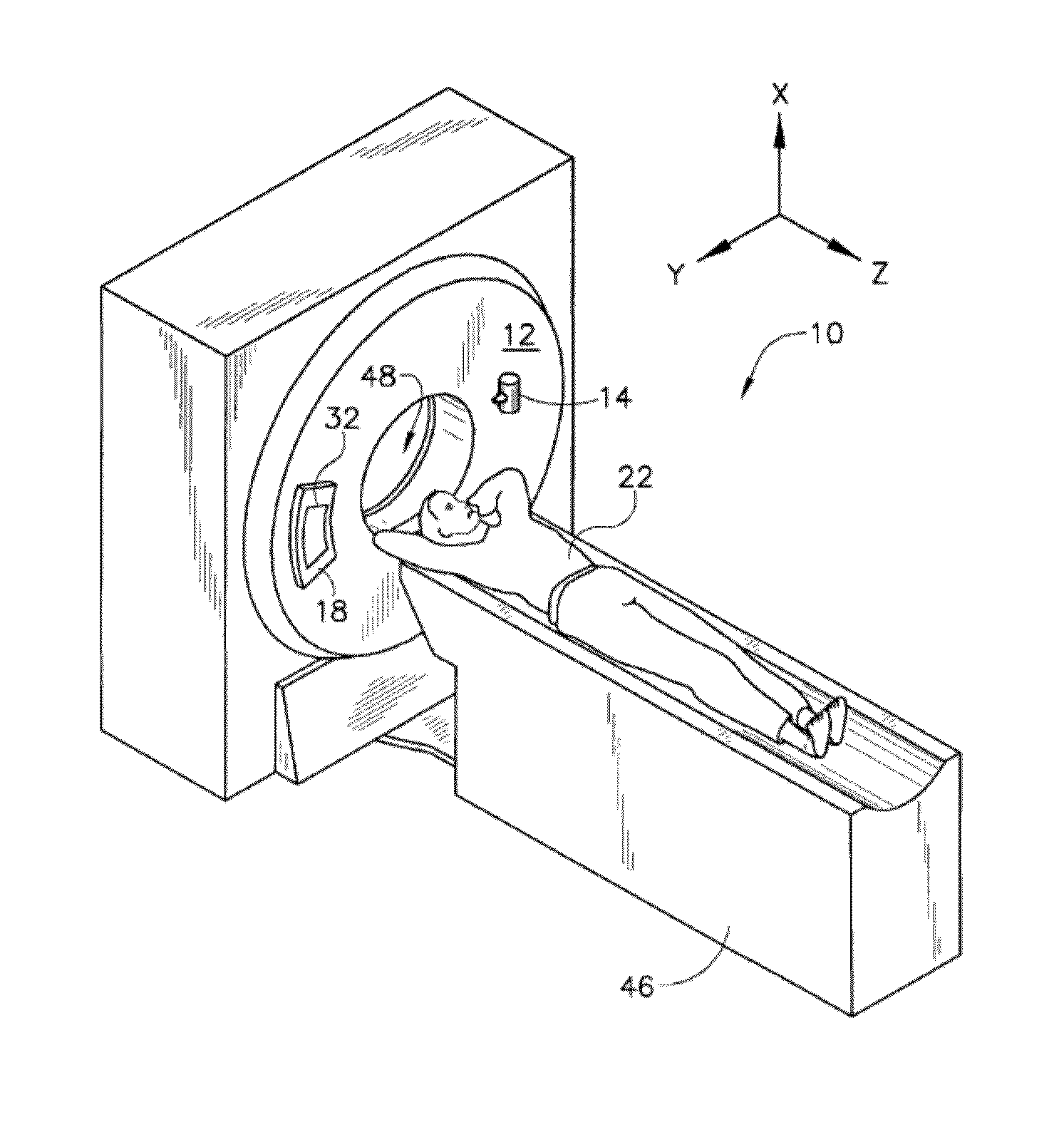
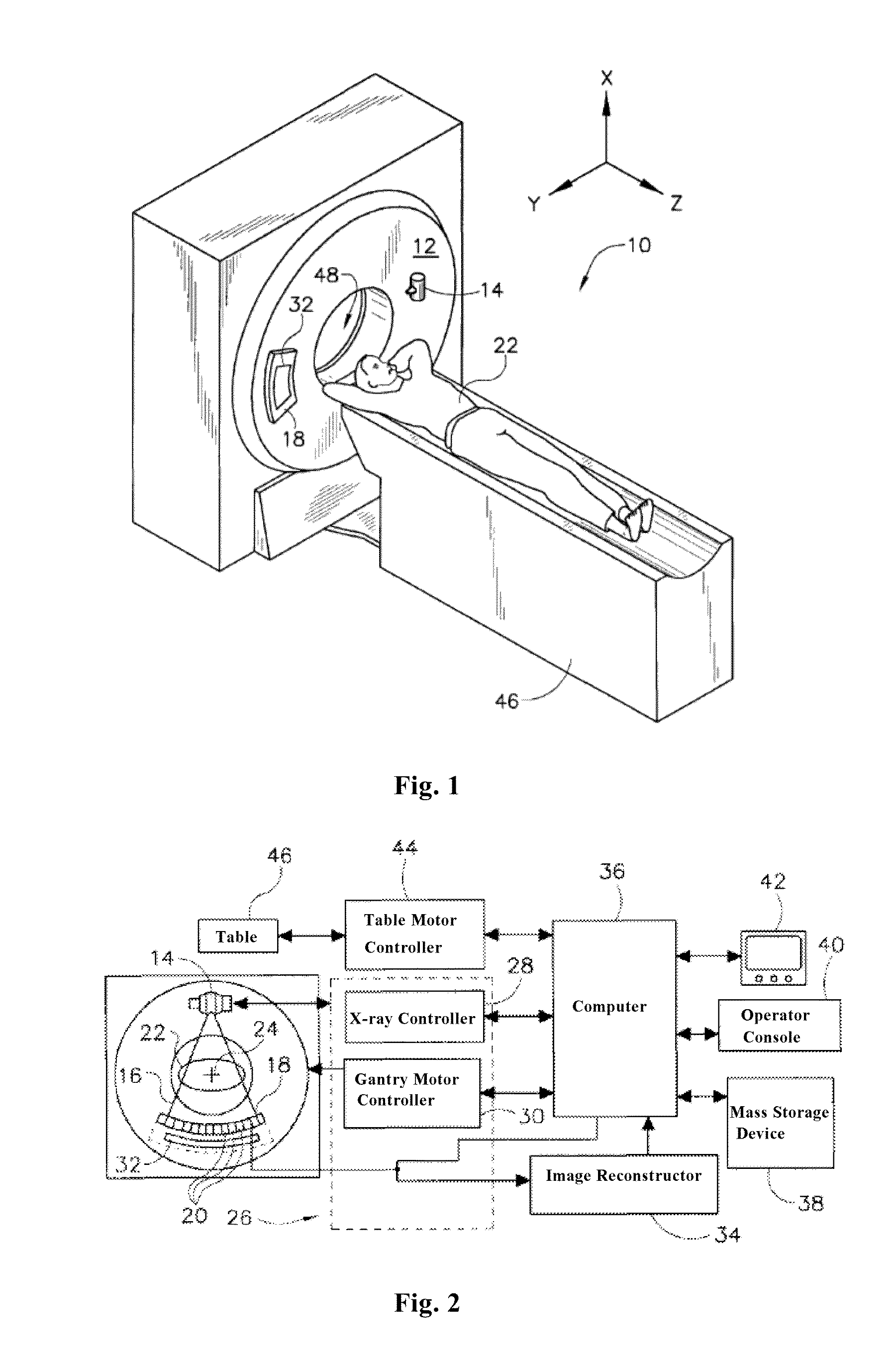
[0025]Referring to FIGS. 1 and 2, a CT imaging system 10 is shown as including a gantry 12. In an embodiment, the system 10 comprises a “third generation” CT scanner. The gantry 12 contains an X-ray source 14 that ...


PUM


Abstract

The present invention provides a method and apparatus for reducing artifacts in CT image reconstruction. The method comprises obtaining an original reconstructed image and an original sinogram and determining a proportion of metal pixels in the original reconstructed image. If the proportion is greater than a first threshold value, then generating an expanded metal template based on the original reconstructed image, generating a metal-free, metal artifact reduced (MAR) image based on the expanded metal template and the original sinogram, and generating a final image based on the expanded metal template and the metal-free, MAR image. However, if the proportion of metal pixels is less than a second threshold value, then generating an expanded metal template based on the original reconstructed image, generating a metal-free, metal artifact reduced image based on a treatment, and generating a final image based on the expanded metal template and the metal-free, MAR image.

Description

TECHNICAL FIELD[0001]The present invention relates generally to computed tomography (CT) and, more particularly, to a method and apparatus for reducing artifacts in CT reconstruction image.BACKGROUND[0002]Auxiliary diagnostic apparatus comprise magnetic resonance (MR) systems, ultrasonic systems, computed tomography (CT) systems, positron emission tomography (PET) systems, nuclear medicine and other types of imaging systems.[0003]For example, during CT imaging of a patient by a CT system, X-rays are used for imaging features of inner structures and regions of interest (ROI) of the patient body. The imaging is performed by a CT scanner. In operation, a photograph object is scanned for collection of raw data, and then an image is reconstructed based on the raw data.[0004]An object having high X-ray absorption characteristics such as metal materials and so on can cause artifacts in the reconstructed CT image and thus influence the results of diagnosis. These artifacts can be reduced us...

Claims


Application Information

Patent Timeline
IPC IPC(8): G06T11/00
CPCG06T11/003G06T2207/30004G06T2207/10072G06T2207/10004G06T11/008G06T11/006
Inventor DONG, SHUQINDONG, JIAQINLI, SHOU
Owner GE MEDICAL SYST GLOBAL TECH CO LLC
Why Eureka
Try Eureka