
Method and apparatus for trabeculectomy and suprachoroidal shunt surgery

a trabeculectomy and suprachoroidal shunt technology, applied in the field of trabeculectomy and suprachoroidal shunt surgery, can solve the problems of significant side effects, blindness if untreated, and patients may suffer substantial, irreversible vision loss,

Active Publication Date: 2015-04-23
PEREZ GROSSMANN RODOLFO ALFREDO
View PDF2 Cites 26 Cited by
  • Summary
  • Abstract
  • Description
  • Claims
  • Application Information

AI Technical Summary

Benefits of technology

The patent describes a bent surgical device with a cannula that has a notch through its wall, which can be used to create a channel in the eye for the purpose of directing aqueous humor from the anterior chamber to the suprachoroidal space. The device is solid material proximal to the bend and can be used in a method for performing a trabeculectomy with suprachoroidal derivation. The technical effects of this device include improved precision and control during surgery, as well as reduced risk of complications and improved outcomes for patients.

Problems solved by technology

Glaucoma causes pathological changes in the optic nerve, visible on the optic disk, and it causes corresponding visual field loss, resulting in blindness if untreated.
Patients may suffer substantial, irreversible vision loss prior to diagnosis and treatment.
However, these drug therapies for glaucoma are sometimes associated with significant side effects, such as headache, blurred vision, allergic reactions, death from cardiopulmonary complications, and potential interactions with other drugs.

Method used


Image

  • Method and apparatus for trabeculectomy and suprachoroidal shunt surgery
  • Method and apparatus for trabeculectomy and suprachoroidal shunt surgery
  • Method and apparatus for trabeculectomy and suprachoroidal shunt surgery

Examples


Embodiment Construction

[0022]As used herein, the term “proximal” refers to the handle-end of a device held by a user, and the term “distal” refers to the opposite end.

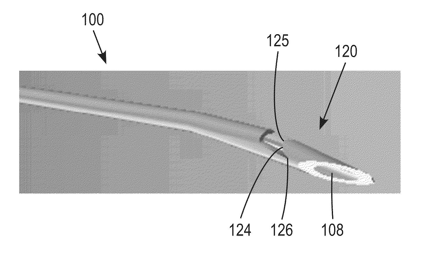
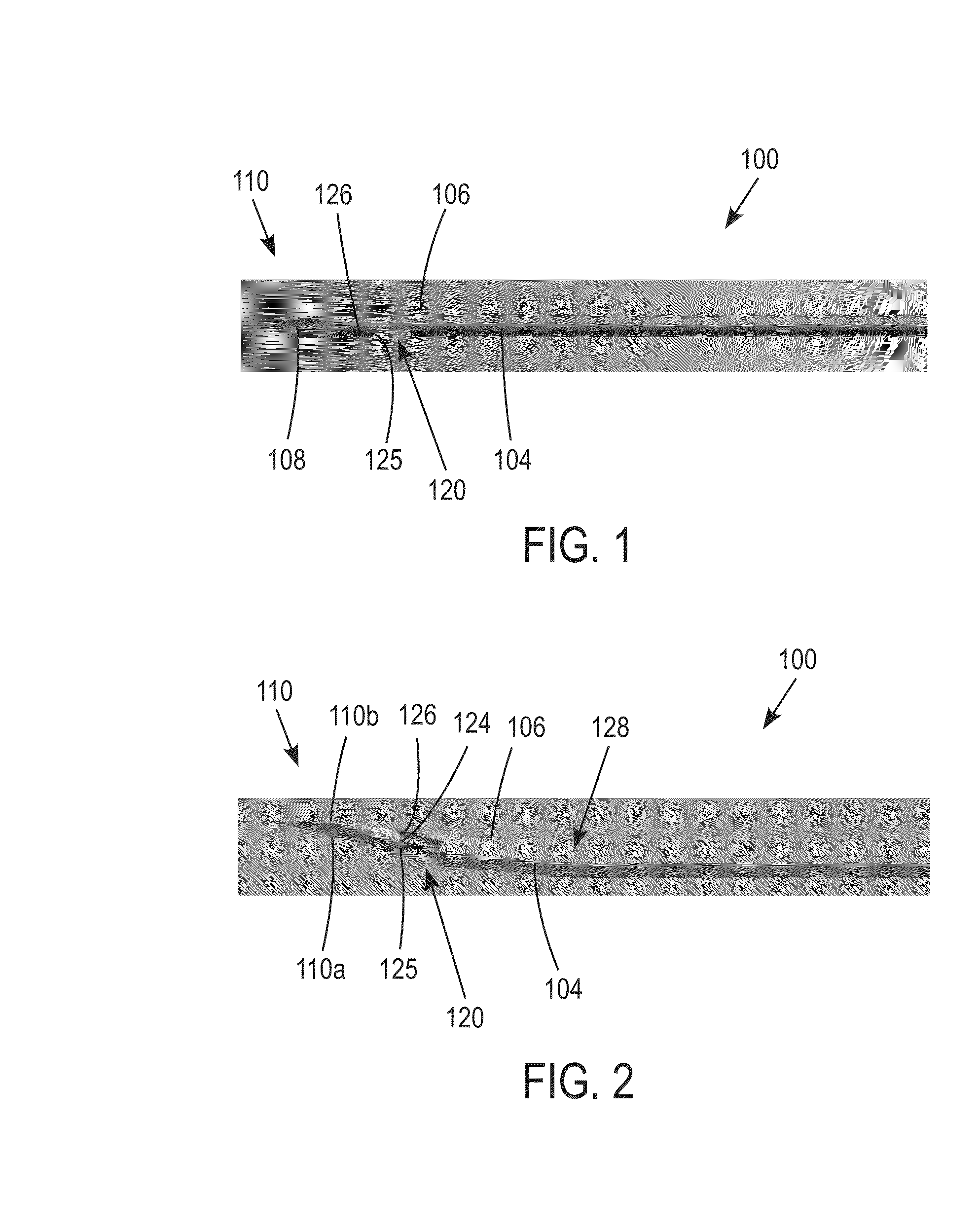
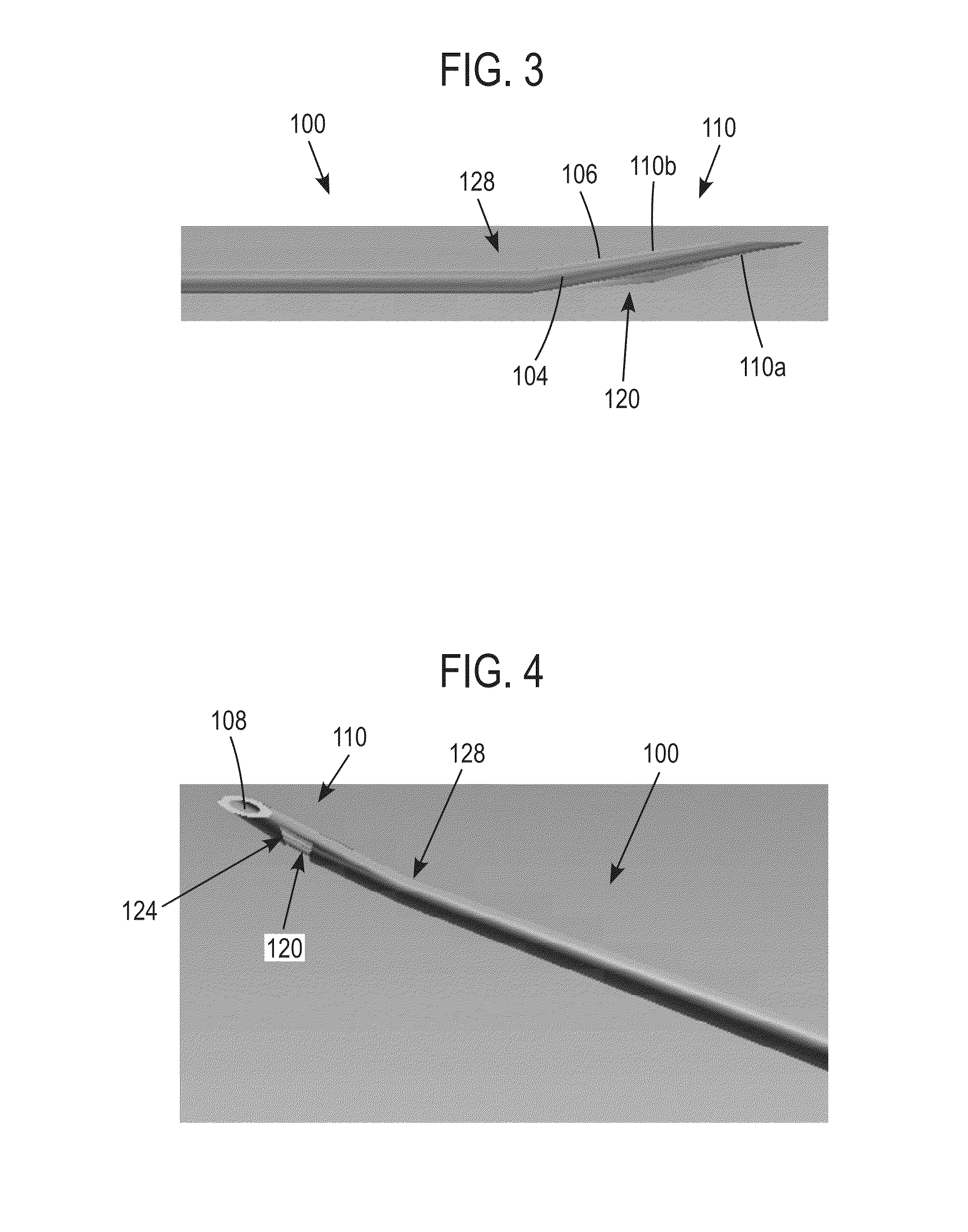
[0023]One embodiment of the surgical device is described with references to FIGS. 1-5. As shown in the top plan view of FIG. 1, the device 100 includes an elongate cannula 104 at the distal end. The cannula includes a cannula wall 106 that defines a cannula lumen 108. A distal end 110 of the cannula 104 is beveled, including a long side 110a substantially parallel with a section of the central longitudinal axis of the cannula 104 and extending to its distal-most tip end. A short side 110b of the beveled distal end is opposite the long end 110a. As shown in FIGS. 1-5, a notch 120 is disposed proximally adjacent to the distal beveled end 110 and is generally centered in longitudinal alignment at a point about half-way between the long beveled end side 110a and the short beveled end side 110b. In preferred embodiments, the notch 120 is defined ...


PUM


Abstract

A medical device and surgical procedure for the treatment of glaucoma in patients with primary open angle glaucoma, secondary open angle glaucoma, closed angle glaucoma, and refractory glaucoma. The device is inserted between two scleral flaps, cutting the trabecular meshwork and the sclera. When the device is removed, a proximal-facing cutting edge forms a tunnel allowing the aqueous humor to flow out of the anterior chamber into the suprachoroidal space.

Description

FIELD OF THE INVENTION[0001]This invention relates to a novel medical device and surgical procedure for the treatment of glaucoma in patients with primary and secondary open angle glaucoma, closed-angle glaucoma, and refractory glaucoma.BACKGROUND[0002]The present invention generally relates to medical devices and methods for reducing intraocular pressure in the eye of mammals. More particularly, the present invention relates to the treatment of glaucoma via the use of a bent surgical device to surgically create a channel thereby permitting aqueous humor to flow out of the anterior chamber into the suprachoroidal space.[0003]The human eye is a specialized sensory organ capable of light reception and able to receive visual images. The trabecular meshwork serves as a drainage channel and is located in anterior chamber angle formed between the iris and the cornea. The trabecular meshwork maintains a balanced pressure in the anterior chamber of the eye by draining aqueous humor from the...

Claims


Application Information

Patent Timeline
Patent Type & Authority Applications(United States)
IPC IPC(8): A61F9/00A61B17/3205
CPCA61B17/3205A61F9/00A61F9/00781A61B17/34
Inventor PEREZ GROSSMANN, RODOLFO ALFREDO
Owner PEREZ GROSSMANN RODOLFO ALFREDO
Learn More