Elastography Using Ultrasound Imaging of a Thin Volume

a thin volume, ultrasound technology, applied in the field of medical imaging, can solve the problems of computationally intensive techniques, complex computing hardware, and existing real-time ultrasound elastography systems

Active Publication Date: 2014-11-06
THE UNIV OF BRITISH COLUMBIA
View PDF9 Cites 35 Cited by
  • Summary
  • Abstract
  • Description
  • Claims
  • Application Information

AI Technical Summary

Benefits of technology

[0014]The vibration source can be an electromagnetic voice coil and be mounted on an adjustable arm. The arm can be mounted on an ultrasound machine, a patient bed, or a portable pole. Alternatively, the vibration source can be configured as a hand-held device. Also, the vibration source and the ultrasound probe can be integrated together into single device.

Problems solved by technology

However, existing real-time ultrasound elastography systems that provide imaging of tissue properties use techniques that are computationally intensive and which require sophisticated and expensive computing hardware, or which acquire images only in a 2D imaging plane.
Such 2D measurements introduce errors or diminish the ability to measure the absolute value of elasticity, and instead measure just relative variations throughout an image.
Therefore, existing ultrasound elastography systems using probes which only acquire data in a 2D imaging plane tend to produce inaccurate measurements.

Method used


Image

  • Elastography Using Ultrasound Imaging of a Thin Volume
  • Elastography Using Ultrasound Imaging of a Thin Volume
  • Elastography Using Ultrasound Imaging of a Thin Volume

Examples


Embodiment Construction

[0040]Directional terms such as “top”, “bottom”, “upwards”, “downwards”, “vertically” and “laterally” are used in the following description for the purpose of providing relative reference only, and are not intended to suggest any limitations on how any element is to be displayed during use or relative to an environment.

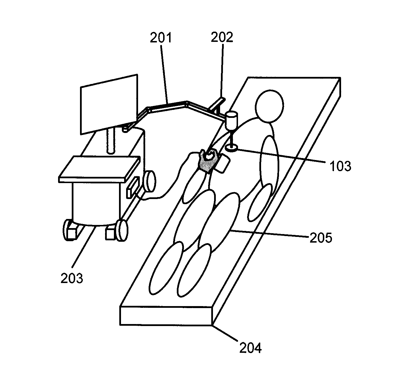
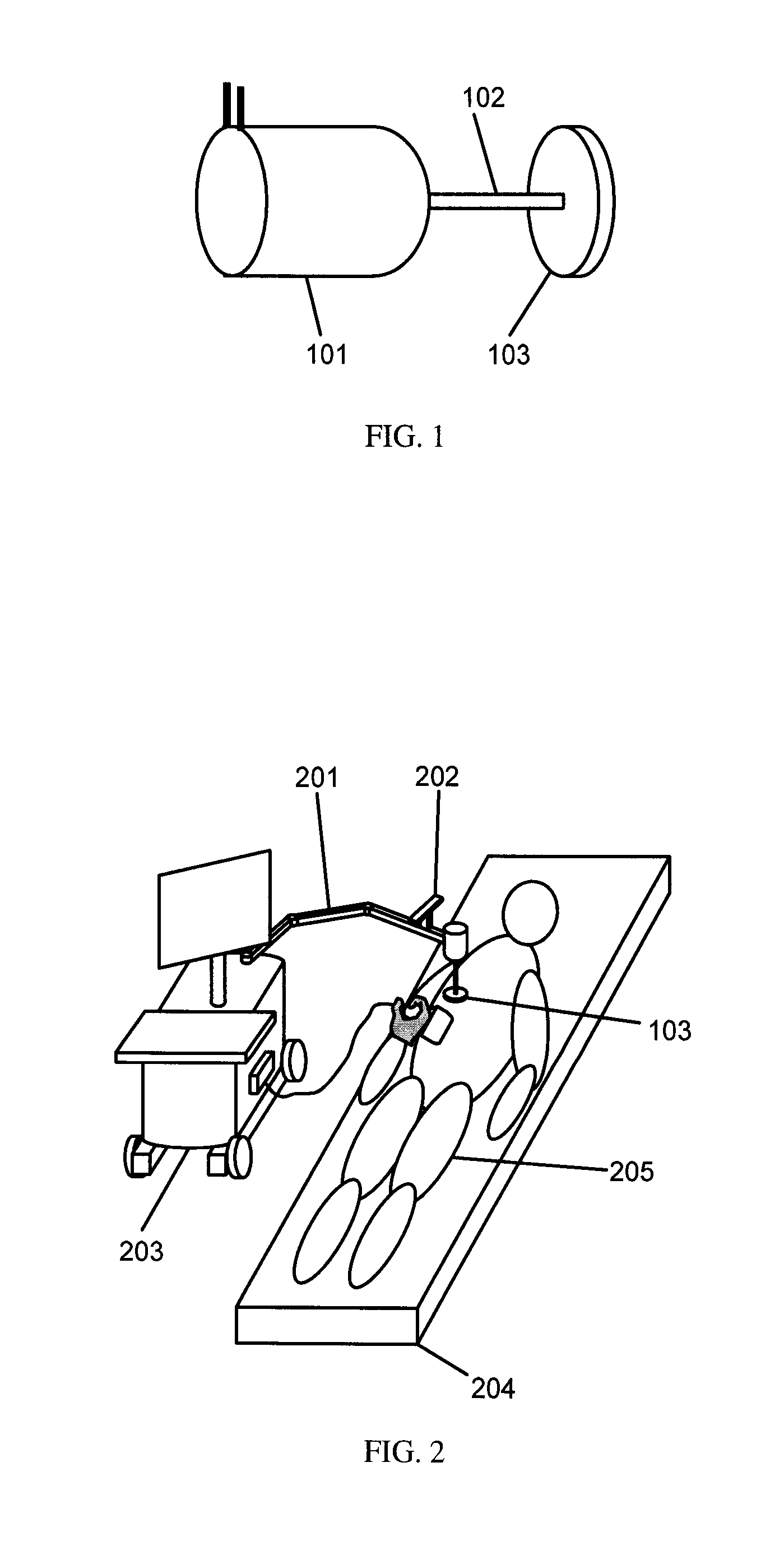
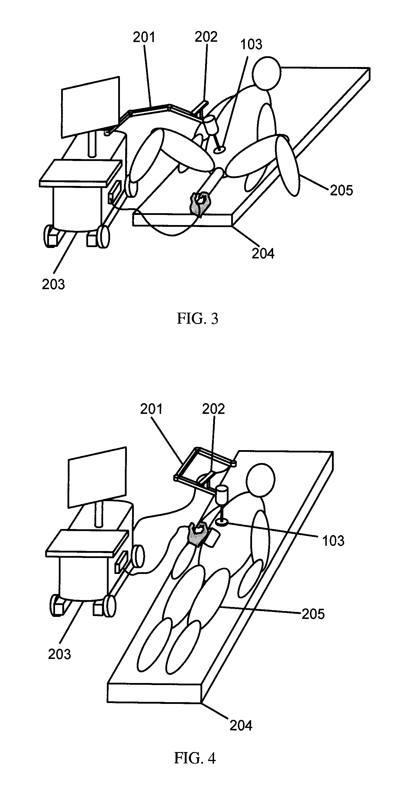
[0041]The embodiments described herein relate generally to an elastography method and system for obtaining ultrasound images of an excited tissue over a certain time period, then computationally determining one or more mechanical properties of the tissue within a real time refresh rate. This method can perform elastography in real time as only a thin volume of the excited tissue is imaged and processed. The thin volume includes a desired cross-sectional plane of the tissue and at least two planes that are adjacent to the desired cross-sectional plane. A maximum number of adjacent planes is selected so that a computer system is capable of computationally determining me...


PUM


Abstract

The embodiments described herein relate generally to an elastography method and system for obtaining ultrasound images of an excited tissue over a certain time period, then computationally determining one or more mechanical properties of the tissue within a real time refresh rate. This method can perform elastography in real time as only a thin volume of the excited tissue is imaged and processed. The thin volume includes a desired cross-sectional plane of the tissue and at least two adjacent planes that are adjacent to the desired cross-sectional plane. A maximum number of adjacent planes is selected so that a computer system is capable of computationally determining mechanical properties within a real time refresh rate.

Description

CROSS-REFERENCE TO RELATED APPLICATIONS[0001]This application claims priority to a U.S. provisional application having serial number 6,1525,378, filed on Aug. 19, 2011, the entirety of which is herein incorporated by reference.FIELD OF INVENTION[0002]This invention relates generally to medical imaging, and in particular to elastography using ultrasound imaging of a thin volume of a subject such as tissue to determine visco-elastic properties of the subject.BACKGROUND[0003]Elastography generally refers to methods of imaging mechanical properties of tissue such as elasticity, viscosity, relaxation time, shear modulus, porosity, etc. Elastography is generally carried out in four steps:[0004]1. exciting the tissue by causing some form of deformation or movement in the tissue;[0005]2. observing and recording with a medical imaging device a series of images depicting the motion of different locations in the tissue over an interval of time;[0006]3. estimating the displacements of the tissu...

Claims


Application Information

Patent Timeline
Patent Type & Authority Applications(United States)
IPC IPC(8): A61B8/08A61B8/00
CPCA61B8/485A61B8/5223A61B8/463A61B8/483A61B8/0825A61B8/12
Inventor BAGHANI, ALIESKANDARI, HANIROHLING, ROBERT N.SALCUDEAN, SEPTIMU E.
Owner THE UNIV OF BRITISH COLUMBIA
Why Eureka
Try Eureka