
Method and ekg trigger device for correcting an ekg signal in magnetic resonance image acquisition

Inactive Publication Date: 2014-10-30
SIEMENS HEALTHCARE GMBH
View PDF8 Cites 4 Cited by
  • Summary
  • Abstract
  • Description
  • Claims
  • Application Information

AI Technical Summary

Benefits of technology

The patent text describes a method for improving the detection of an R-spike, which is a critical trigger for a magnetic resonance device. This method uses a cine-imaging technique that eliminates interference signals caused by gradient jumps in the EKG signal, which requires no additional hardware or communication. The method does not require pre-calibration and can use the parameters that are always present in the sequence, such as the repetition time, to acquire a reference signal. The method also uses a specific time window, called the "dead time," to start the magnetic resonance sequence only after the R-spike is detected, which further increases the reliability of the method. Overall, this method provides a more robust and accurate triggering mechanism for magnetic resonance imaging.

Problems solved by technology

However, within the scope of magnetic resonance measurement problems occur in the measurement of the EKG signal because two sources exist that generate unwanted interference signals.
In some sequences, interference signals due to gradient jumps can be strong enough to overlap the actual EKG signal, such that the detection algorithm fails.
A first approach proposes to use alternative trigger mechanisms in addition to EKG triggering, for example optical pulse triggering or acoustic triggering which, however, disadvantageously require additional hardware.
In addition, further disadvantages exist in comparison to EKG measurement, in particular a method-related, non-negligible delay from the R-spike peak to the generated trigger signal, which makes this method unsuitable for high-quality cine-imaging in which the triggering should occur before the beginning of the heart contraction.
In spite of all this research work, methods are known in which the success rate of the method is approximately 95%, for example, which means that—according to this example—a triggering error can currently occur in 5% of patients, which can lead to approximately one failure per day in a cardiac center, for example.
In this method, however, a considerable delay also occurs from the peak of the R-spike to the point in time of the trigger signal, such that it is unsuitable for cine-imaging.
A high cost, and high demands on the electronics implementing the method, consequently are associated with this method.

Method used


Image

  • Method and ekg trigger device for correcting an ekg signal in magnetic resonance image acquisition
  • Method and ekg trigger device for correcting an ekg signal in magnetic resonance image acquisition
  • Method and ekg trigger device for correcting an ekg signal in magnetic resonance image acquisition

Examples


Embodiment Construction

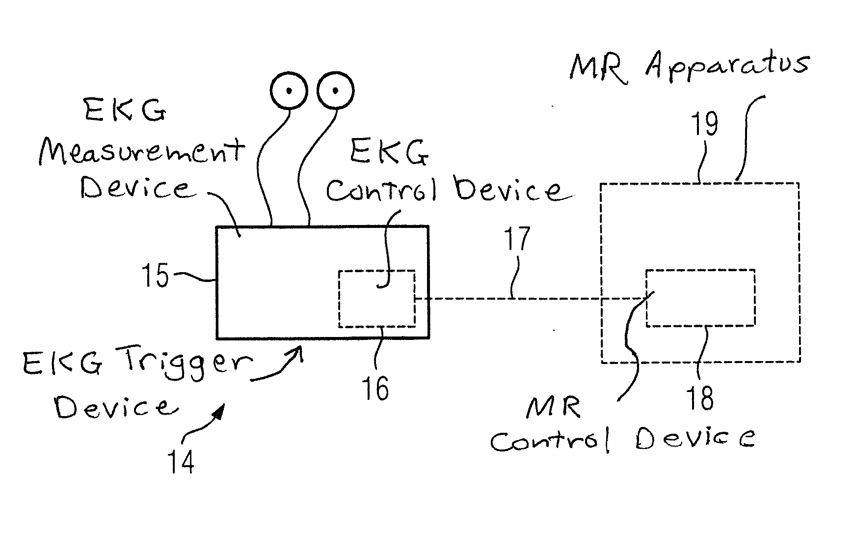
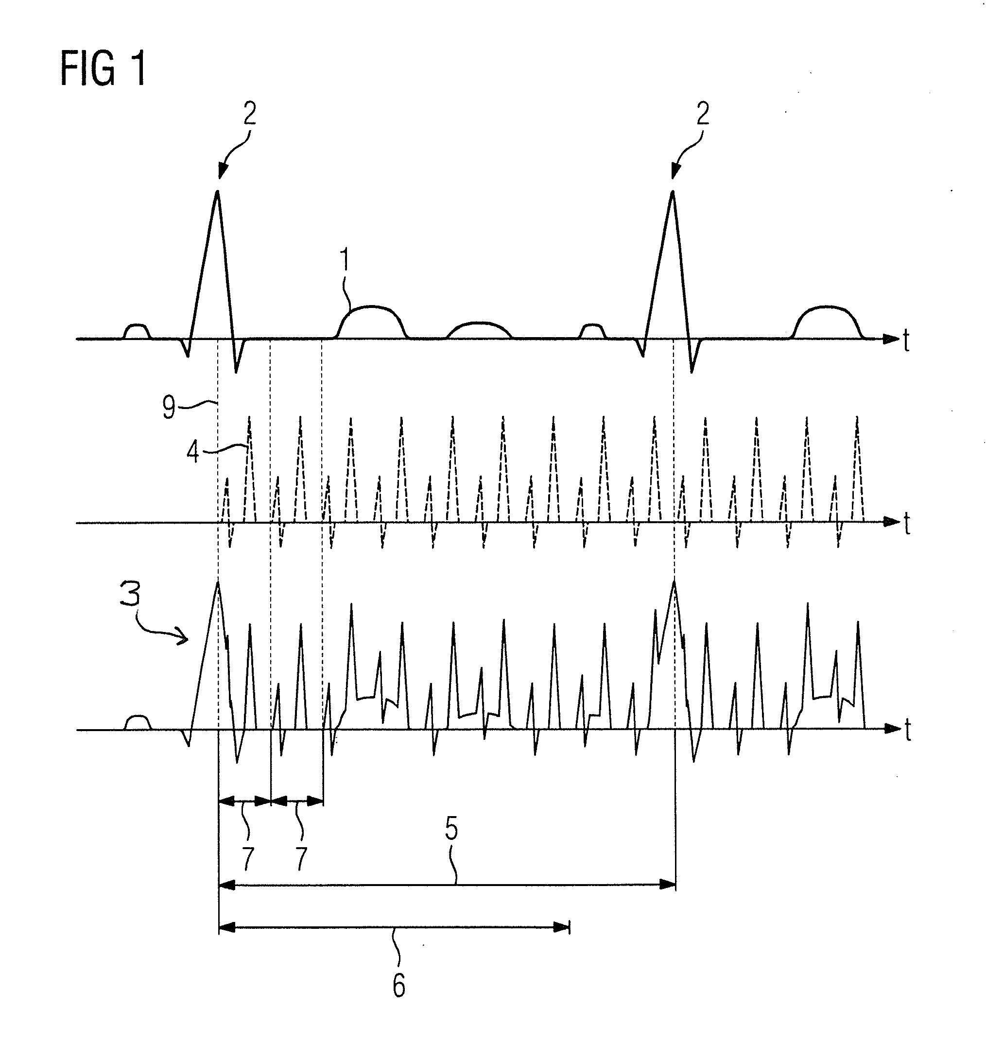
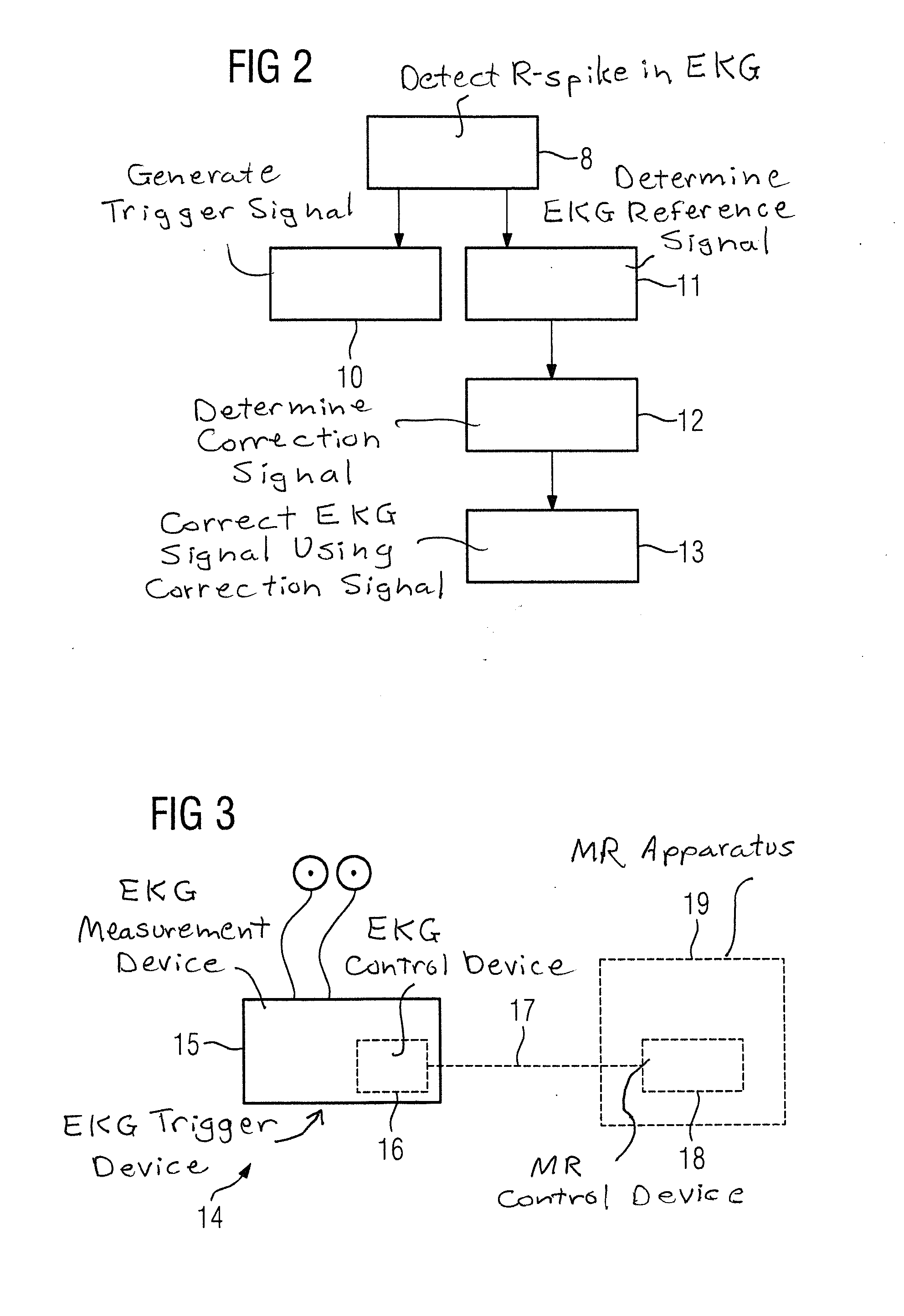
[0034]FIG. 1 shows in detail the problem forming the basis of the invention, and the requirements for its solution via the method according to FIG. 2. In the upper part of FIG. 1, an undistorted EKG signal 1 is schematically depicted over time. The different spikes occurring in the pure EKG signal 1 are known in principle, wherein the most prominent spike (and consequently the spike used for triggering) is the R-spike 2. The R-spike 2 in an actual acquired EKG signal 3 should be detected via a detection algorithm. Whenever the R-spike 2 is detected, a trigger signal is generated, which can be stored and evaluated with acquired magnetic resonance data and / or can equally be used to control the image acquisition operation, for example in order to switch from a “dry run” of the magnetic resonance sequence into an actual acquisition mode in which echoes (MR signals) are acquired. All of this is known in principle to those skilled in the field of MR imaging.

[0035]The detection of the R-sp...


PUM


Abstract

In a method to correct an EKG signal, the EKG signal is acquired, and used for R-spike triggering, during a magnetic resonance (MR) image acquisition sequence that produces interference signals in the EKG signal generated by gradient jumps, wherein the gradient jumps repeat at a fixed time interval, and wherein the duration of a cardiac cycle measured via the EKG signal is at least five times the time interval. During at least a first cardiac cycle, immediately after detection of the R-spike in the EKG signal no detection of the R-spike for triggering takes place for a dead time that is shorter than the duration of the cardiac cycle and during which the MR sequence is already running, and the EKG signal is acquired in that dead time as a reference signal. The reference signal is analyzed to extract interference signals that respectively repeat after the time interval, which are used to determine a correction signal having a duration equal to the time interval. During the further execution of the MR sequence, the correction signal, which is synchronously repeated in the time interval, at least outside of the dead time, is used to correct the measured EKG signal.

Description

BACKGROUND OF THE INVENTION[0001]1. Field of the Invention[0002]The present invention concerns: a method for correcting an EKG signal, acquired during an image acquisition with a magnetic resonance (MR) device according to a magnetic resonance sequence, for interference signals generated by magnetic gradient field jumps that occur during the magnetic resonance sequence, as well as an EKG trigger device for a magnetic resonance measurement.[0003]2. Description of the Prior Art[0004]Magnetic resonance imaging is an established imaging modality that is used especially often in medicine. In many applications, it is the case that the image acquisition must be synchronized with physiological processes, either in order to achieve an optimal image quality or to allow an evaluation of the acquired magnetic resonance data. With regard to cardiac imaging, it is necessary to synchronize the image acquisition with the beating heart. For this purpose, it is typical to measure the EKG signal of th...

Claims


Application Information

Patent Timeline
IPC IPC(8): A61B5/055A61B5/00A61B5/352
CPCA61B5/7289A61B5/055A61B5/7203A61B5/7285A61B5/352
Inventor SPEIER, PETER
Owner SIEMENS HEALTHCARE GMBH
Learn More