
Imaging agent for detection of diseased cells

a technology for detecting cells and imaging agents, applied in the direction of diagnostic recording/measuring, diagnostic recording/measuring, ultrasonic/sonic/infrasonic diagnostics, etc., can solve the problems of local tumor recurrence, reduced survival rate, increased likelihood of metastases,

Active Publication Date: 2014-10-09
LUMICELL
View PDF4 Cites 29 Cited by
  • Summary
  • Abstract
  • Description
  • Claims
  • Application Information

AI Technical Summary

Benefits of technology

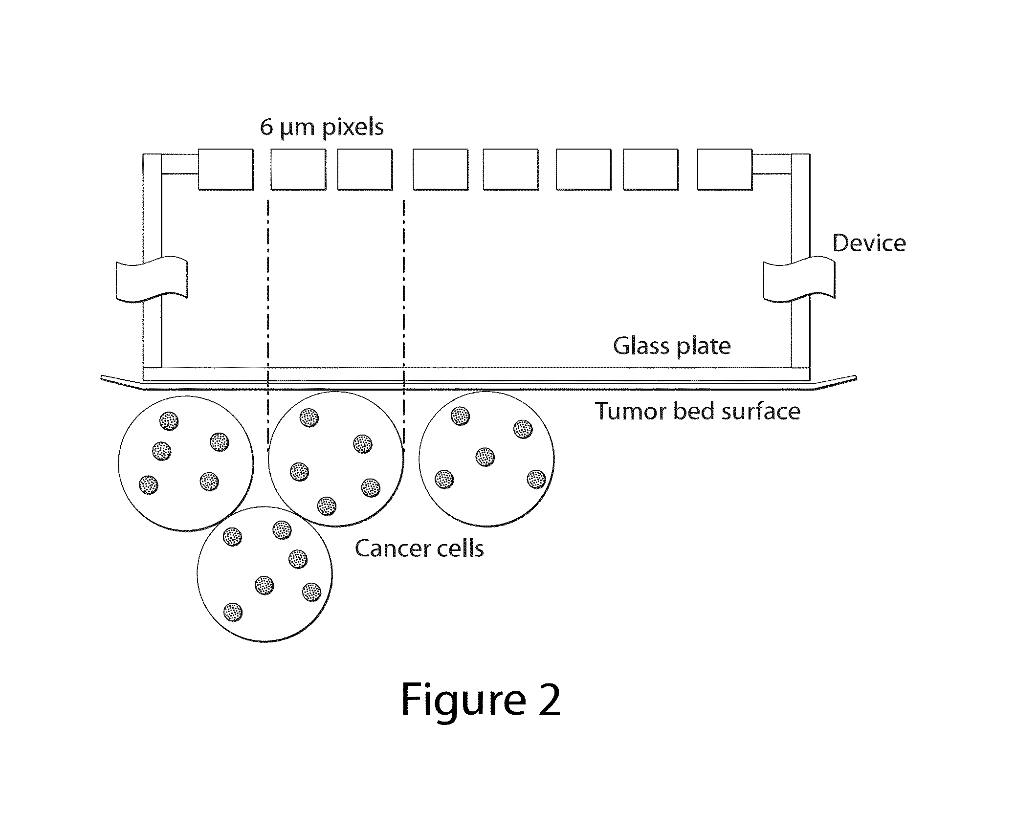
The present invention provides compounds of formula I, pharmaceutically acceptable salts thereof, and compositions thereof, which are useful as imaging agents. These compounds have desirable characteristics for the same and can be used to spatially determine tissue heterogeneity of a subject by administering a composition to the subject and obtaining an in-situ image of the tissue. The compounds can distinguish between disease cells and healthy cells within 5 mm, 4 mm, 3 mm, 2 mm, or even 1 mm from the surface of the tissue. The invention also provides a method to image the exposed tissue bed left behind after tumor resection.

Problems solved by technology

Residual cancer in the tumor bed is a leading risk factor for local tumor recurrence, reduced survival rates and increased likelihood of metastases.
This poor performance of pathology is primarily due to a sampling error since only a small fraction of the entire tumor is analyzed.
However this pathology exercise is a time intensive procedure and often takes days for final results to be sent to the physician.
First, the pathology process relies on sampling a given tumor at certain spatial intervals which may result in missing a critical portion of the tumor and is therefore not a very reliable source of information.
In addition, the process disrupts the surgical workflow since the physician and the patient have to wait for the pathology report to return prior to finishing the surgery or return for a second surgery should the pathology process exceed the time window for the first surgery.

Method used


Image

  • Imaging agent for detection of diseased cells
  • Imaging agent for detection of diseased cells
  • Imaging agent for detection of diseased cells

Examples


examples

Imaging Agent

[0061]Composition A comprises compounds of formula I, which comprise a PEGylated heptapeptide covalently linked to both a fluorophore (Cy5) and a dark quencher (QSY21). The central heptapeptide provides both cathepsin substrate specificity and proper spacing of the fluorophore and quencher. The QSY21 and Cy5 functionalities are covalently attached to the α-amino terminus of the peptide and to the ε-amino group of lysine, respectively. An amino-ethoxyethoxyacetyl group is incorporated into the amide backbone and the carboxy terminus of the heptapeptide is further modified by attachment of a 20 kDa methoxypolyethylene glycol (mPEG) molecule to the side chain of cysteine.

[0062]In certain embodiments, composition A is supplied lyophilized, in bulk powder form as an acetate salt. The 20 kDa methoxy-polyethylene glycol moiety has a variable molecule weight of 10%. One of ordinary skill in the art will understand that composition A comprises multiple compounds that vary in the...


PUM

PropertyMeasurementUnit
Lengthaaaaaaaaaa
Lengthaaaaaaaaaa
Lengthaaaaaaaaaa

Abstract

The present invention provides compositions and methods for imaging, for example, tumor resections.

Description

RELATED APPLICATIONS[0001]This application claims priority under 35 U.S.C. §119(e) to U.S. Provisional Application Ser. No. 61 / 781,601, entitled “IMAGING AGENT FOR DETECTION OF DISEASED CELLS” filed on Mar. 14, 2013 and U.S. Provisional Application Ser. No. 61 / 785,136, entitled “IMAGING AGENT FOR DETECTION OF DISEASED CELLS” filed on Mar. 14, 2013, which are herein incorporated by reference in their entirety.BACKGROUND OF THE INVENTION[0002]There are over one million cancer surgeries per year performed in the United States and nearly 40% of them miss resecting the entire tumor according to the National Cancer Institute Surveillance Epidemiology and End Results report. For example in breast cancer lumpectomies, failure to remove all the cancer cells during the primary surgery (positive margins) occurs approximately 50% of the time and requires second surgeries. Residual cancer in the tumor bed is a leading risk factor for local tumor recurrence, reduced survival rates and increased l...

Claims


Application Information

Patent Timeline
IPC IPC(8): A61K49/00
CPCA61K49/0032A61K49/0056A61B2560/0431A61B5/4312A61B2505/05A61B90/361A61B5/6886A61B2090/309A61B2090/306A61B2090/3616A61B2090/3941A61B5/0091A61B10/0041A61B5/0071A61B5/0075A61B5/0086
Inventor LEE, W. DAVIDBAWENDI, MOUNGI G.FERRER, JORGE
Owner LUMICELL
Learn More