
Method and apparatus for medical image display, and user interface screen generating method

a technology of image display and user interface, applied in the field of medical image display, can solve the problem of inability to accurately identify the timing of strain between segments of a heart wall, and achieve the effect of fast and accurate check-up

Active Publication Date: 2014-04-17
SAMSUNG MEDISON
View PDF7 Cites 2 Cited by
  • Summary
  • Abstract
  • Description
  • Claims
  • Application Information

AI Technical Summary

Benefits of technology

The patent provides a way to display medical images that can help doctors quickly and accurately analyze the movement of segments of a heart wall to diagnose heart disease. This method is particularly useful for checking whether different parts of the heart move simultaneously. The patent also includes a method for generating a user interface screen for this purpose.

Problems solved by technology

However, when the degree of strain of each segment of a heart wall is indicated by colors on a 2D image as in the conventional technology, strain timing between segments of a heart wall may not be accurately identified.
Accordingly, in addition to the 2D image corresponding to a heart, separate medical examination material needs to also be checked, which is inconvenient.

Method used


Image

  • Method and apparatus for medical image display, and user interface screen generating method
  • Method and apparatus for medical image display, and user interface screen generating method
  • Method and apparatus for medical image display, and user interface screen generating method

Examples


Embodiment Construction

[0026]The terms used in the present specification are for explaining a specific exemplary embodiment of the disclosure, and are not limiting of the present inventive concepts. Thus, the expression of singularity in the present specification includes the expression of plurality unless clearly specified otherwise in context. Unless defined otherwise, all terms used herein including technical or scientific terms have the same meanings as those generally understood by those having ordinary skill in the art to which the present inventive concepts may pertain. Terms defined in generally used dictionaries are construed to have meanings matching that in the context of related technology and, unless clearly defined otherwise, are not construed to be ideally or excessively formal.

[0027]When a part may “include” a certain constituent element, unless specified otherwise, it may not be construed to exclude other constituent elements but may be construed to further include other constituent eleme...


PUM


Abstract

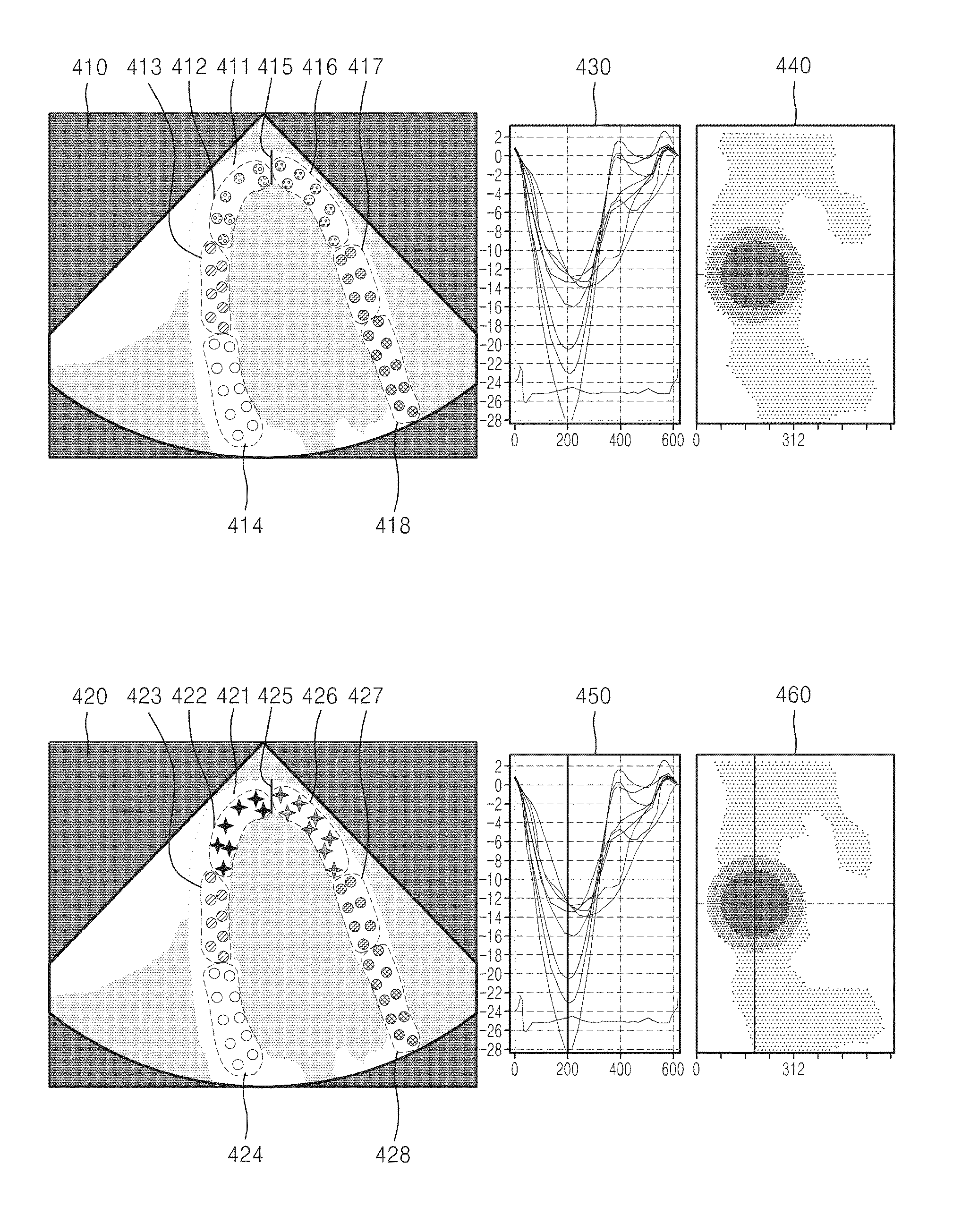
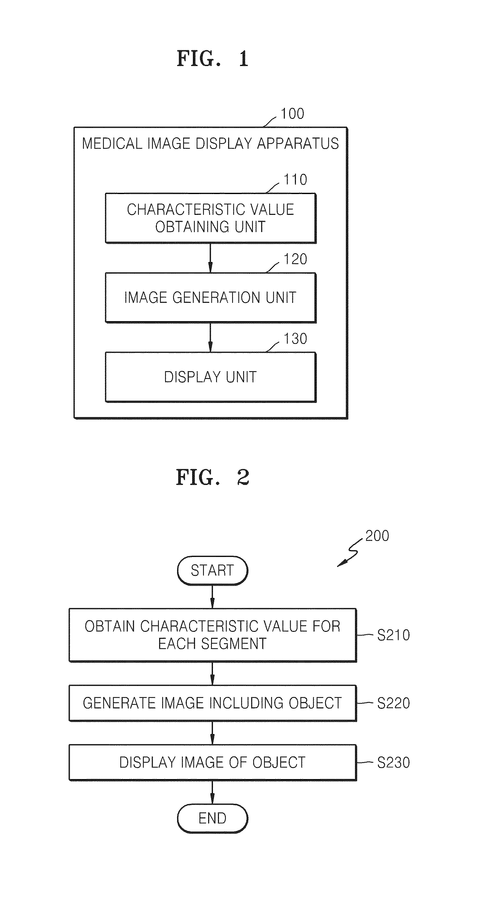
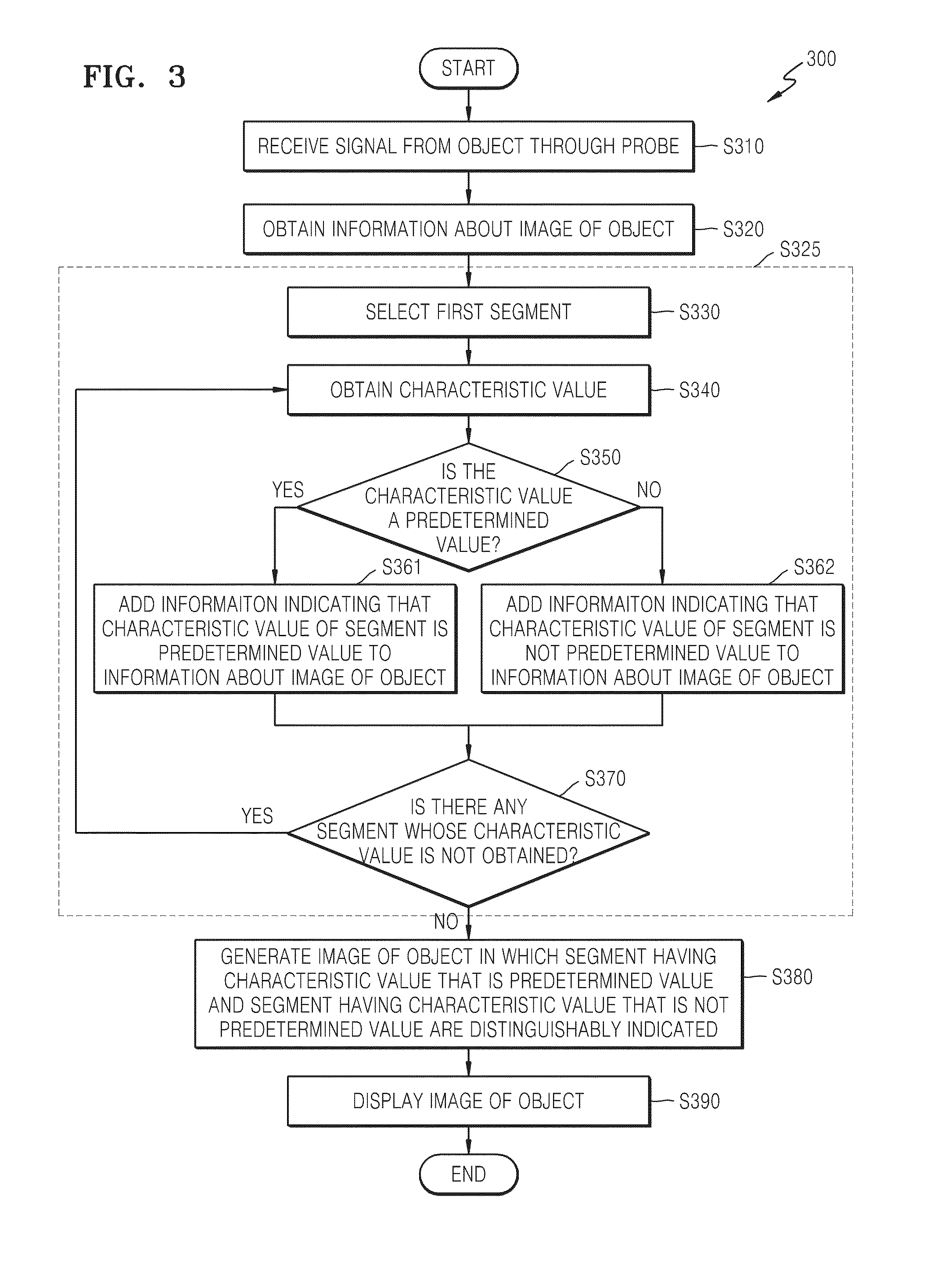
A cardiac ultrasound image display to analyze a heart wall motion, and a method and apparatus for displaying a cardiac ultrasound image. A method of displaying a medical image includes obtaining a characteristic value for each of one or more segments included in an object, generating an image of the object indicating the segment of which the characteristic value is a predetermined value, and displaying the image.

Description

CROSS-REFERENCE TO RELATED PATENT APPLICATION[0001]This application claims the benefit of Korean Patent Application No. 10-2012-0113036, filed on Oct. 11, 2012, in the Korean Intellectual Property Office, the disclosure of which is incorporated herein in its entirety by reference.BACKGROUND OF THE DISCLOSURE[0002]1. Field of the Disclosure The present disclosure relates to a method and apparatus for a medical image display, and more particularly, to a cardiac ultrasound image display to analyze a heart wall motion, and a method and apparatus for displaying a cardiac ultrasound image.[0003]2. Description of the Related Art[0004]In the ultrasound image display field, displaying a tissue deformation characteristic such as strain and a strain rate of a local portion of a target object provides a direct and quantitative measurement standard about the ability of a muscle that strains and relaxes.[0005]In particular, displaying a real-time strain rate of a muscle as an image may be applied...

Claims


Application Information

Patent Timeline
IPC IPC(8): G06T11/20
CPCA61B8/5215A61B2576/023A61B8/0883A61B8/463A61B8/485G06T7/0012G06T2207/30048G06T11/206G06T11/20G16H50/20A61B8/02G06T7/20
Inventor SONG, JOO-HYUNKIM, JONG-SIKLEE, JIN-YONGJO, JAE-MOON
Owner SAMSUNG MEDISON
Learn More