
Apparatus, system and method for providing an imaging device for medical applications

an imaging device and medical technology, applied in the field of imaging devices, can solve the problems of unsafe use and expired imaging devices

Inactive Publication Date: 2014-01-09
OLIVE MEDICAL CORP
View PDF10 Cites 16 Cited by
  • Summary
  • Abstract
  • Description
  • Claims
  • Application Information

AI Technical Summary

Benefits of technology

This patent describes an imaging device that is fully covered in a plastic material with a heat sink to ensure it meets industry standards. The device can be stamped with the current time and will not function if powered off for a set amount of time. The device also displays a message if it has been used more than once per sterilization cycle to protect patients and doctors from unsafe or invalid use. Additionally, the text describes a connection system that ensures the device is secure and can communicate with other devices without causing any damage or malfunction.

Problems solved by technology

The imaging device may display an onscreen message telling the user that the camera has already been used and will not allow current operation.
If the imaging device is older than the required date, an onscreen warning will tell the user that the imaging device has expired and is unsafe for use.

Method used


Image

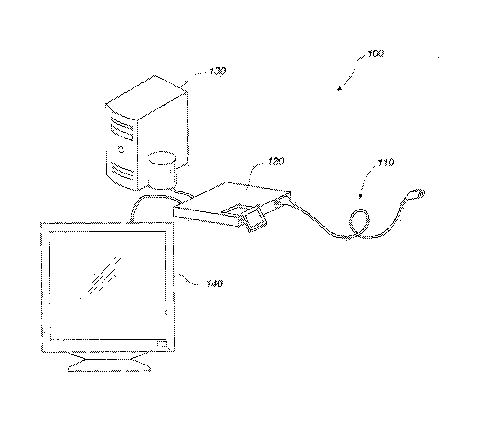
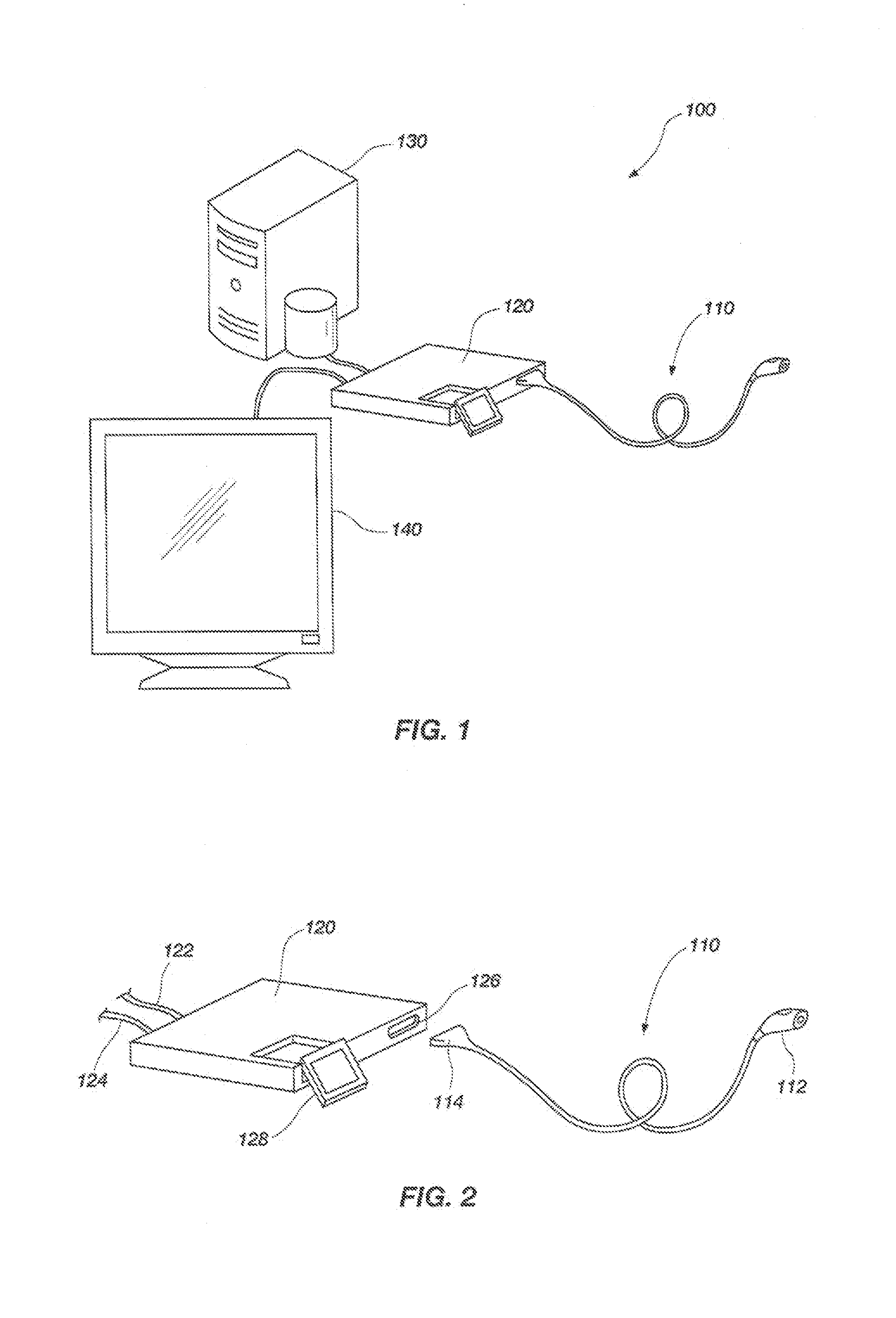
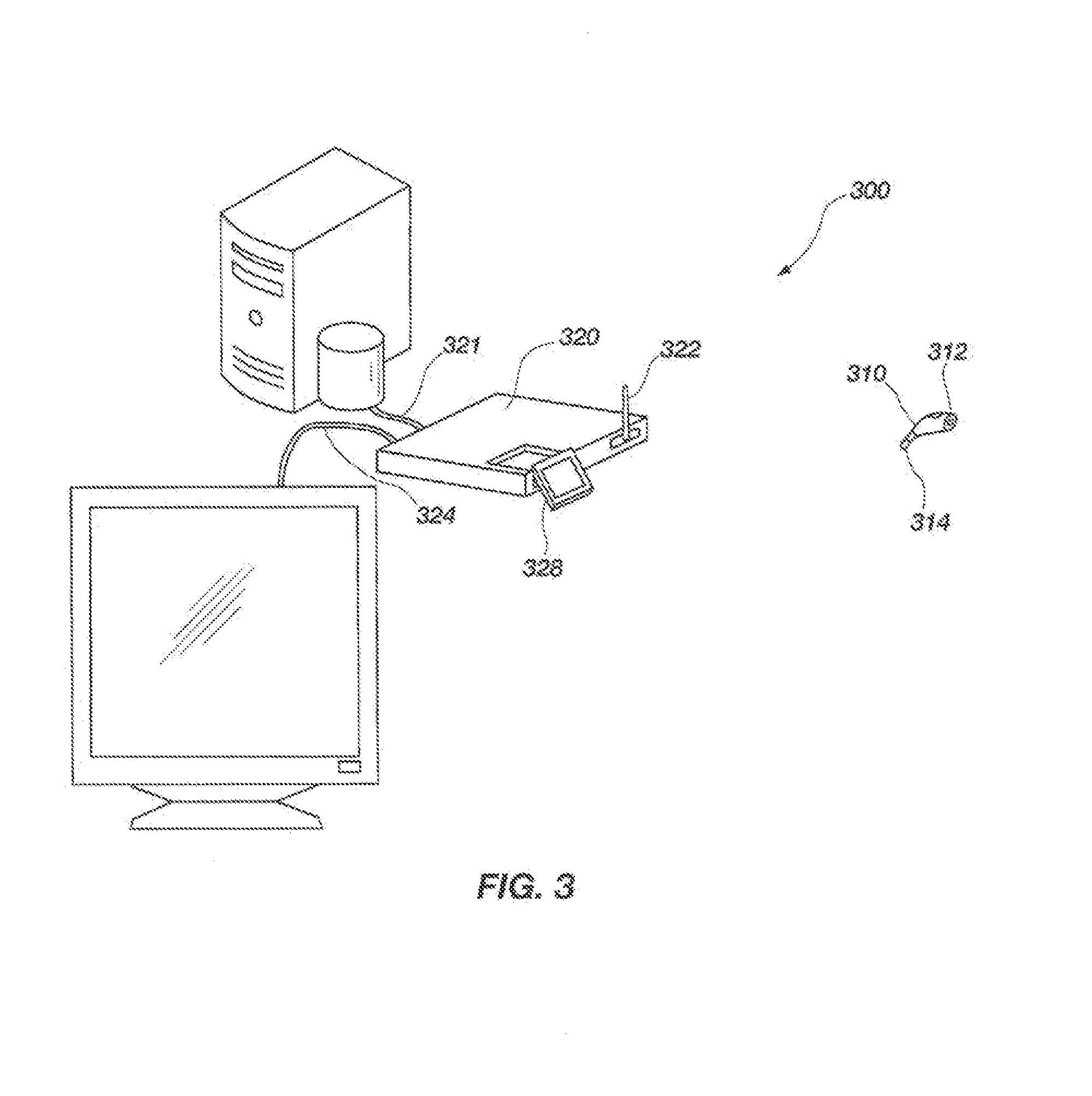
  • Apparatus, system and method for providing an imaging device for medical applications
  • Apparatus, system and method for providing an imaging device for medical applications
  • Apparatus, system and method for providing an imaging device for medical applications

Examples


Embodiment Construction

[0046]For the purposes of promoting an understanding of the principles in accordance with the disclosure, reference will now be made to the embodiments illustrated in the drawings and specific language will be used to describe the same. It will nevertheless be understood that no limitation of the scope of the disclosure is thereby intended. Any alterations and further modifications of the inventive features illustrated herein, and any additional applications of the principles of the disclosure as illustrated herein, which would normally occur to one skilled in the relevant art and having possession of this disclosure, are to be considered within the scope of the disclosure claimed.

[0047]Before the devices, systems, methods and processes for providing and reclaiming single use imaging devices are disclosed and described, it is to be understood that this disclosure is not limited to the particular embodiments, configurations, or process steps disclosed herein as such embodiments, conf...


PUM


Abstract

An apparatus, system and methods for providing and reclaiming a single use imaging device for sterile environments is disclosed and described. The system may include a single use high definition camera used for general purpose surgical procedures including, but not limited to: arthroscopic, laparoscopic, gynecologic, and urologic procedures, may comprise an imaging device that is a sterile and designed to ensure single use. The imaging device may have a single imaging sensor, either CCD or CMOS, encased in a housing.

Description

CROSS-REFERENCE TO RELATED APPLICATIONS[0001]This application claims the benefit of U.S. Provisional Application No. 61 / 580,138, filed Dec. 23, 2011, which is hereby incorporated by reference herein in its entirety, including but not limited to those portions that specifically appear hereinafter, the incorporation by reference being made with the following exception: In the event that any portion of the above-referenced provisional application is inconsistent with this application, this application supercedes said above-referenced provisional application.STATEMENT REGARDING FEDERALLY SPONSORED RESEARCH OR DEVELOPMENT[0002]Not Applicable.BACKGROUND[0003]The disclosure relates generally to imaging devices used during surgical procedures to visualize a surgical area, and more particularly, but not necessarily entirely, to an imaging device for use, and communicating, with a control unit and a system, method and process of communicating between an imaging device and a control unit.[0004...

Claims


Application Information

Patent Timeline
Patent Type & Authority Applications(United States)
IPC IPC(8): A61B5/00
CPCA61B5/0077A61B1/0002A61B1/00059A61B1/00103A61B1/00124A61B1/042A61B2560/028A61B2562/08
Inventor TALBERT, JOSHUA D.HENLEY, JEREMIAH D.WICHERN, DONALD M.WICHERN, CURTIS L.
Owner OLIVE MEDICAL CORP
Learn More