
Positron emission tomogrpahy detector for dual-modality imaging

a dual-modality imaging and detector technology, applied in the field of imaging systems, can solve the problems of reducing the imaging effectiveness of either the mri imaging system or the pet imaging system, generating heat that may affect and affecting the operation of the photodiod

Inactive Publication Date: 2013-10-31
GENERAL ELECTRIC CO
View PDF4 Cites 33 Cited by
  • Summary
  • Abstract
  • Description
  • Claims
  • Application Information

AI Technical Summary

Benefits of technology

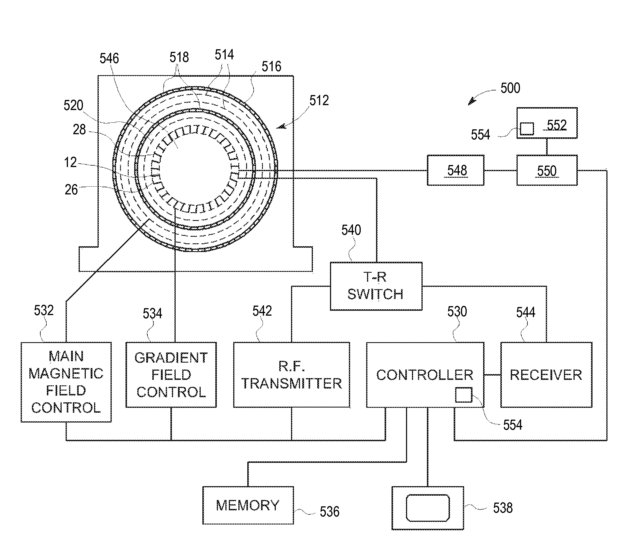
The patent text describes a positron emission tomography (PET) detector assembly including a cold plate and a readout electronics section. Additionally, an RF body coil assembly and a dual-modality imaging system are also described. The technical effects of the PET detector assembly include improved image quality and reduced noise, while the dual-modality imaging system provides better spatial resolution and improved detection of small lesions. These improvements can lead to improved accuracy and reliability in medical diagnosis and treatment.

Problems solved by technology

In operation, the readout electronics generate heat that may affect the operation of the photodiodes.
However, conventional cooling systems may create an adverse interaction with the gradient magnetic fields generated by the MRI system.
As a result, the addition of the PET detector within the MRI imaging system may reduce the imaging effectiveness of either the MRI imaging system or the PET imaging system.

Method used


Image

  • Positron emission tomogrpahy detector for dual-modality imaging
  • Positron emission tomogrpahy detector for dual-modality imaging
  • Positron emission tomogrpahy detector for dual-modality imaging

Examples


Embodiment Construction

[0023]Various embodiments will be better understood when read in conjunction with the appended drawings. To the extent that the figures illustrate diagrams of the functional blocks of various embodiments, the functional blocks are not necessarily indicative of the division between hardware circuitry. Thus, for example, one or more of the functional blocks (e.g., processors, controllers or memories) may be implemented in a single piece of hardware (e.g., a general purpose signal processor or random access memory, hard disk, or the like) or multiple pieces of hardware. Similarly, the programs may be stand alone programs, may be incorporated as subroutines in an operating system, may be functions in an installed software package, and the like. It should be understood that the various embodiments are not limited to the arrangements and instrumentality shown in the drawings.

[0024]As used herein, an element or step recited in the singular and proceeded with the word “a” or “an” should be ...


PUM


Abstract

A Positron Emission Tomography (PET) detector assembly includes a cold plate having a first side and an opposite second side, the cold plate being fabricated from a thermally conductive and electrically non-conductive material, a plurality of PET detector units coupled to the first side of the cold plate, and a readout electronics section coupled to the second side of the cold plate. A radio frequency (RF) body coil assembly and a dual-modality imaging system are also described herein.

Description

BACKGROUND OF THE INVENTION[0001]The subject matter disclosed herein relates generally to imaging systems, and more particularly to a positron emission tomography (PET) detector for a dual-modality imaging system.[0002]Magnetic resonance imaging (MRI) is a medical imaging modality that generates images of the inside of a human body without using x-rays or other ionizing radiation. MRI uses a magnet to create a strong, uniform, static magnetic field (i.e., the “main magnetic field”) and gradient coils to produce smaller amplitude, spatially varying magnetic fields when a current is applied to the gradient coils. RF coils are used to create pulses of RF energy at or near the resonance frequency of the hydrogen nuclei, also referred to herein as the Larmour frequency. The RF coils transmit RF excitation signals and receive MR signals used to form the images.[0003]It may be desirable to incorporate the functionality of a PET imaging system and the functionality of the MRI imaging system...

Claims


Application Information

Patent Timeline
IPC IPC(8): G01T1/164
CPCG01R33/481G01R33/34076G01R33/34046G01T1/1603
Inventor MCBROOM, GARY V.KIM, CHANG LYONGMCDANIEL, DAVID LEOMALANEY, JAMES LINDGRENPETERSON, WILLIAM TODD
Owner GENERAL ELECTRIC CO
Learn More