
Western blot analytical technique

a western blot and analytical technique technology, applied in the field of western blot analytical technique, can solve the problems of time-consuming and labor-intensive, the method requires labor-intensive staining and destaining steps, and the user cannot readily analyse the results of protein separation

Inactive Publication Date: 2013-05-09
AGILENT TECH INC
View PDF2 Cites 7 Cited by
  • Summary
  • Abstract
  • Description
  • Claims
  • Application Information

AI Technical Summary

Benefits of technology

This patent provides an improved way to do a Western blot analysis, which is a method used to analyze proteins. The method is faster and more efficient than previous methods, and it allows for easier identification and tracking of samples. This helps with good lab practice and makes the analysis process more reliable for diagnostic purposes. Overall, this method provides a better way to do Western blot analysis.

Problems solved by technology

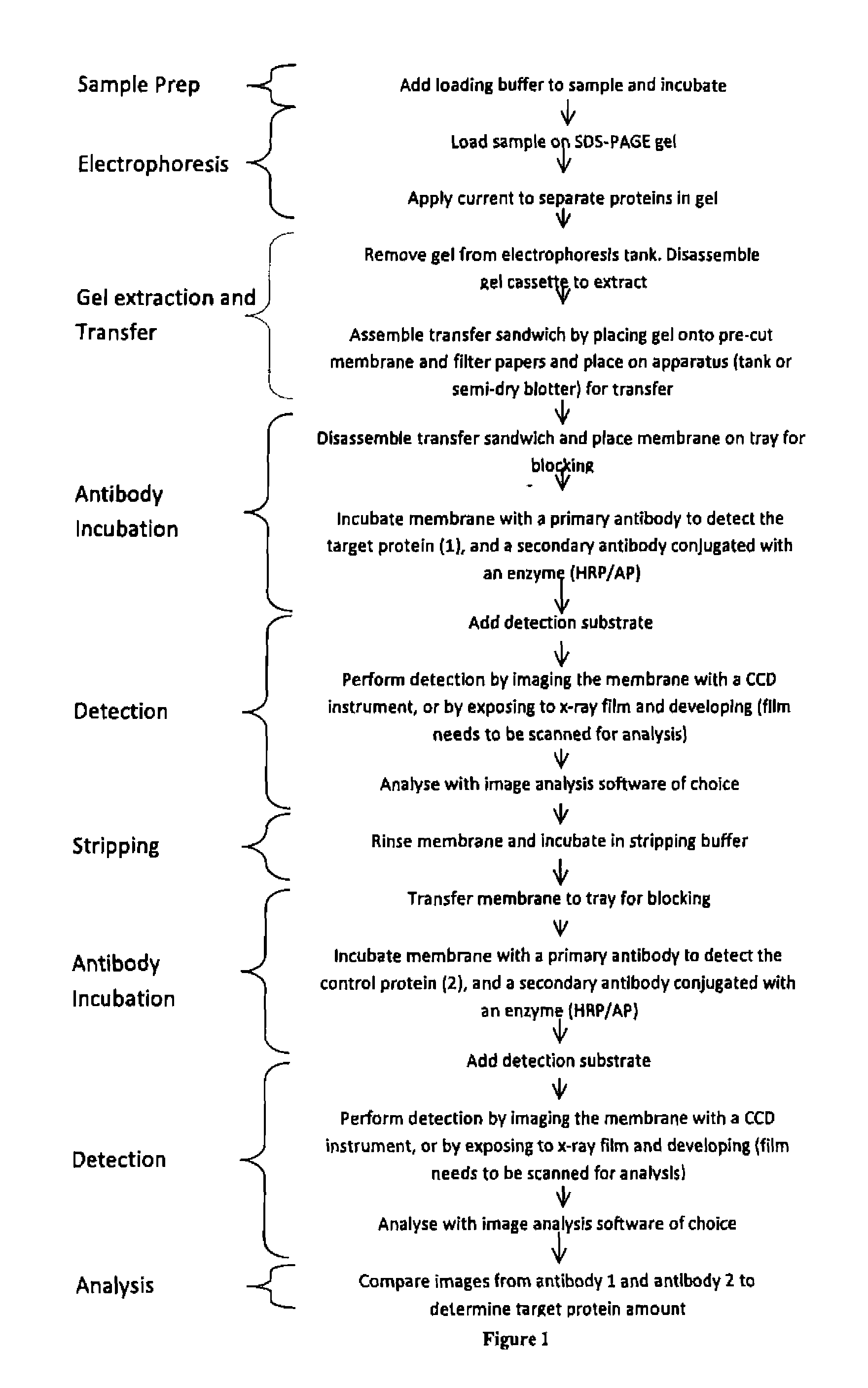
Problems with the standard western blot method include the fact that following electrophoresis the separated protein sample cannot normally be visualised and so a user cannot readily analyse the results of the protein separation.
The separated protein sample could be stained after separation so that a user can visualise the separated proteins, however, this is both labour intensive and time consuming.
This methodology requires labour intensive staining and destaining steps before the proteins are rendered visible.
The proteins revealed using this technique are often faint and lacking the contrast required to capture digital images.
Also, as current methods of immunodetection usually detect the target protein using chemiluminescence and photographic film the image of the total protein from Poceau S staining will not be directly comparable to the image of the target protein.
This method is an unattractive option in many situations especially with valuable samples or where there is only a small sample volume.
Also, as the majority of people load samples into electrophoresis apparatus manual comparison of protein content between different electrophoresis separations can lead to the incorrect interpretation of the result due to inexact loading of the sample.
This demonstrates that the additional process steps required to determine protein expression incur additional costs in the form of reagents and time.
In addition, if a standard western blot method is used, a user cannot readily compare the results from the same separated protein sample before and after immunodetection.
Another problem with the standard western blot method is that it comprises multiple steps involving several different apparatuses thereby creating barriers to integration, including a separate device being needed to image the final sample after it has been probed.
Additionally, many of these multiple steps involve wet chemistry which can lead to a lack of reproducibility.
The photographic film processing units required to develop the exposed film are expensive and often difficult to maintain.
Another difficulty of using photographic film is that obtaining an unsaturated image of the target protein suitable for quantitative analysis requires time consuming trial and error.
Overall, the standard western blot method is labour intensive, time consuming and uses a significant quantity of reagents.

Method used


Image

  • Western blot analytical technique
  • Western blot analytical technique
  • Western blot analytical technique

Examples


example 1

Gel Electrophoresis

[0083]A sample comprising proteins was prepared as follows:[0084]a. Incubating a 20 μl protein sample with 20 μl fluorescent stain at 75° C. for 7 minutes; and[0085]b. Adding 40 μl of a loading buffer, mixing and incubating again at 75° C. for 5 minutes.

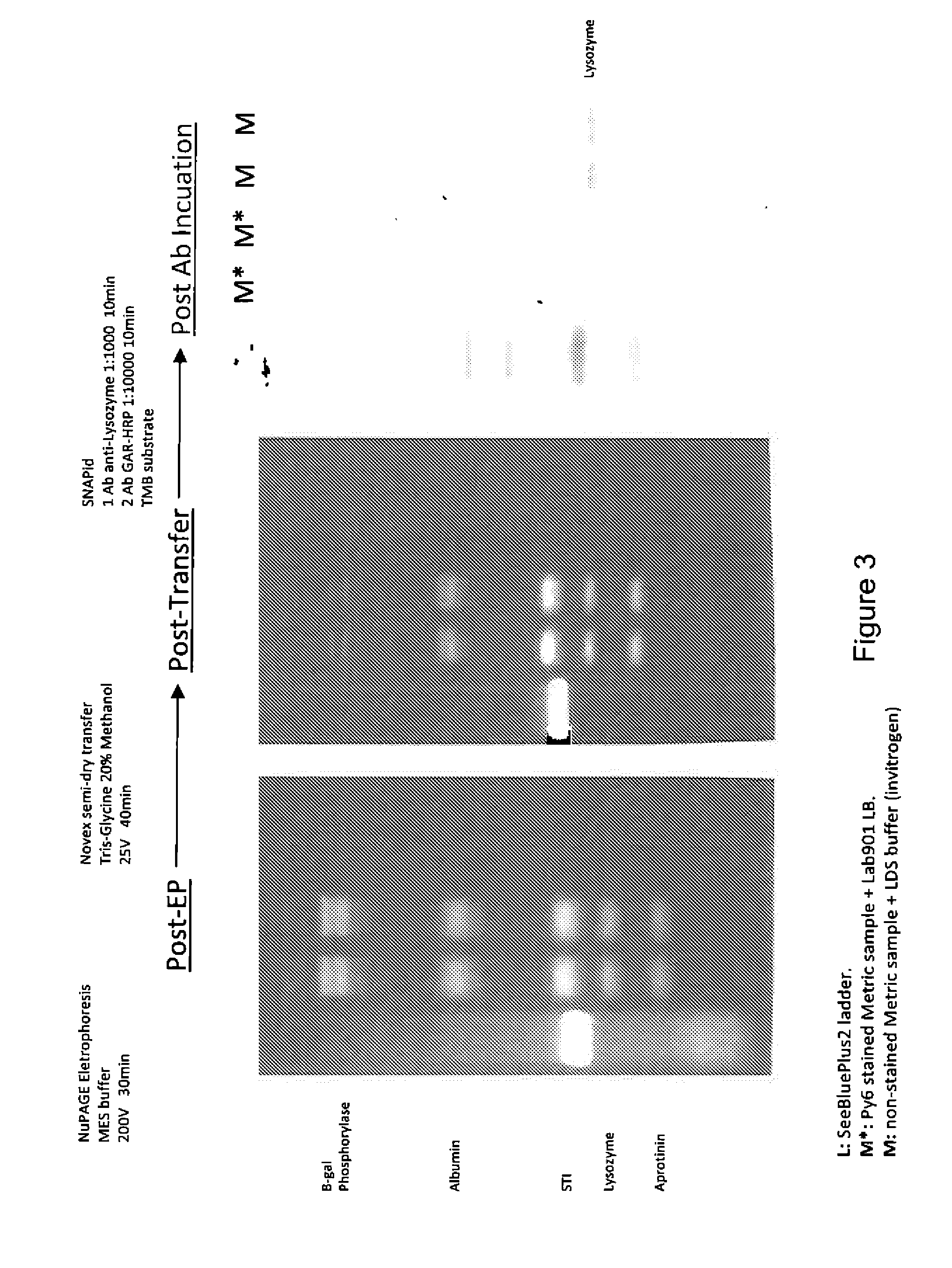
[0086]A control sample was also prepared as follows:[0087]a. Mixing a 20 μl protein sample with 10 μl of a reducing agent and 4×25 μl of Invitrogen's LDS buffer; and[0088]b. Incubating at 75° C. for 5 minutes.

[0089]Both samples were loaded onto a NuPAGE® electrophoresis gel and run according to the manufacturer's standard protocol to separate the proteins and then an image of the gel was captured using a UV transilluminator and a digital camera.

Transferring the Separated Samples onto Membranes

[0090]The gel comprising the separated proteins was separated into two halves and then one half was transferred onto a PVDF membrane and the other half was transferred onto a nitrocellulose membrane. The separated proteins wer...

example 2

Gel Electrophoresis

[0102]A sample comprising proteins was prepared as follows:[0103]a. Incubating a 2 μl protein sample with 2 μl fluorescent stain at 75° C. for 7 minutes;[0104]b. Adding 4 μl of a loading buffer, mixing and incubating again at 75° C. for 5 minutes; and[0105]c. Adding 2 μl of in-lane marker.

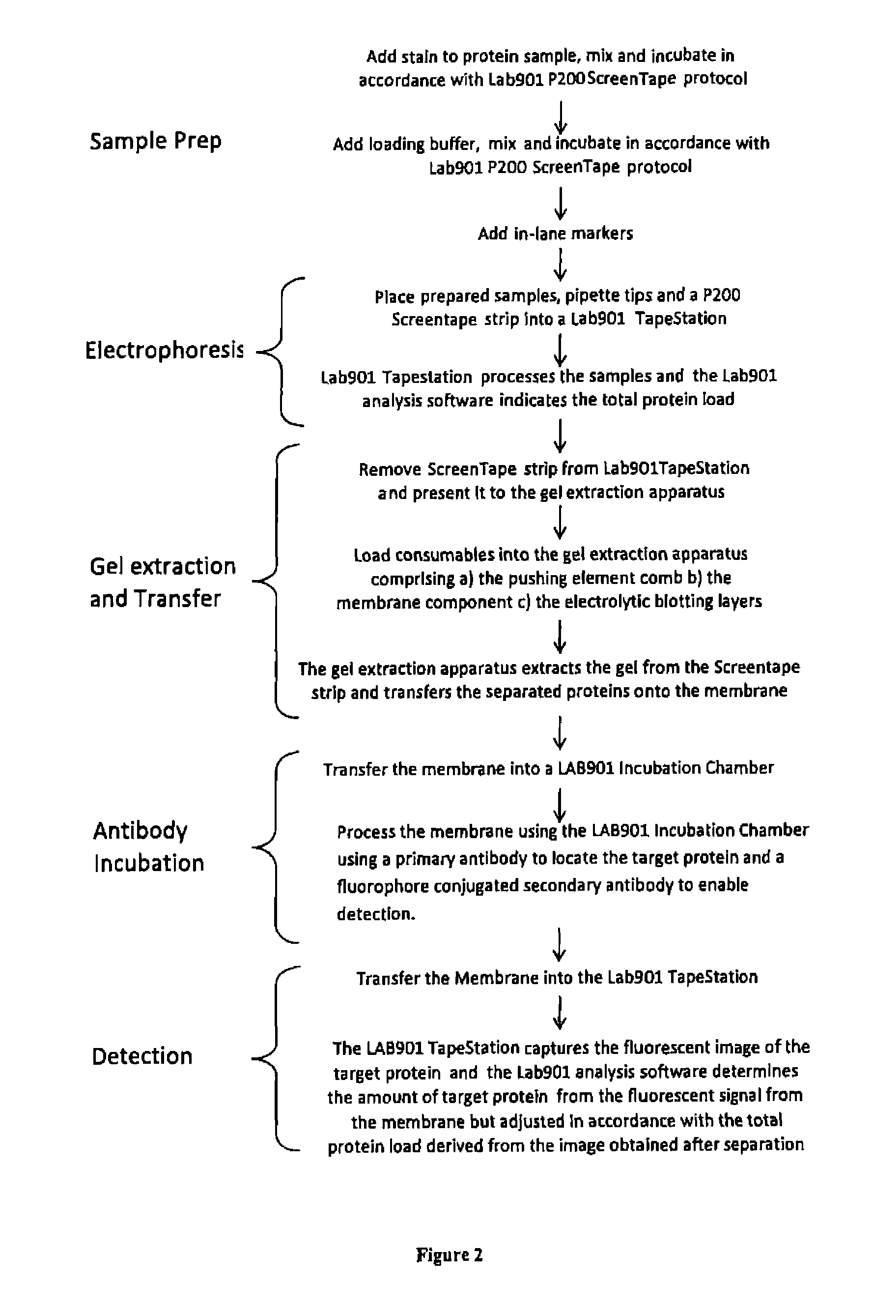
[0106]The samples were loaded onto a Lab901 P200 ScreenTape® electrophoresis gel and run according to the manufacturer's standard protocol to separate the proteins. The used ScreenTape® was imaged using the Lab901 TapeStation® (see “P200” in FIG. 5a).

Transferring the Separated Samples onto Membranes

[0107]The used ScreenTape® comprising the separated proteins was recovered from the TapeStation®, its carrier layer was removed and two blades were used to cut away the top and bottom of the ScreenTape® exposing the top and bottom of the gel columns contained within 16 sub-containers. A comb comprising 16 gel pushing elements was used to push against the gel within each of the sub-cont...

example 3

Gel Electrophoresis

[0119]A sample comprising proteins was prepared as follows:[0120]a. Incubating a 2 μl protein sample with 2 μl fluorescent stain at 75° C. for 7 minutes;[0121]b. Adding 4 μl of a loading buffer, mixing and incubating again at 75° C. for 5 minutes; and[0122]c. Adding 2 μl of in-lane marker.

[0123]The samples were loaded onto a Lab901 P200 ScreenTape® electrophoresis gel and run according to the manufacturer's standard protocol to separate the proteins. The used ScreenTape® was imaged using the Lab901 TapeStation® (see “P200” in FIG. 6a).

Transferring the Separated Samples onto Membranes

[0124]The used ScreenTape® comprising the separated proteins was recovered from the TapeStation®, its carrier layer was removed and two blades were used to cut away the top and bottom of the ScreenTape® exposing the top and bottom of the gel columns contained within 16 sub-containers. A comb comprising 16 gel pushing elements was used to push against the gel within each of the sub-cont...


PUM

PropertyMeasurementUnit
gel electrophoresisaaaaaaaaaa
molecular weightaaaaaaaaaa
intensity of fluorescenceaaaaaaaaaa

Abstract

In accordance with an embodiment of the present invention, there is provided a method of performing the western blot analytical technique, wherein the method comprises carrying out the following steps in the following order: a), pre-staining the proteins within a sample; b). separating the proteins using gel electrophoresis; c). analysing the separated proteins to determine the total protein load and / or at least one housekeeping protein load; d). transferring the proteins onto at least one membrane; e). probing the separated proteins to detect a target protein; and f). analysing the probed proteins to determine the target protein's molecular weight and / or load.

Description

[0001]This application claims the benefit of the filing date of GB1008517.3 filed 21 May 2010 and of GB1100092.4 filed 5 Jan. 2011, the disclosure of which is hereby incorporated herein by reference.FIELD OF THE INVENTION[0002]This patent application relates to an improved method of performing the western blot analytical technique.TECHNOLOGICAL BACKGROUND[0003]Western blotting is a labour intensive laboratory analysis method that is widely used in the life sciences to determine whether a target protein is present in a complex sample and to determine the relative quantity of the target protein. The phrase “target protein” is used to refer to a protein that a user of an analysis method wishes to identify within a complex sample. The relative quantity of a protein is used to measure changes in protein expression (i.e. up regulation and down regulation).[0004]Determining whether a particular protein is present is achieved by connecting two variables: the molecular weight of the protein ...

Claims


Application Information

Patent Timeline
Patent Type & Authority Applications(United States)
IPC IPC(8): G01N33/559G01N33/566
CPCG01N27/44726G01N33/6803G01N27/44739
Inventor MACNAMARA, KENNETH G.POLWART, STUART
Owner AGILENT TECH INC
Learn More