
System and method for automated disease assessment in capsule endoscopy

a capsule endoscopy and automated technology, applied in the field of systems and methods of processing images from the capsule endoscopy, can solve the problems of wasting time, limited clinical utility of the capsule endoscopy, and wasting time in the evaluation process

Inactive Publication Date: 2012-12-13
THE JOHN HOPKINS UNIV SCHOOL OF MEDICINE
View PDF2 Cites 137 Cited by
  • Summary
  • Abstract
  • Description
  • Claims
  • Application Information

AI Technical Summary

Benefits of technology

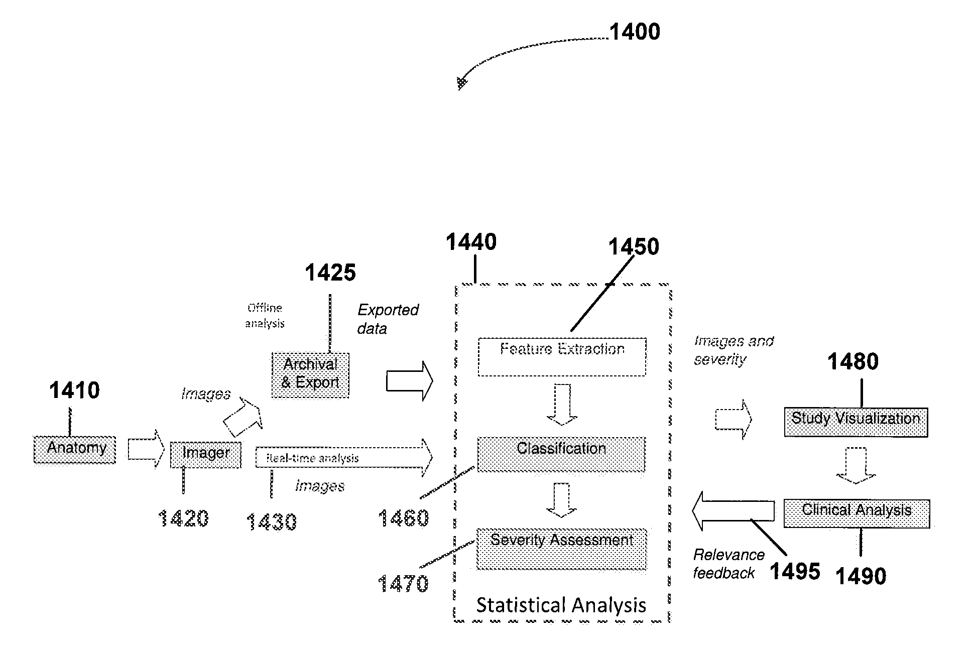
[0015]In yet another embodiment of the current invention, a computer readable medium stores executable instructions for execution by a computer having memory. The medium stores instructions for receiving one or more endoscopic images, processing each of the endoscopic images to determine whether at least...

Problems solved by technology

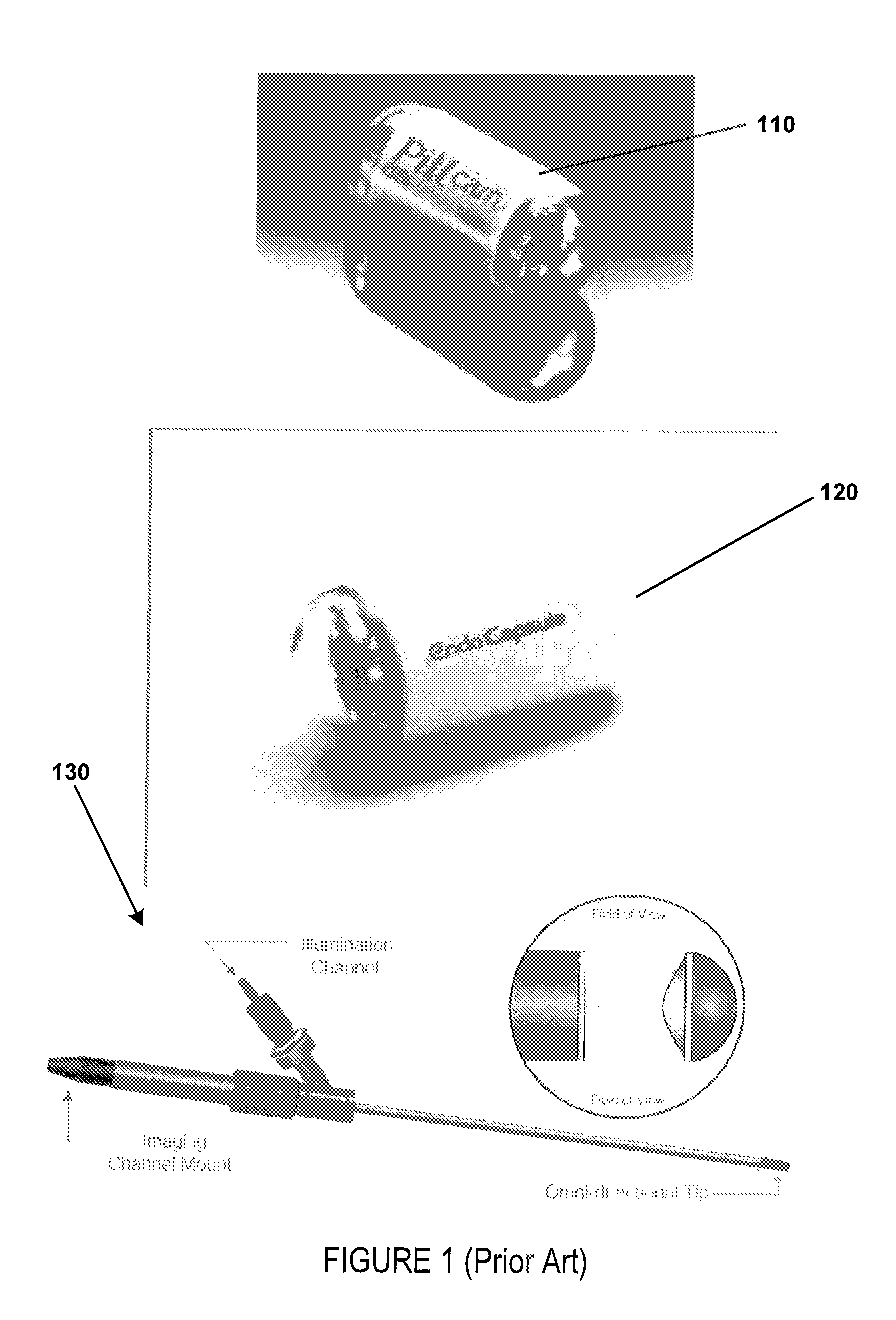
The clinical utility of these capsules has been limited due to the lack of accurate anatomical localization and visualization.
This makes evaluation of data a tedious and time consuming (usually 1-2 hours) process.
However, due to the low frame rate, neighboring images may not necessarily contain the same areas of interest and the clinician is typically left toggling between images in the sequence, thus making the process even more time consuming.
In addition to being a tedious and time consuming process, detection rates may also vary among clinicians, especially for early stage pathology.
However, these have proven to be of limited benefit.

Method used


Image

  • System and method for automated disease assessment in capsule endoscopy
  • System and method for automated disease assessment in capsule endoscopy
  • System and method for automated disease assessment in capsule endoscopy

Examples


examples

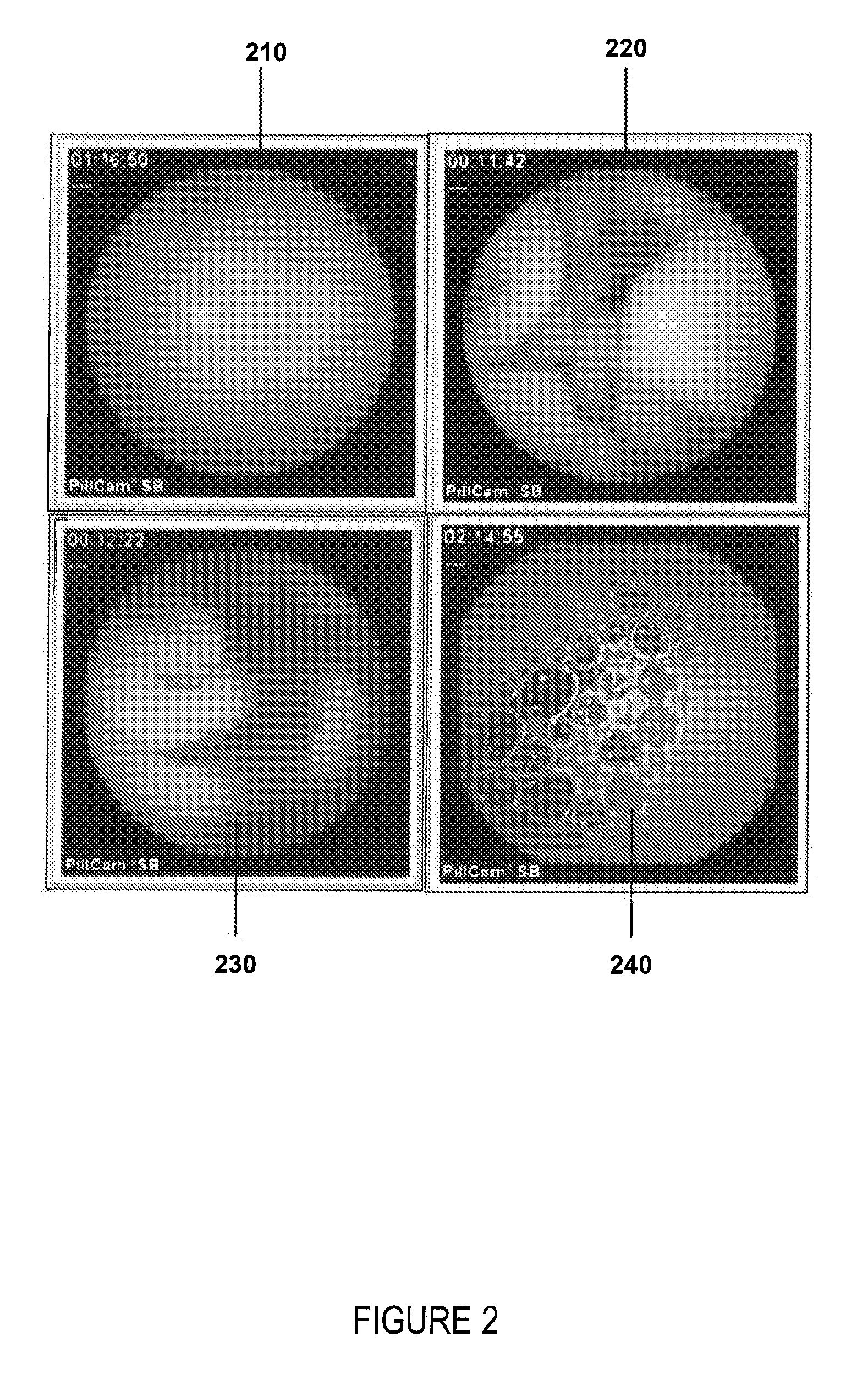
[0080]in one embodiment, support vector machines (SVM) may be used to classify CE images into lesion (L), normal tissue, and extraneous matter (food, bile, stool, air bubbles, etc). FIG. 4 depicts example normal tissue 410; air bubbles 420; floating matter, bile, food, and stool 430; abnormalities such as bleeding, polyps, non-Chrohn's lesions, darkening old blood 440; and rated lesions from severe, moderate, to mild 450. In addition to lesions other attributes of interest may include blood, bleeding, inflammation, mucosal inflammation, submucosal inflammation, discoloration, an erosion, an ulcer, stenosis, a stricture, a fistulae, a perforation, an erythema, edema, or a boundary organ

[0081]SVM has been used previously to segment the GI tract boundaries in CE images (M. Coimbra, P. Campos, J. P. Silva Cunha; “Topographic segmentation and transit time estimation for endoscopic capsule exams”, in Proc. IEEE ICASSP, 2006). SVM may use a kernel function to transform the input data into ...

embodiment

[0099]In one embodiment, lesions as well as data for other classes for interest may be selected and assigned a global ranking (e.g., for example, mild, moderate, or severe) based upon the size, and severity of lesion and any surrounding inflammation, for example. Lesions may be ranked into three categories: mild, moderate or severe disease. FIG. 5, 510 shows a typical Crohn's disease lesion with the lesion highlighted. As a lesion may appear in several images, data representing 50 seconds, for example, of recording time around the selected image frame may also be reviewed, annotated, and exported as part of a sequence. In addition, a number of extra image sequences not containing lesions may be exported as background data for training of statistical methods.

[0100]Global lesion ranking may be used to generate the required preference relationships. For example, over 188,000 pairwise relationships may be possible in a dataset of 600 lesion image frames that have been assigned a global ...


PUM


Abstract

A system and method for automated image analysis which may enhance, for example, capsule endoscopy diagnosis. The system and methods may reduce the time required for diagnosis, and also help improve diagnostic consistency using an interactive feedback tool. Furthermore, the system and methods may be applicable to any procedure where efficient and accurate visual assessment of a large set of images is required.

Description

CROSS-REFERENCE TO RELATED APPLICATION[0001]This application claims priority to U.S. Provisional Application No. 61 / 223,585 filed Jul. 7, 2009, the entire content of which is hereby incorporated by reference.FEDERAL FUNDING[0002]This invention was made with U.S. Government support of Grant No. 5R21EB008227-02, awarded by National Institutes of Health. The U.S. Government has certain rights in this invention.BACKGROUND[0003]1. Field of Invention[0004]The current invention relates to systems and methods of processing images from an endoscope, and more particularly automated systems and methods of processing images from an endoscope.[0005]2. Discussion of Related Art[0006]The contents of all references, including articles, published patent applications and patents referred to anywhere in this specification are hereby incorporated by reference.[0007]There have been several capsules developed for “blind” collection of diagnostic data in the GI tract. For example the Medtronic Bravo (rece...

Claims


Application Information

Patent Timeline
IPC IPC(8): A61B6/00H04N7/18
CPCA61B1/00009A61B1/041G06T2207/30032G06T2207/10016G06T2207/10068G06T7/0012A61B1/000094A61B1/000096
Inventor KUMAR, RAJESHDASSOPOULOS, THEMISTOCLESGIRGIS, HANIHAGER, GREGORY
Owner THE JOHN HOPKINS UNIV SCHOOL OF MEDICINE
Learn More