
Method and System for Heart Isolation in Cardiac Computed Tomography Volumes for Patients with Coronary Artery Bypasses

a computed tomography volume and heart technology, applied in image analysis, image enhancement, instruments, etc., can solve the problems of providing a significant challenge to heart segmentation algorithms, and achieve the effect of effectively isolated hear

Active Publication Date: 2012-05-31
SIEMENS HEALTHCARE GMBH
View PDF0 Cites 30 Cited by
  • Summary
  • Abstract
  • Description
  • Claims
  • Application Information

AI Technical Summary

Benefits of technology

[0004]The present invention provides a method and system for automatically isolating the heart in 3D medical image data, such as cardiac CT volumes. Embodiments of the present invention automatically generate a heart isolation mask that effectively isolates the heart in cardiac CT volumes for patients with coronary artery bypasses.

Problems solved by technology

This provides a significant challenge to heart segmentation algorithms.

Method used


Image

  • Method and System for Heart Isolation in Cardiac Computed Tomography Volumes for Patients with Coronary Artery Bypasses
  • Method and System for Heart Isolation in Cardiac Computed Tomography Volumes for Patients with Coronary Artery Bypasses
  • Method and System for Heart Isolation in Cardiac Computed Tomography Volumes for Patients with Coronary Artery Bypasses

Examples


Embodiment Construction

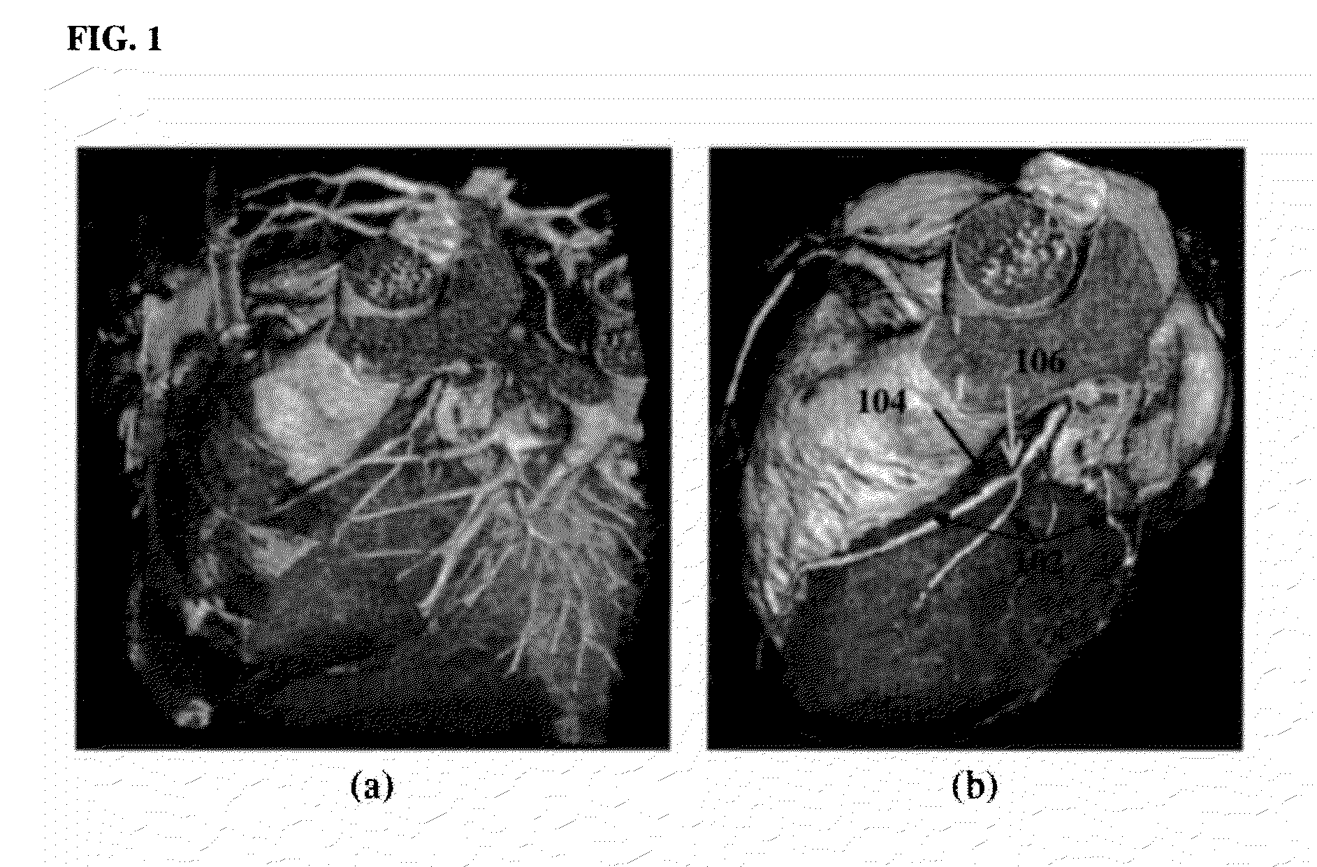
[0023]The present invention is directed to a method and system for heart isolation in 3D medical images, such as 3D cardiac CT volumes. Embodiments of the present invention are described herein to give a visual understanding of the heart isolation method. A digital image is often composed of digital representations of one or more objects (or shapes). The digital representation of an object is often described herein in terms of identifying and manipulating the objects. Such manipulations are virtual manipulations accomplished in the memory or other circuitry / hardware of a computer system. Accordingly, it is to be understood that embodiments of the present invention may be performed within a computer system using data stored within the computer system.

[0024]Heart Isolation is highly relevant to several applications. For example, after separating the heart from tissues in proximity to the heart (e.g., lung, liver, and rib cage), the coronary arteries can be clearly visualized in 3D. FI...


PUM


Abstract

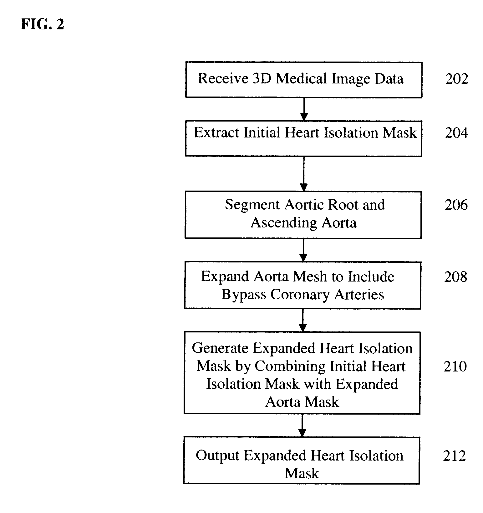
A method and system for isolating the heart in a 3D volume, such as a cardiac CT volume, for patients with coronary artery bypasses is disclosed. An initial heart isolation mask is extracted from a 3D volume, such as a cardiac CT volume. The aortic root and ascending aorta are segmented in the 3D volume, resulting in an aorta mesh. The aorta mesh is expanded to include bypass coronary arteries. An expanded heart isolation mask is generated by combining the initial heart isolation mask with an expanded aorta mask defined by the expanded aorta mesh.

Description

[0001]This application claims the benefit of U.S. Provisional Application No. 61 / 417,602, filed Nov. 29, 2011, the disclosure of which is herein incorporated by reference.BACKGROUND OF THE INVENTION[0002]The present invention relates to isolating the heart in medical image data, and more particularly, to isolating the heart in medical imaging data for patients with coronary artery bypasses.[0003]Heart isolation refers separating the heart in medical image data from other tissues in close proximity to the heart, such as the lungs, liver, and rib cage. Most previous work relating heart segmentation focuses on segmenting heart chambers, especially the left ventricle. Segmenting the heart as a whole, or heart isolation, has relevance to several applications. For example, after separating the heart from the proximity tissues (e.g., lung, liver, and rib cage) in a 3D computed tomography (CT) volume, the coronary arteries can be clearly visualized in 3D. According to statistics from the Un...

Claims


Application Information

Patent Timeline
IPC IPC(8): G06K9/00
CPCG06T7/0081G06T7/0089G06T2207/30048G06T2207/20124G06T2207/10081G06T7/11G06T7/149
Inventor ZHENG, YEFENGVEGA-HIGUERA, FERNANDOCOMANICIU, DORIN
Owner SIEMENS HEALTHCARE GMBH
Learn More