Eureka AIR delivers breakthrough ideas for toughest innovation challenges, trusted by R&D personnel around the world.

Multi-Modal Imaging for Diagnosis of Early Stage Epithelial Cancers

a multi-modal imaging and early stage technology, applied in the direction of diagnostic recording/measuring, catheters, applications, etc., can solve the problems of not being able to perform large organ scanning, substantially etc., and achieve the effect of increasing the length of the procedur

Inactive Publication Date: 2012-04-26
PHYSICAL SCI
View PDF6 Cites 11 Cited by
  • Summary
  • Abstract
  • Description
  • Claims
  • Application Information

AI Technical Summary

Benefits of technology

The patent describes a method for early detection of epithelial cancer using a combination of fluorescence imaging and optical coherence tomography (OCT). The method involves staining tissue with a cancer-targeting agent and using an endoscope to deliver radiation for fluorescence imaging and OCT imaging. The use of a guidance tool, such as fluorescence imaging, can help highlight suspicious areas for diagnosis. The invention provides a more efficient and reliable method for detecting cancer in its early stages, reducing the need for multiple biopsies and minimizing drug spreading in other organs. The imaging system can also provide faster diagnostic results, decrease patient anxiety, and provide an alternative to virtual colonoscopy.

Problems solved by technology

While Optical Coherence Tomography (OCT) can be optimized as a diagnostic tool for detection and characterization of epithelial cancer, it typically has a limited field of view, on the order of millimeters, and thus scanning of large organs is not very practical because it involves the time-consuming analysis of very large data sets.
In addition, it can substantially increase the length of the procedure.

Method used


Image

  • Multi-Modal Imaging for Diagnosis of Early Stage Epithelial Cancers
  • Multi-Modal Imaging for Diagnosis of Early Stage Epithelial Cancers
  • Multi-Modal Imaging for Diagnosis of Early Stage Epithelial Cancers

Examples


Embodiment Construction

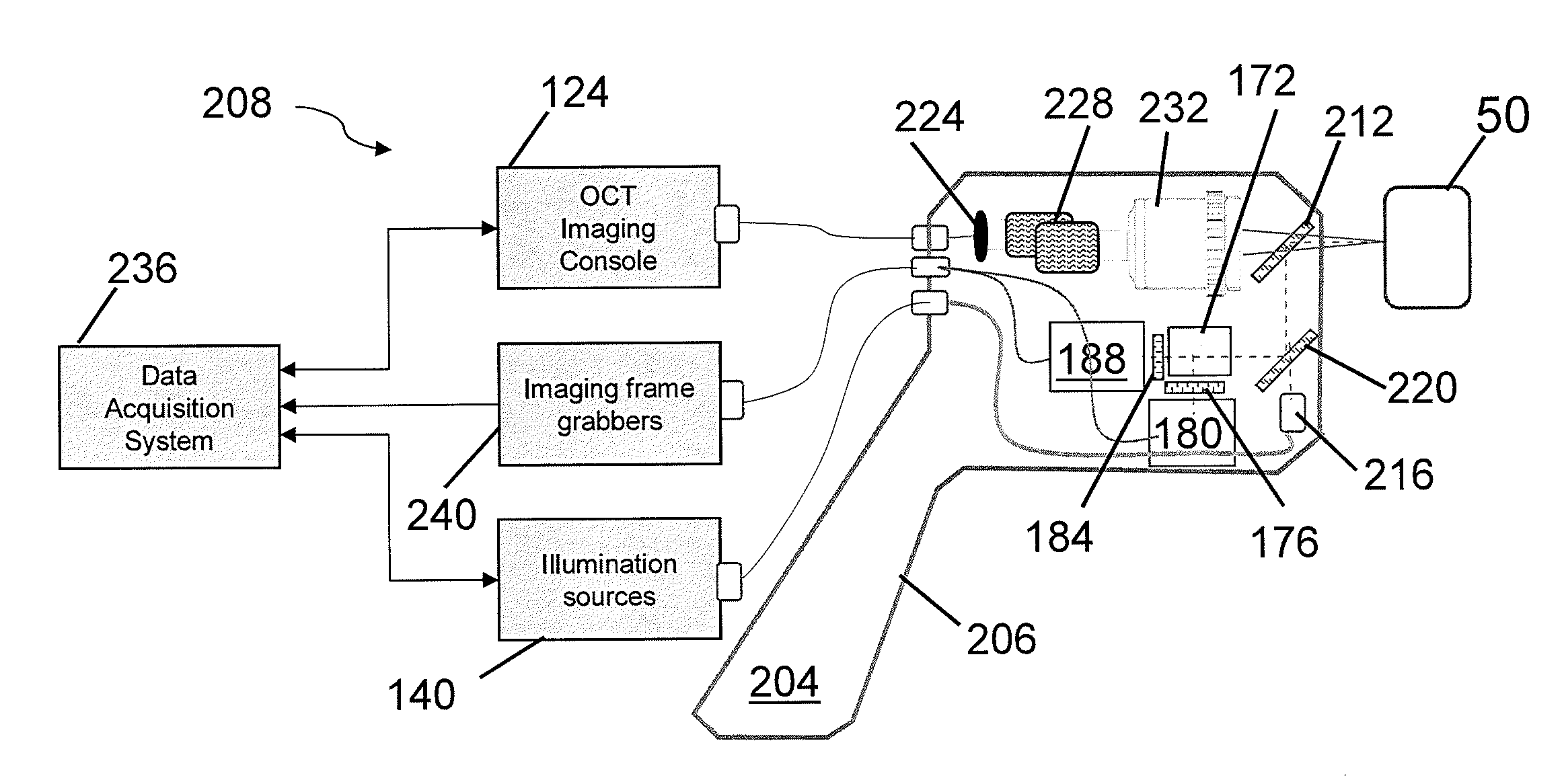
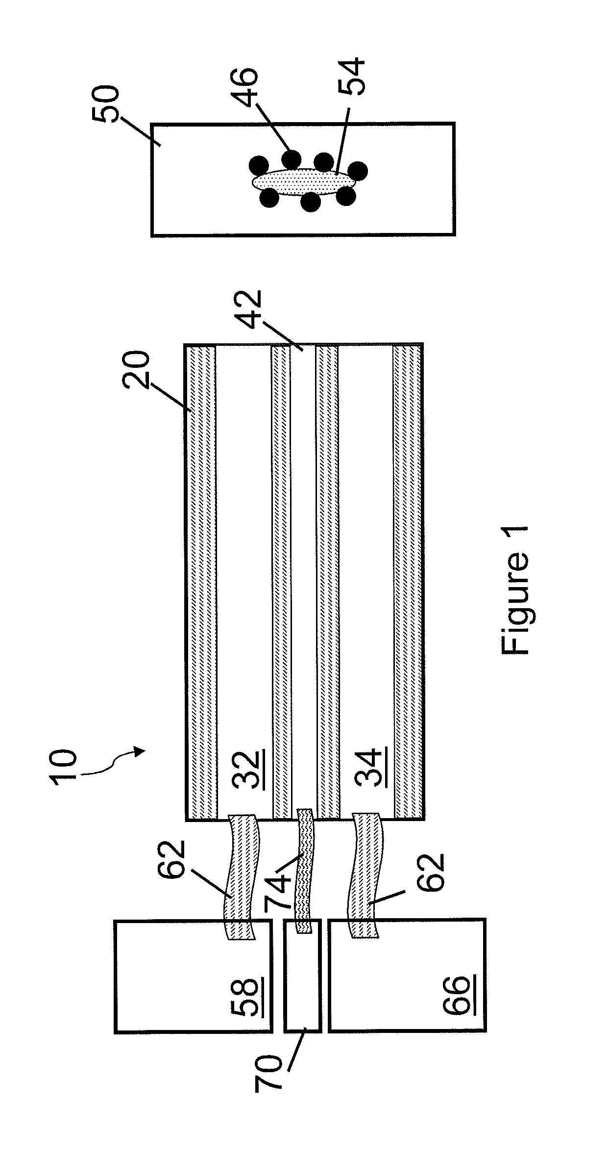
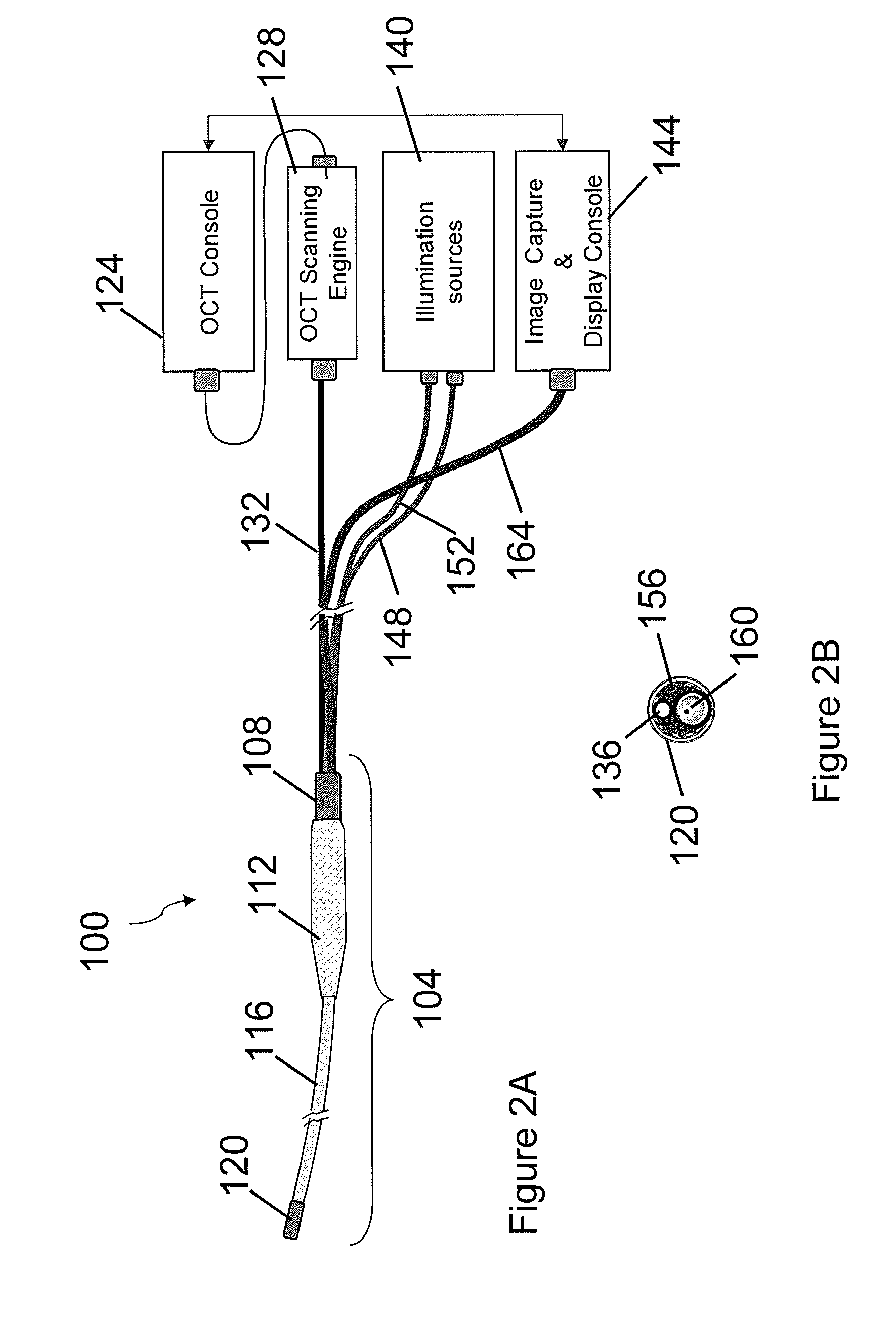
[0028]FIG. 1 shows an exemplary probe 10 including a body 20 defining a first channel 32 for fluorescence imaging and a second channel 34 for OCT imaging. The first channel 32 can be used for bright field imaging, or a separate channel can be used for bright field imaging. The endoscopic probe 10 can define a third channel 42 used for delivery of a cancer targeting agent 46, such as the RGD-functionalized Au-PCL microparticles or nanoparticles, to tissue 50. The cancer targeting agent 46 can stain the tissue 50 containing a potentially cancerous lesion 54. Fluorescence imaging is used to identify the potentially cancerous lesion, and OCT imaging of the potentially cancerous lesion is used for cancer diagnosis. The endoscopic probe 10 need not include the third channel 42. In certain embodiments, the cancer targeting agent 46 can be introduced through alternative means, such as direct delivery or by a catheter.

[0029]The first channel 32 can be coupled to a fluorescence imaging system...


PUM

PropertyMeasurementUnit
diameteraaaaaaaaaa
diameteraaaaaaaaaa
volumeaaaaaaaaaa

Abstract

Epithelial cancer screening can include a staining tissue with a cancer targeting agent, identifying a potentially cancerous lesion using fluorescence imaging, and imaging the potentially cancerous lesion for a cancer diagnosis using optical coherence tomography.

Description

CROSS-REFERENCE TO RELATED APPLICATION[0001]This application claims the benefit of and priority to U.S. Provisional Patent Application No. 61 / 405,059 filed Oct. 20, 2010, which is owned by the assignee of the instant application and the disclosure of which is incorporated herein by reference in its entirety.GOVERNMENT RIGHTS[0002]The invention was made with government support from the National Institute of Health under grant number 5R41 CA 132256-02. The government may have certain rights in the invention.FIELD OF THE INVENTION[0003]The invention relates generally to endoscopic diagnosis of early stage epithelial cancers, and more particularly to cancer screening using a multi-modal imaging system and a cancer targeting molecular probe.BACKGROUND[0004]Traditional imaging modalities, such as computed invention (CT) scans, magnetic resonance (MR), and endoscopy can be used for cancer screening. Due to lack of resolution, they can only detect lesions that are already formed, which are ...

Claims


Application Information

Patent Timeline
Patent Type & Authority Applications(United States)
IPC IPC(8): A61B6/00A61K49/00B82Y15/00
CPCA61B5/0035A61B5/0066A61B5/0071A61B5/0084B82Y30/00A61K49/0056A61K49/0091A61K49/0093B82Y5/00A61B2560/0406
Inventor IFTIMIA, NICUSOR V.LUE, NIYOMHAMMER, DANIEL X.MUJAT, MIRCEAFERGUSON, R. DANIEL
Owner PHYSICAL SCI
Why Eureka
Eureka Blog
Learn More