Ultrasonic diagnostic apparatus and elastic image display method

a diagnostic apparatus and elastic technology, applied in the field of ultrasonic diagnostic apparatus and elastic image display method, can solve the problems of inability to acquire inability to uniformly press the tissue of the object, and inability to achieve elastic image suitable for diagnosis, etc., to achieve easy diagnosis

Inactive Publication Date: 2011-12-08
HITACHI LTD
View PDF6 Cites 27 Cited by
  • Summary
  • Abstract
  • Description
  • Claims
  • Application Information

AI Technical Summary

Benefits of technology

[0024]According to the invention, the examiner can acquire an elastic image suitable for diagnosis easily without depending on the examiner's pressure technique.

Problems solved by technology

However, although the method in which an examiner operates an ultrasonic probe manually to press the object is advantageous in terms of being real time, simplicity, resolution, and cost, there is a problem that an elastic image suitable for diagnosis is not acquired if the pressure is not properly performed and this depends on the examiner's pressure technique.
For example, if the object is pressed by an ultrasonic probe inclined from the surface of the object along the scanning direction, tissue of the object is not uniformly pressed.
Accordingly, an elastic image may not be suitable for diagnosis.

Method used


Image

  • Ultrasonic diagnostic apparatus and elastic image display method
  • Ultrasonic diagnostic apparatus and elastic image display method
  • Ultrasonic diagnostic apparatus and elastic image display method

Examples


embodiment 1

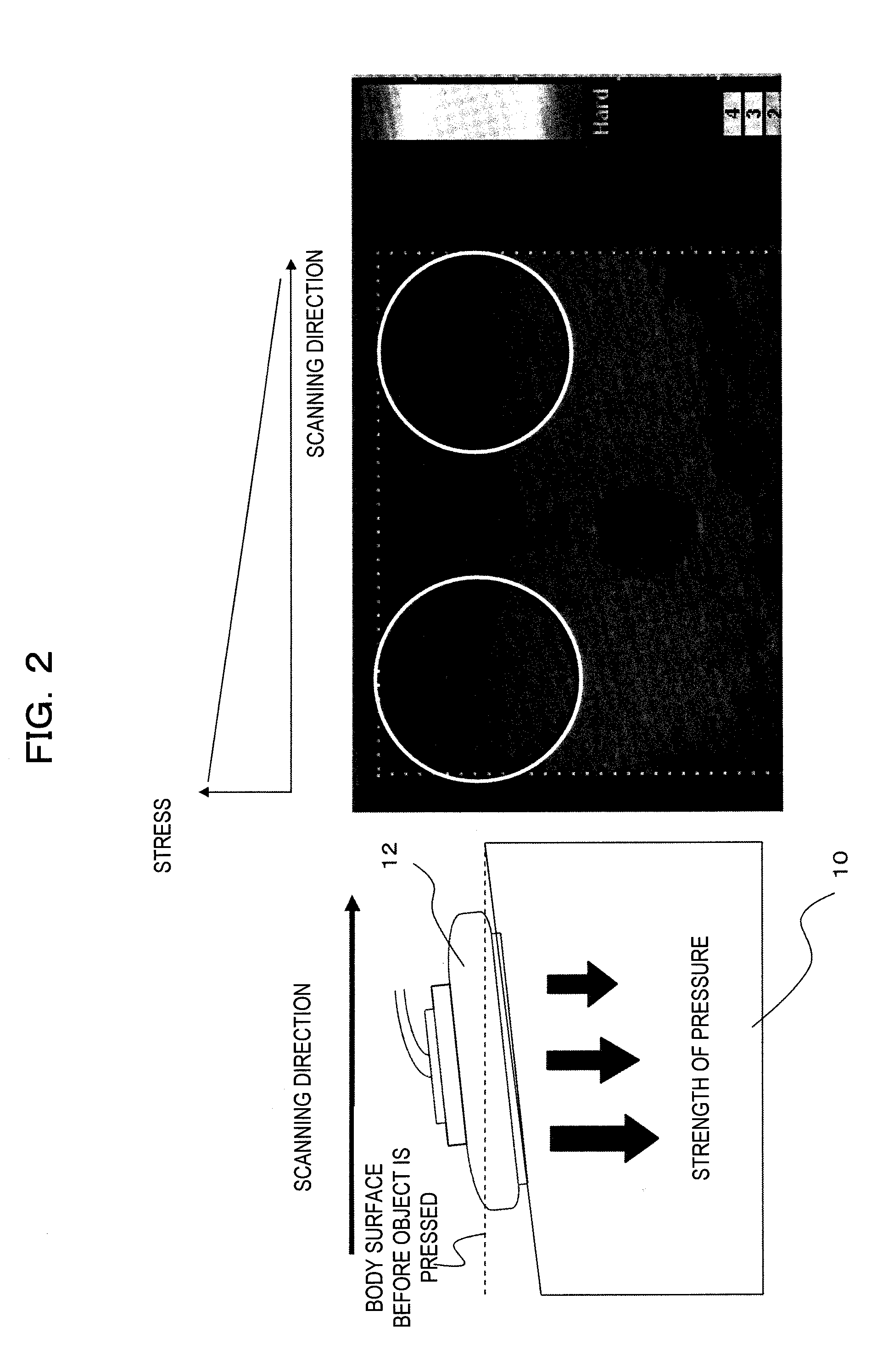
[0063]FIG. 3 is a view illustrating processing of the gradient detecting unit 40 and the like in a first embodiment. FIG. 3(a) is a view showing the detailed configuration of the gradient detecting unit 40. As shown in FIG. 3(a), the gradient detecting unit 40 is configured to include: a displacement gradient calculating circuit 60 which calculates a displacement gradient by calculating the average displacement of each beam line of the displacement frame output from the displacement measuring unit 30; a correction coefficient calculating circuit 62 which calculates a correction coefficient of each beam line on the basis of the calculated displacement gradient based on the average displacement or the like; and a displacement frame correcting circuit 64 which corrects a displacement frame on the basis of the correction coefficient. In addition, the displacement gradient calculating circuit 60 has a feature value calculation region setting unit 61 which sets a region where the feature ...

embodiment 2

[0068]FIG. 4 is a view illustrating processing of the gradient detecting unit 40 and the like in a second embodiment. The detailed configuration of the gradient detecting unit 40 is the same as that in the first embodiment. Hereinafter, processing in the second embodiment which is executed by the displacement gradient calculating circuit 60, the correction coefficient calculating circuit 62, the displacement frame correcting circuit 64, and the like will be described. In addition, explanation regarding the same units as in the first embodiment will be omitted. In this example, the displacement gradient calculating circuit 60 calculates the displacement gradient 50 as shown in FIG. 4(c) and then corrects the displacement gradient on the basis of the average of the feature value of displacement of each beam line as shown in FIG. 4(c-2). For example, the displacement gradient calculating circuit 60 calculates the average (Disp mean) of the displacement feature value of each beam line b...

embodiment 3

[0070]FIG. 5 is a view illustrating processing of the gradient detecting unit 40 and the like in a third embodiment. The detailed configuration of the gradient detecting unit 40 is the same as that in the first embodiment. Hereinafter, processing in the third embodiment which is executed by the displacement gradient calculating circuit 60, the correction coefficient calculating circuit 62, the displacement frame correcting circuit 64, and the like will be described. In addition, explanation on the same unit as in the first and second embodiments will be omitted. In this example, the displacement gradient calculating circuit 60 calculates the displacement gradient 54 corrected as shown in FIG. 5(c-2) and then corrects this displacement gradient further on the basis of a stress distribution coefficient 56 in the scanning direction of an ultrasonic probe set in advance. For example, the displacement gradient calculating circuit 60 calculates a displacement gradient by multiplying the c...


PUM


Abstract

An elastic image suitable for diagnosis is easily acquired without depending on the examiner's pressure technique. The displacement of tissue in a tomographic portion of an object is measured on the basis of a pair of RF signal frame data items regarding the tomographic portion, which are obtained at different times by transmitting and receiving an ultrasonic wave to and from the object (b). The average displacement of each beam line (d0 to dn) of an ultrasonic wave is calculated (c), and a displacement gradient (50) in the scanning direction of an ultrasonic probe is calculated on the basis of the displacement average (d). A correction coefficient (52) with respect to the displacement of each beam line is calculated on the basis of the calculated displacement gradient (e), and the displacement distribution measured on the basis of the calculated correction coefficient is corrected (f). In this manner, even if the object is pressed by an ultrasonic probe inclined along the scanning direction, an elastic image generated by pressing the object with the ultrasonic probe not inclined along the scanning direction can be generated in a pseudo manner by correcting the displacement distribution on the basis of the correction coefficient.

Description

TECHNICAL FIELD[0001]The present invention relates to an ultrasonic diagnostic apparatus and an elastic image display method and in particular, to an ultrasonic diagnostic apparatus and an elastic image display method for displaying an elastic image showing the hardness or softness of tissue in a tomographic portion of an object.BACKGROUND ART[0002]The ultrasonic diagnostic apparatus transmits an ultrasonic wave from an ultrasonic probe into the object, receives a reflected echo signal of the ultrasonic wave corresponding to the structure of body tissue from the inside of the object, forms a tomographic image, for example, an ultrasonic tomographic image or the like, and displays it for diagnosis.[0003]In recent years, it has been disclosed that an ultrasonic reception signal can be measured by pressing the object with an ultrasonic probe using a manual or mechanical method, displacement of each portion of the body occurring due to the pressure is calculated on the basis of frame da...

Claims


Application Information

Patent Timeline
Patent Type & Authority Applications(United States)
IPC IPC(8): A61B8/14
CPCA61B8/08A61B8/485
Inventor WAKI, KOJI
Owner HITACHI LTD
Why Eureka
Try Eureka