
Methods and compositions for detecting genetic material

a technology of genetic material and composition, applied in the field of methods and compositions for detecting genetic material, can solve the problems of unstable partitions, unstable partitions,

Inactive Publication Date: 2011-06-30
BIO RAD LAB INC
View PDF107 Cites 171 Cited by
  • Summary
  • Abstract
  • Description
  • Claims
  • Application Information

AI Technical Summary

Benefits of technology

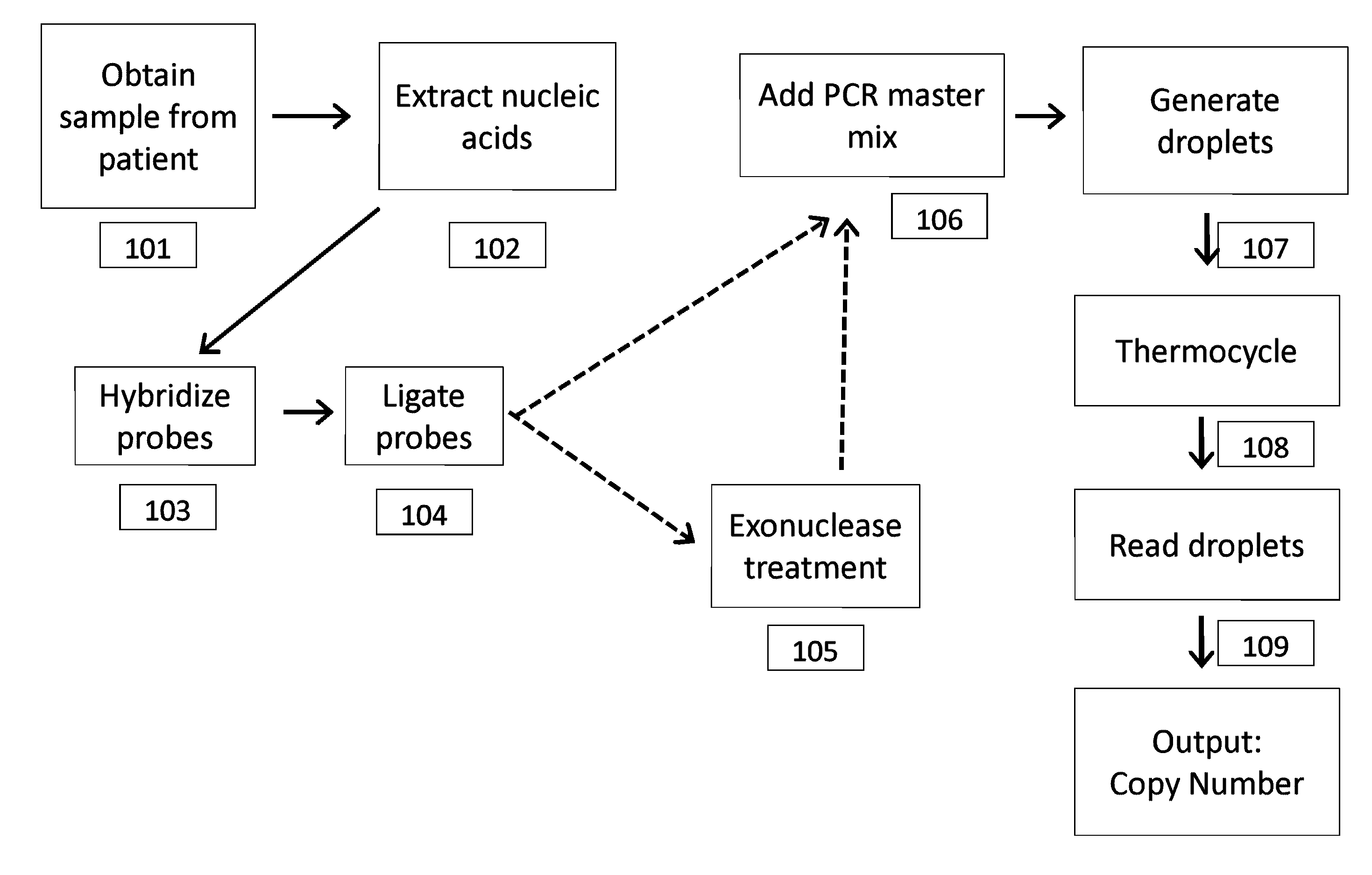
[0032]The microcapsule may comprise one or more of said ligation probes (e.g., padlock probe, molecular inversion probe, ligation detection reaction (LDR) probe, circular probe, etc.). The microcapsules may comprise a linear probe obtained by linearizing a probe previously circularized after a ligation reaction. In some cases, the microcapsules comprise circularized ligation probe. In some cases, the microcapsules contain linear products of a ligation detection reaction (LDR).
[0033]The compositions and method provided herein may also relate to a water-in-oil mixture comprising two or more aqueous droplets, wherein at least one of said two or more aqueous droplets comprises a first ligation probe directed to a first target polynucleotide and at least one of said two or more aqueous droplets

Problems solved by technology

The partitions may also be stable or unstable.

Method used


Image

  • Methods and compositions for detecting genetic material
  • Methods and compositions for detecting genetic material
  • Methods and compositions for detecting genetic material

Examples


example 1

Detection of Fetal DNA Using a Two-Color Detection Scheme

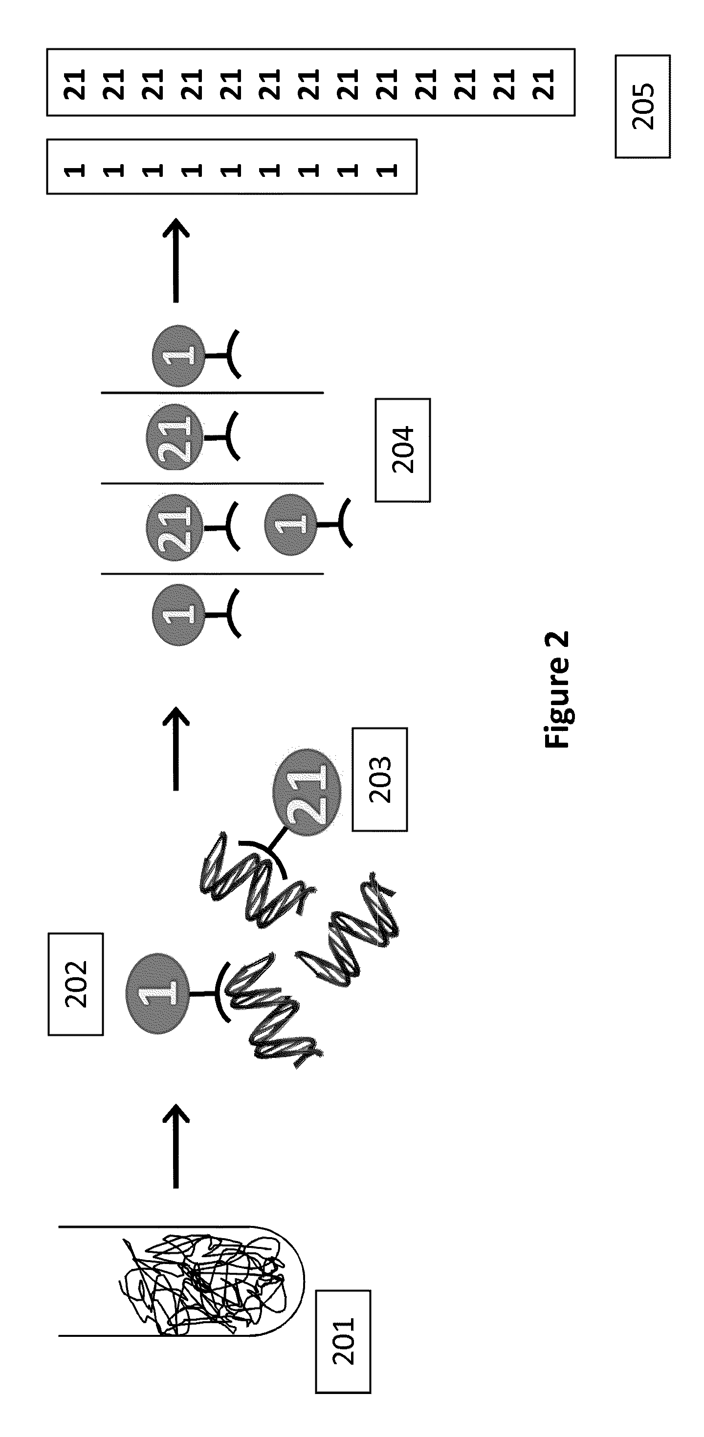
[0213]In this example, detection of a trisomy 21 fetal aneuploidy is described, where there is 3% fetal DNA in a maternal plasma sample. There are 1000 genome equivalents (GE) per mL in the maternal plasma, and a maternal blood volume of 20 mL is collected.

[0214]Plasma is isolated from the maternal blood sample by centrifugation, and the nucleic acids are purified and concentrated to a volume of 50 μL. The sample is mixed with an equal volume of PCR reagent containing the multiplexed assay components. The entire 100 μL sample is partitioned into 100,000 aqueous droplets having a volume of 1 nL per droplet. For an ideal positive droplet percentage for quantitation of 75%, this would mean 1.47 copies of target sequence per droplet, based on Poisson distribution, which translates to 147,000 targets that need to be compartmentalized into 100,000 1 nL droplets. The number of primer sets required to reach this is 147,000 GE / 10,000 G...

example 2

Detection of Fetal DNA Using a One-Color Detection Scheme

[0215]The conditions for Example 1 are used here, except that rather than using different colored target and reference probes, the sample is split (e.g. in half), then two set of droplets are generated, amplified and separately analyzed, with one half using a target probe and the other half using a reference probe.

example 3

Detecting Fetal DNA Using MIP-ddPCR

[0216]Cell-free plasma is isolated from a maternal blood sample by centrifugation. The nucleic acids are then purified and concentrated using a cell free DNA kit (Qiagen). The purified genomic DNA is then mixed with 1000 chromosome-sequence specific oligonucleotide probes (e.g., MIP probe) to Chromosome 21 (MIP-21Chr), and 1000 chromosome-sequence specific oligonucleotide probes (e.g., MIP probe) to Chromosome 1 (MIP-1Chr). Ligase, polymerase and other reaction components are added to the mix. The sample is incubated at 20° C. for 4 minutes. The sample is then incubated at 95° C. for 5 minutes to promote denaturation, and then at 60° C. for 15 minutes in order to promote annealing of the MIP probes to the genomic DNA. A gap fill reaction is then performed in order to circularize the MIP probes. (In some cases, the ends may be directly ligated without a gap fill reaction). Nucleotides are added to the sample, which is then incubated at 60° C. for 10...


PUM

PropertyMeasurementUnit
Temperatureaaaaaaaaaa
Fractionaaaaaaaaaa

Abstract

This invention provides compositions and methods for detecting differences in copy number of a target polynucleotide. In some cases, the methods and compositions provided herein are useful for diagnosis of fetal genetic abnormalities, when the starting sample is maternal tissue (e.g., blood, plasma). The methods and materials described apply techniques for allowing detection of small, but statistically significant, differences in polynucleotide copy number.

Description

CROSS-REFERENCE TO RELATED APPLICATIONS[0001]This application claims the benefit under 35 U.S.C. §119(e) of U.S. Provisional Application No. 61 / 264,591, filed on Nov. 25, 2009; U.S. Provisional Application No. 61 / 309,837, filed on March 2, 2010; U.S. Provisional Application No. 61 / 309,845, filed on Mar. 2, 2010; U.S. Provisional Application No. 61 / 317,635, filed on Mar. 25, 2010; U.S. Provisional Application No. 61 / 317,639, filed on Mar. 25, 2010; U.S. Provisional Application No. 61 / 317,684, filed on Mar. 25, 2010; U.S. Provisional Application No. 61 / 341,065, filed on Mar. 25, 2010; U.S. Provisional Application No. 61 / 341,218, filed on Mar. 25, 2010; U.S. Provisional Application No. 61 / 380,981, filed on Sep. 8, 2010; U.S. Provisional Application No. 61 / 409,106, filed on Nov. 1, 2010; U.S. Provisional Application No. 61 / 409,473, filed on Nov. 2, 2010; and U.S. Provisional Application No. 61 / 410,769, filed on Nov. 5, 2010, each of which is incorporated herein by reference in its entir...

Claims


Application Information

Patent Timeline
IPC IPC(8): C12Q1/68C07H21/00
CPCC12Q1/6827C12Q1/6806C12Q2537/157C12Q2563/161C12Q2533/107C12Q2521/319
Inventor HINDSON, BENAJAMINSAXONOV, SERGEBELGRADER, PHILLIPNESS, KEVINLUCERO, MICHAELCOLSTON, BILLY
Owner BIO RAD LAB INC
Learn More