Method, Implant & Instruments for Percutaneous Expansion of the Spinal Canal

Inactive Publication Date: 2010-07-01
INNOVATIVE SURGICAL DESIGNS
View PDF54 Cites 85 Cited by
  • Summary
  • Abstract
  • Description
  • Claims
  • Application Information

AI Technical Summary

Benefits of technology

[0017]The present invention comprises novel instruments for cutting the spinal pedicles in precise and reproducible location without injury to the surrounding delicate nerve tissue or fluid filled nerve sac. Also disclosed are novel spinal implants and devices capable of aligning the pedicle cut precisely to the spinal implant, performing the distraction of the pedicle cut and fixating the cut in the expanded state to allow bony healing to occur, thus permanently expanding the spinal canal. Further provided are spinal implants capable of gaining bony purchase at the site of the pedicle cuts to provide adequate grip of bony structure to allow pedicle distraction to be achieved.
[0018]In addition, a novel mechanism is provided, allowing for connection between the upper (proximal) and lower (distal) portions of the implant, even in the setting of misalignment which may occur during the pedicle lengthening process. Also disclosed is a simple, reproducible means of performing the pedicle lengthening procedure in the human spine through a percutaneous technique to relieve pressure on the spinal nerves.

Problems solved by technology

Spinal stenosis affects millions of people world wide and leads to symptoms of back and leg pain, weakness, numbness and trouble walking Spinal stenosis is a particularly common problem among older individuals and can result in severe disability and lack of normal mobility.
Although laminectomy, laminotomy and intraspinous spacer devices may all be successful for patients with spinal stenosis, each of these approaches has significant limitations in a significant proportion of patients with spinal stenosis.
For instance, laminectomy generally is a major spinal operation requiring general anesthesia which can lead to complications especially in older patients.
Several important disadvantages have been identified with the use of laminectomy to treat spinal stenosis such as damage to back muscles, destabilization of the spine and scarring around the nerve roots.
In some cases, destabilization of the spine may cause a serious forward slippage of one vertebra on the adjacent vertebra requiring a major revision surgery called spinal fusion.
Also, laminectomy requires a large surgical incision, leading to the risk of major bleeding and the need for general anesthesia.
Because most patients with spinal stenosis are elderly, major surgery such as laminectomy may lead to medical complications, making this approach suboptimal for the older, medically fragile patient.
In addition, laminectomy may not provide a permanent cure for spinal stenosis, which recur causing the need for further major surgery in the future.
However, laminotomy still requires major open spinal surgery and general anesthesia.
It also is more technically difficult to perform compared to laminectomy and may not adequately relieve the pressure on the spinal nerves.
In addition, there is a risk that with time, the spinal stenosis may recur, leading to the need for additional surgery.
Unfortunately, intraspinous devices only provide a slight expansion of the spinal canal compared to laminectomy and laminotomy.
Thus, intraspinous spacers are only useful in the subset of spinal stenosis patients with relatively mild stenosis.
Also, because the narrowed spinal canal is not significantly enlarged, and because the narrowing of the spinal canal worsens with time, intraspinous process device may only provide temporary relief of the symptoms of spinal stenosis.
Thus, many patients treated with laminectomy may ultimately require a laminectomy as their condition worsens.
Also, intraspinous process spacers are not able to be used in patients whose spinous processes are weakened by osteoporosis or absent due to a prior laminectomy procedure.
However, the approach of Bloemer has certain limitations and disadvantages that severely limit its usefulness for treating spinal stenosis.
First, no cutting method or tools disclosed by Bloemer allow the bone cuts to be performed.
In addition, the bone of the pedicle has thick and thin regions which make the cutting task a substantial challenge for which no instruments or tools have been previously known to the art of spine and bone surgery.
Second, the implants disclosed by Bloomer fail to gain purchase within the pedicle bone cut, but rather rely on bony purchase within the pedicle bore.
Third, the device of Bloemer does not provide for the geometric offset that tends to occur during pedicle lengthening.
This potential problem was not anticipated by Bloemer, and thus no mechanism to contend with geometric offset was disclosed by Bloemer.
Fourth, the disclosure of Bloemer provides no means to precisely align the pedicle cut with the portion of the implant that performs the pedicle lengthening maneuver.
Proper alignment of the implant with the pedicle cut is crucial for the pedicle lengthening device to work correctly and yet no means to achieve this alignment was disclosed.
For all these reasons, Bloemer fails to provide a workable concept to achieve pedicle lengthening for the correction of spinal stenosis.
Not surprisingly, the work of Bloemer has not been reduced to practice within the field of spine surgery, nor has research on the technique been disclosed in the public domain.

Method used


Image

  • Method, Implant & Instruments for Percutaneous Expansion of the Spinal Canal
  • Method, Implant & Instruments for Percutaneous Expansion of the Spinal Canal
  • Method, Implant & Instruments for Percutaneous Expansion of the Spinal Canal

Examples


Embodiment Construction

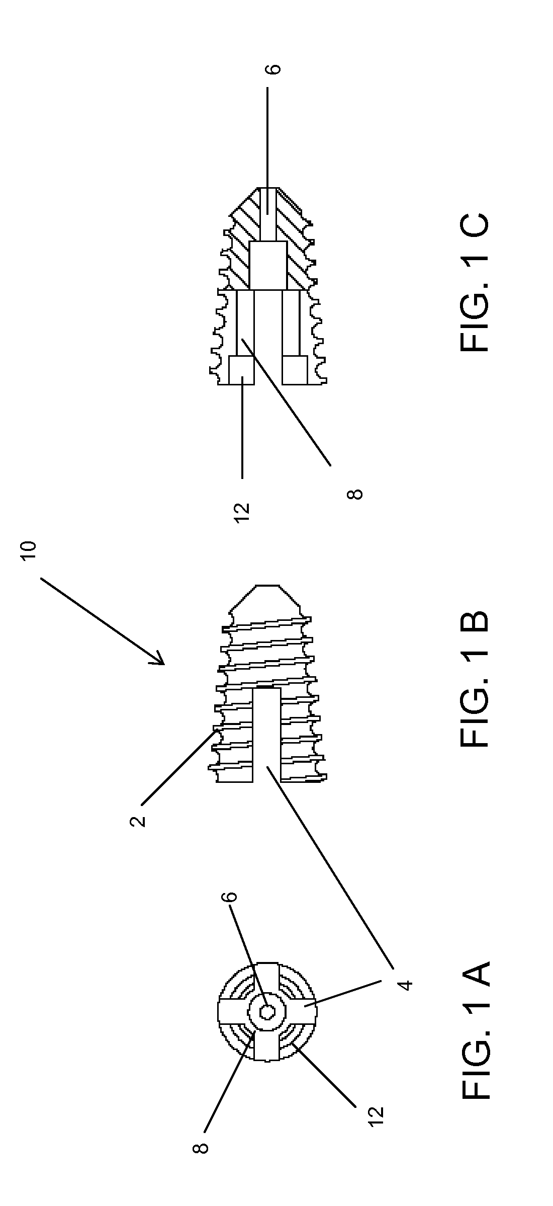
[0101]Referring now to the drawings, where like numerals indicate like elements, there is shown in FIGS. 1A-1C, a top view 1A, side view 1B, and cross-sectional view 1C, of a lower or distal shell portion 10 of a pedicle lengthening implant of the present invention. The lower implant shell 10 has outer threads 2 and hollow inner spaces 8, 12, along with side wall slots 4 and a cannulation hole 6.

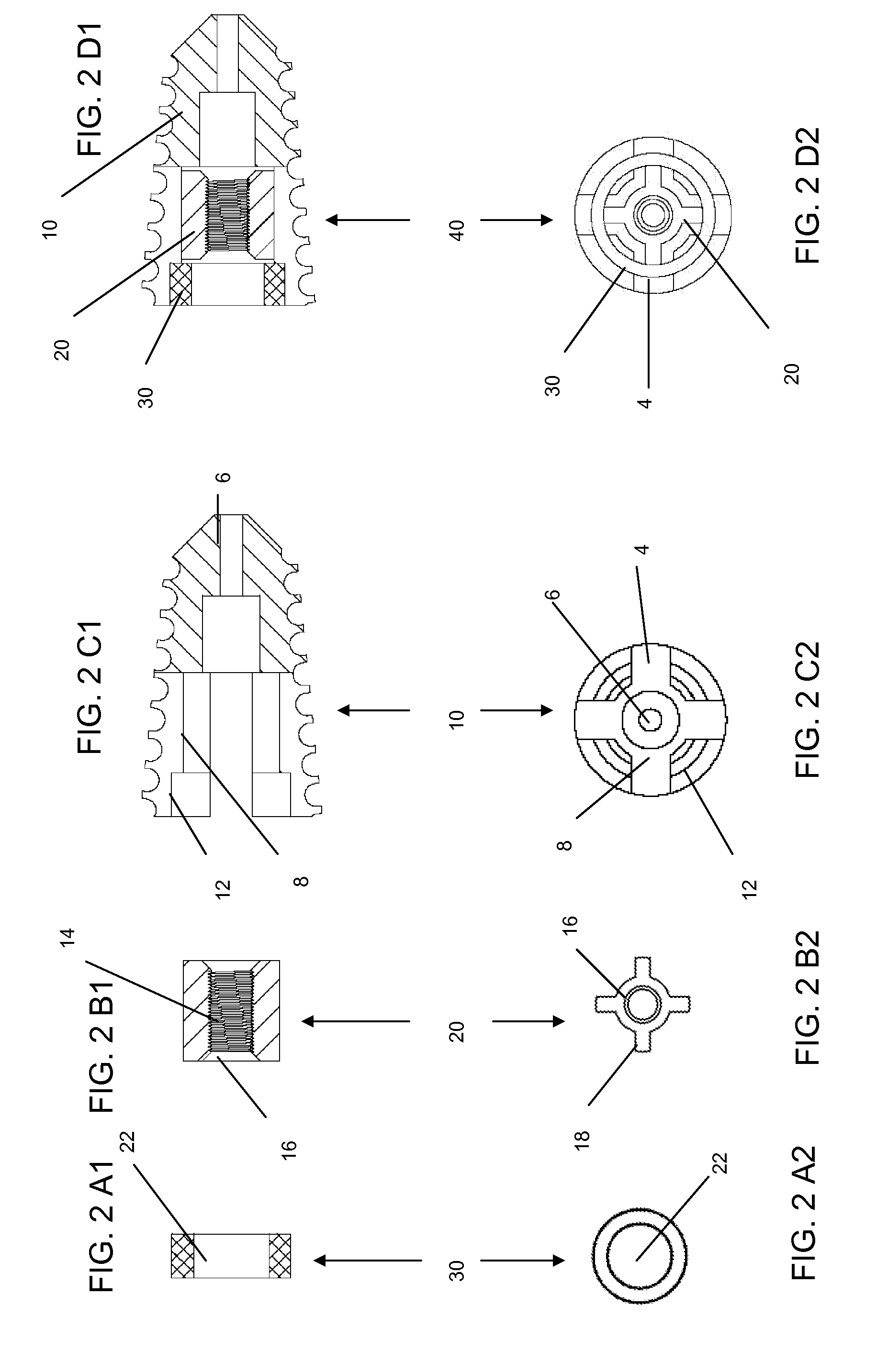
[0102]FIGS. 2A1-2D2 comprises lower implant components, including the lower implant shell 10, a floating nut 20 and a locking ring 30. The locking ring 30 is shown in cross-section, and by top view, in FIGS. 2A1 and 2A2, respectively. The locking ring 30 comprises a substantially circular shape and has a central passage 22. The floating nut 20 is shown in cross-section and by top view in FIGS. 2B1 and 2B2, respectively. The floating nut 20 contains a plurality of flanges 18 on its' outer surface, and a tapered entrance 16 to an inner threaded portion 14.

[0103]An assembled lower implant porti...


PUM


Abstract

A method for correcting spinal stenosis involves cannulating a passage in a vertebra, and placing a distal portion of an implant therein. Then, a circumferential vertebral cut is performed from within the passage, using a proximal end of the distal portion of the implant to align the vertebral cut. Then, a proximal portion of the implant is placed into the passage, positioned against the distal portion at the vertebral cut. Operation of the distal and the proximal portions of the implant relative to one another widens the vertebral cut and expands the spinal canal. The proximal portion can then be secured to the distal portion to stabilize the vertebral cut, allowing vertebral healing with the spinal canal expanded. Flanges may exist that radially extend outward from the implant and into the vertebral cut, or into the passage walls, to assist vertebral cut widening and stabilization.

Description

RELATED APPLICATIONS[0001]This application is a continuation-in-part (CIP) of U.S. Ser. No. 11 / 656,790, filed Jan. 22, 2007; which application is a continuation of U.S. Ser. No. 10 / 102,525, filed Mar. 19, 2002 (now U.S. Pat. No. 7,166,107). This application also claims benefit of U.S. Provisional Application Ser. No. 61 / 117,726, filed Nov. 25, 2008, entitled “Method, Implant & Instruments for Percutaneous Expansion of the Spinal Canal.” The above-identified related applications are incorporated herein by reference.FIELD OF THE INVENTION[0002]The present invention relates generally to spinal surgery, and more particularly to instruments and a device for cutting and lengthening the spinal pedicles to correct spinal canal narrowing or spinal stenosis and to relieve pressure on spinal nerves.BACKGROUND OF THE INVENTION[0003]Spinal stenosis is a condition or disease causing narrowing of the spinal canal and compression of the spinal nerves. Spinal stenosis affects millions of people worl...

Claims


Application Information

Patent Timeline
IPC IPC(8): A61B17/00A61B17/56A61M29/00
CPCA61B17/1617A61B17/1631A61B17/1637A61B17/1671A61B17/7071A61B17/8004A61B17/8685A61B2017/22038A61B17/142
Inventor ANDERSON, D. GREGTURNER, BARRYBEAMS, WAYNE
Owner INNOVATIVE SURGICAL DESIGNS
Why Eureka
Try Eureka