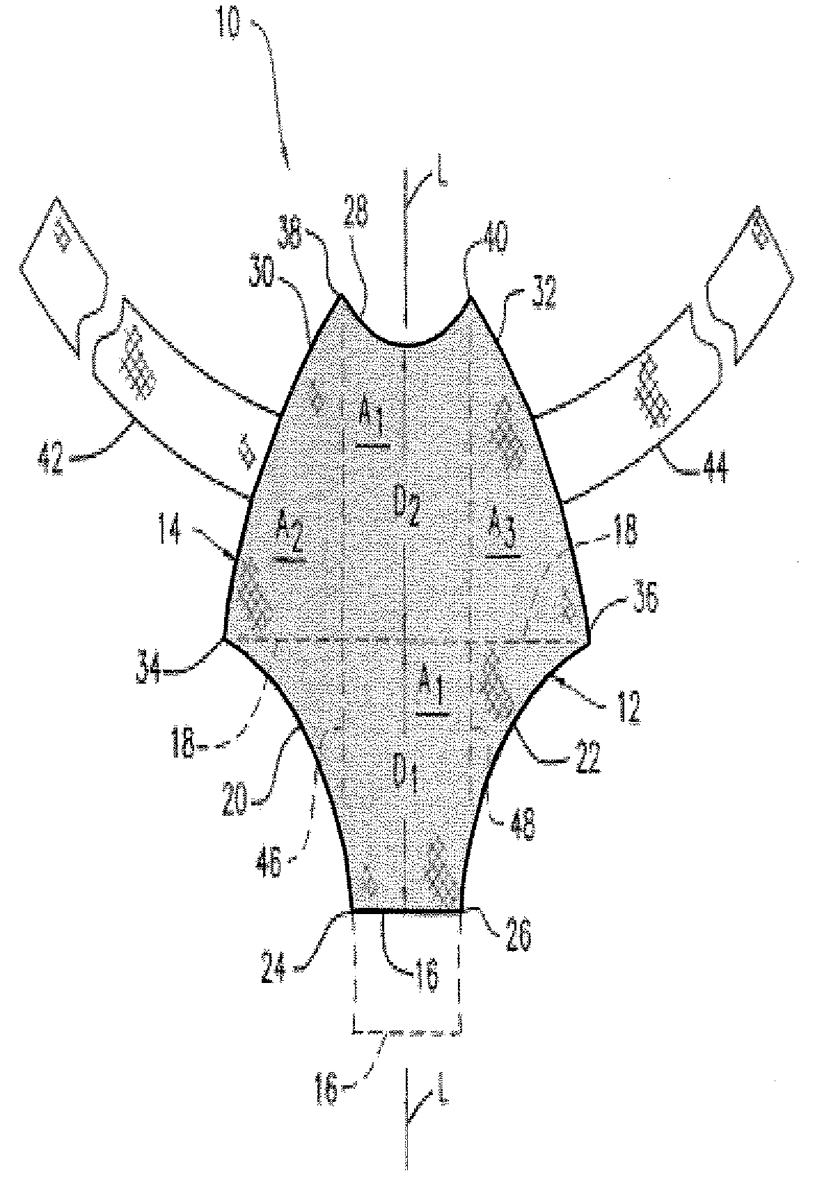
Biocompatible Fiber Based Device for Guided Tissue Regeneration
a biocompatible fiber and tissue technology, applied in the field of tissue repair and regeneration, can solve the problems of herniation of the bladder, severe and negative impact on the physiological and psychological health of patients, and injury of the vesicovaginal fascia
- Summary
- Abstract
- Description
- Claims
- Application Information
AI Technical Summary
Benefits of technology
Problems solved by technology
Method used
Image

Examples
example 1
Fabrication of Tissue Engineering Device for Pelvic Floor Repair by Needlepunching (GYNEMESH PS Mesh+90 / 10 PGA / PLA Nonwoven)
[0069]Nonwoven fiber batts of 90 / 10 (mol %) poly(glycolide-co-lactide) (90 / 10 PGA / PLA) fibers with a density of 2 mg / cm2 were prepared at Concordia Manufacturing, LLC (Coventry, R.I.). A 15 cm×15 cm piece of GYNEMESH PS mesh (Ethicon Inc, Somerville, N.J.) was sandwiched between the nonwoven fiber batts by placing a nonwoven batt on each side of the mesh. The construct was then passed through a needlepunch loom to interlock the fiber batts and hence embedding the mesh within the nonwoven felt. Two needlepunch passes were used to generate the devices. The GYNEMESH PS Mesh+90 / 10 PGA / PLA nonwoven scaffolds were 1.17 mm thick with a density of 75 mg / cc.
[0070]Samples were analyzed by scanning electron microscopy (SEM). The samples were mounted on a microscope stud and coated with a thin layer of gold using the EMS 550 sputter coater (Electron Microscopy Sciences, Ha...
example 2
Fabrication of Tissue Engineering Device for Pelvic Floor Repair by Needlepunching (ULTRAPRO Mesh+90 / 10 PGA / PLA Nonwoven)
[0071]Nonwoven fiber batts of 90 / 10 PGA / PLA fibers with a density of 2 mg / cm2 were prepared at Concordia Manufacturing, LLC (Coventry, R.I.). A 15 cm×15 cm piece of ULTRAPRO mesh (Ethicon Inc, Somerville, N.J.) was sandwiched between the nonwoven fiber batts by placing a nonwoven batt on each side of the mesh. The construct was then passed through a needlepunch loom to interlock the fiber batts and hence embedding the mesh within the nonwoven felt. Two needlepunch passes were used to generate the devices. The Ultrapro mesh+90 / 10 PGA / PLA nonwoven scaffolds were 1.03 mm thick with a density of 71 mg / cc.
[0072]A tissue engineering device prepared as described above was cut to 4 cm×4 cm for burst testing. Burst strength of the devices was evaluated using a Mullen Burst testing apparatus. The sample was placed in the clamp zone of the testing apparatus. The clamp was ac...
example 3
Fabrication of Tissue Engineering Device for Pelvic Floor Repair by Needlepunching (GYNEMESH PS Mesh+Nonwoven Fiber Batt Having a 50:50 Ratio of (90 / 10 PGA / PLA) Fibers:PDO Fibers)
[0074]Nonwoven fiber batts having a 50:50 ratio of 90 / 10 PGA / PLA fibers and polydioxanone (PDO) fibers with a density of 1 mg / cm2 were prepared at Concordia Manufacturing, LLC (Coventry, R.I.). A 15 cm×15 cm piece of GYNEMESH PS mesh (Ethicon Inc, Somerville, N.J.) was sandwiched between the nonwoven fiber batts by placing a nonwoven batt on each side of the mesh. The construct was then passed through a needlepunch loom to interlock the fiber batts and hence embedding the mesh within the nonwoven felt. Two needlepunch passes were used to generate the devices. The GYNEMESH PS Mesh+(50:50) (90 / 10 PGA / PLA):PDO nonwoven scaffolds were 0.97 mm thick with a density of 60 mg / cc.
PUM
| Property | Measurement | Unit |
|---|---|---|
| length | aaaaa | aaaaa |
| length | aaaaa | aaaaa |
| distance D1 | aaaaa | aaaaa |
Abstract
Description
Claims
Application Information
 - R&D
- Intellectual Property
- Life Sciences
- Materials
- Tech Scout
- Unparalleled Data Quality
- Higher Quality Content
- 60% Fewer Hallucinations
Browse by: Latest US Patents, China's latest patents, Technical Efficacy Thesaurus, Application Domain, Technology Topic, Popular Technical Reports.
© 2025 PatSnap. All rights reserved.Legal|Privacy policy|Modern Slavery Act Transparency Statement|Sitemap|About US| Contact US: help@patsnap.com