
Surgical correction of ptosis by polymeric artificial muscles

a polymer artificial muscle and eyelid technology, applied in the field of eyelid function restoration and ptosis correction, can solve the problems of cosmetic failure, non-natural function, etc., and achieve the effect of reliable operation and easy surgical implantability

Active Publication Date: 2010-02-18
SHAHINPOOR MOHSEN +1
View PDF3 Cites 1 Cited by
  • Summary
  • Abstract
  • Description
  • Claims
  • Application Information

AI Technical Summary

Benefits of technology

"The present invention provides a new surgical method and apparatus to restore eyelid function in patients with ptosis. It involves an implantable eye drop activated artificial muscle network that can be easily surgically implanted in the eye and operated over an extended period. The surgical method, artificial muscles, and eye drops correct ptosis by moving the eyelid in a natural motion to restore natural eyelid function. The invention has advantages over previous methods and provides a better solution for patients with ptosis."

Problems solved by technology

While this procedure allows the patient to raise the eyebrow to open the eyelid and therefore see from the eye, it suffers from a number of drawbacks.
Essentially, the procedure restores some eyelid function but that function is not natural.
This procedure is also a cosmetic failure because of the requirement for the patient to raise his or her eyebrow.

Method used


Image

  • Surgical correction of ptosis by polymeric artificial muscles
  • Surgical correction of ptosis by polymeric artificial muscles
  • Surgical correction of ptosis by polymeric artificial muscles

Examples


Embodiment Construction

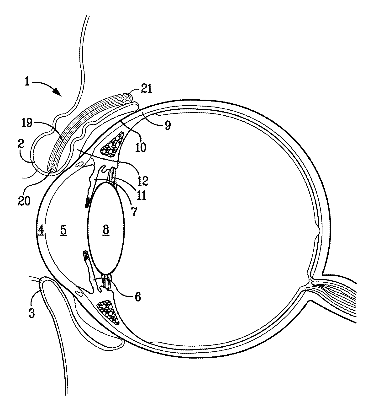
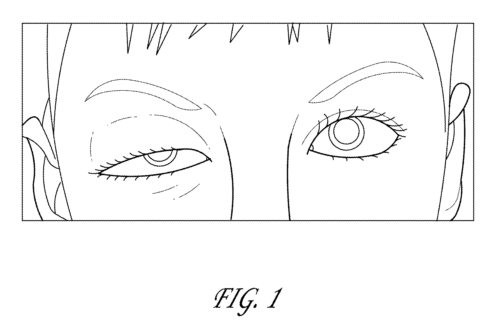
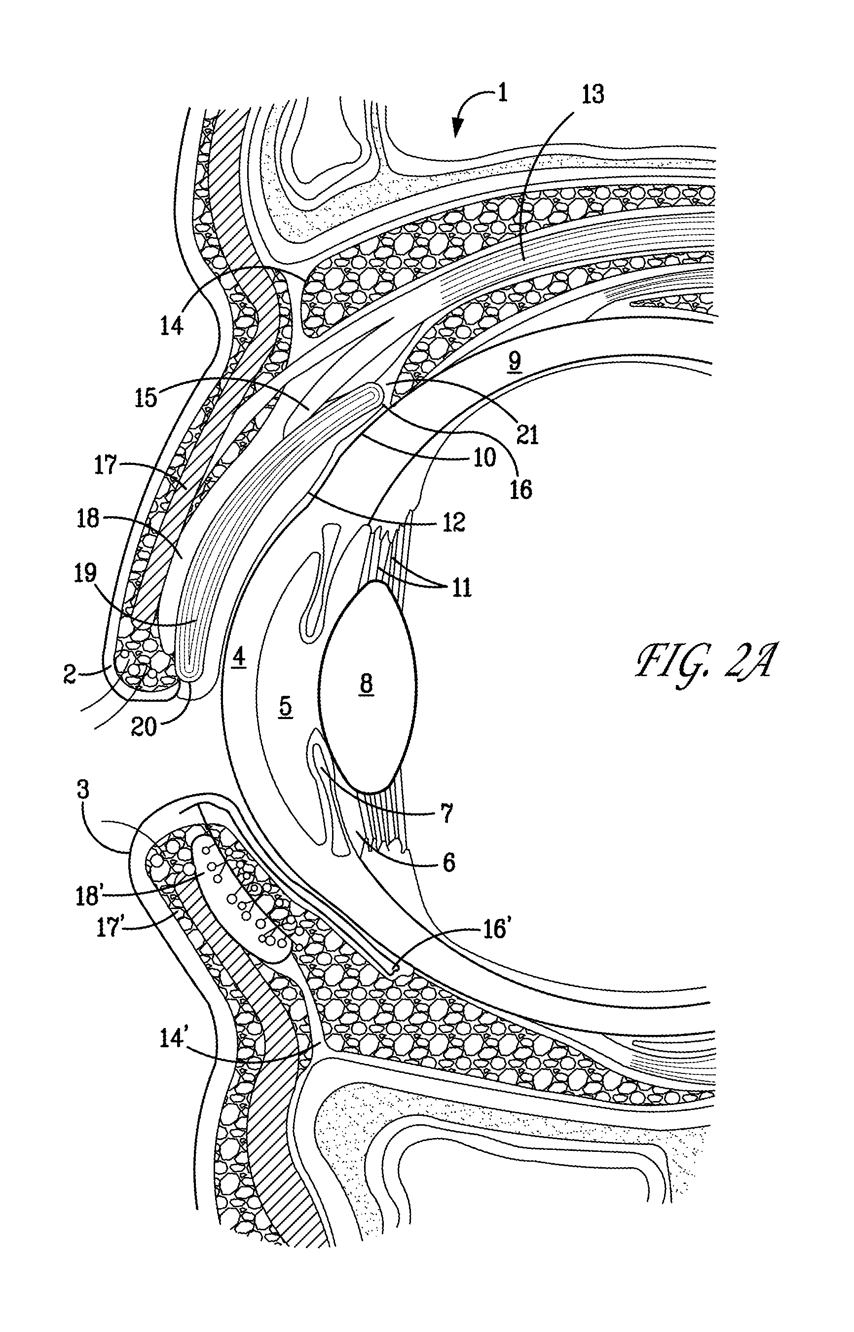
[0019]The current disclosure provides apparatus and methods for correcting ptosis. FIG. 1 shows a patient suffering from ptosis in one eye. As seen in FIG. 1, a typical ptosis manifests itself in the form of upper eyelid droop under normal conditions. Typically, a surgical approach can be implemented to correct the droopy eyelid. According to the present disclosure, an artificial muscle can be surgically implanted into the eyelid. The implant can then be biased in either an open or closed position with application of different biasing means. The biasing means may take the form of different chemical solutions, for example, in the form of eyedrops having different chemical properties such as different acidities.

[0020]As will be described in greater detail below, according to one embodiment of the present invention, biocompatible fibrous contractile and expansive ionic polymeric artificial muscles can be surgically implanted and sutured under the superior palpebral conjunctiva and sutu...


PUM


Abstract

A surgical procedure is described for the restoration of eyelid function in individuals suffering from ptosis or upper eyelid droop syndrome that makes a patient unable to voluntarily fully raise an eyelid. The surgical procedure includes implantation and suturing of eye drop (pH) activated and actuated fibrous contractile and expansive artificial muscles such as pH active hydrogels of polyacrylonitrile (PAN) artificial muscles that are surgically implanted and sutured under the superior palpebral conjunctiva in a serpentine parallel configuration with respect to the tarsal (meibomian) glands of the upper eyelid and anchored to the tissues of superior fornix.

Description

CROSS-REFERENCE TO RELATED APPLICATION[0001]This application is a continuation-in-part of U.S. Application No. 11 / 318,665, filed December 24, 2005, which is incorporated herein by reference.TECHNICAL FIELD[0002]The present invention is generally related to restoration of eyelid function and correction of ptosis and, in particular, to a surgical method for restoring eyelid function in a ptosis patient.BACKGROUND OF THE INVENTION[0003]A drooping of the upper eyelid below its normal level resulting in narrowing of the palpebral opening is known in ophthalmology as ptosis. The muscle controlling lid opening is the levator palpebrae superioris and it is innervated by the third cranial nerve (oculomotor nerve). Ptosis may be congenital or acquired. In congenital ptosis the levator palpebrae superioris muscle that elevates the lid is either absent or not well developed. Acquired ptosis, on the other hand, is usually due to either diseases or injuries of the nerves that control the movement...

Claims


Application Information

Patent Timeline
Patent Type & Authority Applications(United States)
IPC IPC(8): A61F2/08A61B17/06
CPCA61F2002/0894A61F2/08
Inventor SHAHINPOOR, MOHSENSOLTANPOUR, DAVID
Owner SHAHINPOOR MOHSEN
Learn More