


Quick Research
Generate reliable direction feasibility study reports for your R&D in just a few steps.

Technical Q&A
Discover and master advanced knowledge NOW. Basics, ideas, possibilities, all at once.

Find Solutions
As an expert in R&D theories, this can generate solutions to your technical problems instantly.

Evaluate Feasibility
Analyze your overall solution with one click, know your potential R&D risks in advance.

Monitor Landscape
Get weekly tech updates, stay abreast of the latest tech innovations and key insights.
Novel Antigens and Antibodies Associated to Pancreatic Ductal Adenocarcinoma
a technology of pancreatic ductal adenocarcinoma and antigens, which is applied in the field of new means for diagnosing and treating human cancers, can solve the problems of few and poorly reliable markers for early pda diagnosis
- Summary
- Abstract
- Description
- Claims
- Application Information
AI Technical Summary
Benefits of technology
Problems solved by technology
Method used
Image


Examples
example 1
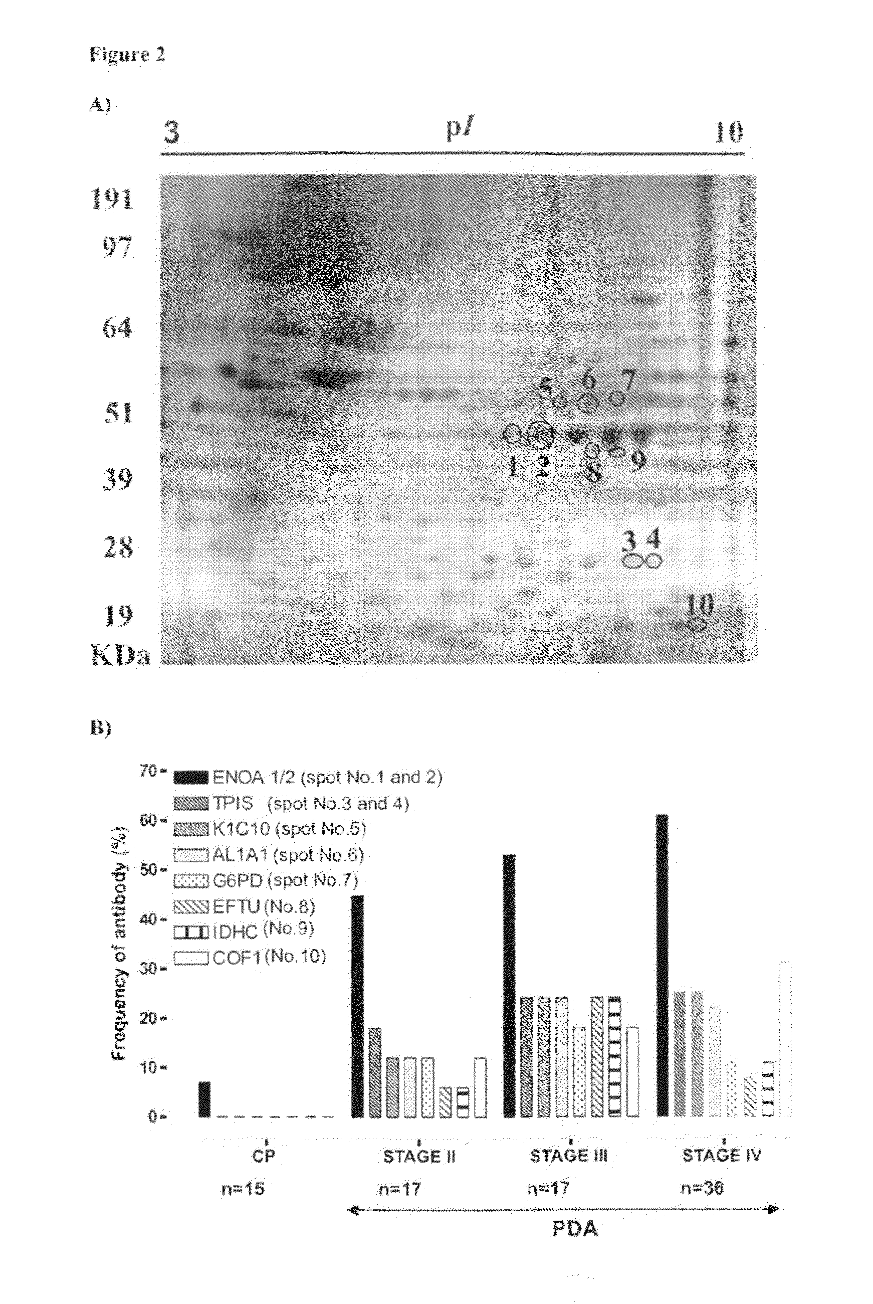
Detection of PDA-Associated Humoral Antigens in the Proteome of CP-PAC-1 Cell Line
Materials & Methods
Human Sera
[0050]Sera samples were isolated from venous blood with the informed consent of patients and healthy donors and the approval from the Ethical Committees of the clinical institutions. Samples were stored at −80° C. until use.
[0051]Sera was obtained from 70 PDA patients (31 men, 39 women; 32-86 range years; age mean±Standard Deviation: 67±11) were and grouped in different clinical stages according to the classification commonly used (Faria S et al., 2004) and to the UICC (Union International Contre le Cancer) classification as follows: 17 at stage II (no metastasis, moderately differentiated), 17 at stage III (no metastasis, poorly differentiated), and 36 at stage 1V (distant metastasis, undifferentiated).
[0052]The reactivity of these sera was compared to that of 40 healthy subjects that were used as controls without a prior history of cancer or autoimmune disease (14 men, 26...
example 2
Analysis of Alpha-Enolase Isoforms Detected by Autoantibodies in CF-PAC-1 Cells
Materials & Methods
[0082]Spots were excised from preparative 2-DE gels and analysed by Matrix-Assisted Laser Desorption Ionization / Time of Flight (MALDI-TOF). Proteins were de-stained overnight with a solution of 0.025 mol / L ammonium bicarbonate and 50% acetonitrile and then digested in gel with trypsin (Promega, Madison, Wis.) as previously described (Hellman U et al., 1995). For MALDI-TOF MS, 0.5 μL of each peptide mixture was applied to a target disk and allowed to air dry. Subsequently, 0.5 μL of matrix solution (1% w / v α-cyano-4 hydroxycinnamic acid in 30% acetonitrile, 0.1% TFA) was applied to the dried sample and again allowed to dry. Spectra were obtained using a Bruker Reflex III MALDI-TOF spectrometer (Bremen, Germany). The MS spectra interpretation of protein digests was done by “peptide mass fingerprinting” (PMF) using MS-Fit software.
Phosphorylation Analysis
[0083]The analysis of hu...
example 3
Effects of Antibodies Binding to Alpha-Enolase on the Growth and Proliferation of Transformed Cell Lines Expressing Phosphorylated Isoforms of Alpha-Enolase
Materials & Methods
Assay for Proliferation of Cell Lines
[0108]The in vitro assay of cell proliferation was performed with the indicated cell lines by seeding 2−10×103 cells / well in 96-well microplates in complete cell culture medium (RPMI-1640 with 10% Fetal Bovine Serum) with or without the anti-alpha-enolase monoclonal antibody 72 / 1 (Moscato S et al., 2000) or mouse IgG1 isotype matched control monoclonal antibody (R&D Systems) used as a negative control.
[0109]After 44 or 68 hours, 20 μl of methyl tetrazolium solution (MTT; 5 mg / ml) was added to each well for a further 4 hours at 37° C. Medium was eliminated and cells were dissolved with DMSO. Plates were read to the spectrophotometer at 540 nanometers.
Results
[0110]The effects of an anti-alpha-enolase antibody on the growth of cell lines presenting different profile of alpha-en...
PUM
| Property | Measurement | Unit |
|---|---|---|
| Volume | aaaaa | aaaaa |
| Volume | aaaaa | aaaaa |
| Volume | aaaaa | aaaaa |
Abstract
Description
Claims
Application Information
 - R&D Engineer
- R&D Manager
- IP Professional
- Industry Leading Data Capabilities
- Powerful AI technology
- Patent DNA Extraction
Browse by: Latest US Patents, China's latest patents, Technical Efficacy Thesaurus, Application Domain, Technology Topic, Popular Technical Reports.
© 2024 PatSnap. All rights reserved.Legal|Privacy policy|Modern Slavery Act Transparency Statement|Sitemap|About US| Contact US: help@patsnap.com