
Noninvasive trans-catheter method and apparatus for remote suture placement such as for septal defect repair, left atrial appendage closure, pacemaker electrode placement, mitral valve repair, and other inner-cardiac and inner-arterial applications

a transcatheter and remote suture technology, applied in the field of non-invasive transcatheter method and remote suture placement, can solve the problems of large amount of oxygen-rich (red) blood leakage from the heart's left side back to the right side, 25% lifetime risk of mortality in unrepaired atrial septal defects, and hole in the atrial septum

Inactive Publication Date: 2009-12-10
ENSION
View PDF33 Cites 58 Cited by
  • Summary
  • Abstract
  • Description
  • Claims
  • Application Information

AI Technical Summary

Benefits of technology

[0074]In one non-limiting embodiment of the present invention the suture line is configured to include

Problems solved by technology

When there is a large defect between the atria, a large amount of oxygen-rich (red) blood leaks from the heart's left side back to the right side.
This is inefficient, because already-oxygenated blood displaces blood that needs oxygen.
Rarely, if the defect is left un-repaired if there's pulmonary hypertension (high blood pressure in the lungs).
There is a 25% lifetime risk of mortality in un-repaired atrial septal defects.
Certain types of ASD's (sinus venosus and primum varieties) have no chance of spontaneous closure, and patients with these types of ASD's are not candidates for transcatheter closure because of the location of the ASD.
If a problem occurs during this process, a hole in the atrial septum may result.
Infants and children with larger, more severe ASDs, however, may possibly show some of the following signs or symptoms: poor appetite, poor growth, fatigue, shortness of breath, lung problems and infections, such as pneumonia.
If an ASD is not treated, health problems can develop later, including an abnormal heart rhythm (known as an atrial arrhythmia) and problems in how well the heart pumps blood.
As children with ASDs get older, they may also be at an increased risk for stroke, since a blood clot that develops can pass through the hole in the wall between the atria and travel to the brain.
The submammary incision may be the most cosmetic, but makes some ASDs difficult to repair.
Defects at the very upper or lower edges of the atrial septum (called ostium primum or sinus venosus) are not good candidates for this procedure.
These complications include the routine risks of cardiac catheterization such as vascular injury (damage to the veins and arteries of the leg), particularly in cases where larger device introducer systems need to be used.
A complication unique to this technology may be the possibility of clot formation on the device itself, with the risk of breakage of the clot causing stroke, or a clot into the vessels of the lung (pulmonary embolus).
The article goes on to suggest that the ASD occluder system (ASDOS) is no longer a widely used device nowadays, but, however, it is implanted in a substantial number of patients.
Microscopy, microbiology, and histology could not establish a proper explanation for the dysfunction; so the article concluded that long-term follow-up investigation may be required in patients with an implanted ASDOS device.
Currently there are no specially designed devices for PFO closure that is approved by the FDA in the United States.
These impulses are relatively rapid and erratic, and are known to not properly control the contractions of the heart.
One of the many problems caused by atrial fibrillation is the pooling of blood in the left atrial appendage during fibrillation.
These blood clots can cause serious problems when the heart resumes proper operation (normal sinus rhythm) and the blood, along with the blood clot(s), is forced out of the left atrial appendage.
Similar problems also occur when a blood clot extending from an atrial appendage into an atrium breaks off and enters the blood supply.
Thus, the blood flow will move the clots into the arteries of the brain and heart which may cause an obstruction in blood flow resulting in a stroke or heart attack.
Consequently, patients with atrial fibrillation also have an increased risk of stroke.
While such treatment can significantly reduce the risk of stroke, it also increases the risk of bleeding and for that reason is inappropriate for many atrial fibrillation patients.
Because of the significant risk and trauma of such procedures, left atrial appendage removal occurs almost exclusively when the patient's chest is opened for other procedures, such as coronary artery bypass or valve surgery.
While such procedures may be performed while the heart remains beating, they still require deflation of the patient's lung and further require that the patient be placed under full anesthesia.
Furthermore, placement of a chest tube is typically required to re-inflate the lung, often requiring a hospitalization for a couple of days.
A thoracoscopic approach which is also suggested by Whayne et al. suffers from the same problems as the thoracoscopic approach suggested by Johnson.
Not all damaged valves are suitable for repair—in some the state of valve disease is too advanced and replacement is necessary.
Often a surgeon can only make a decision of repair versus replace during the actual operation.
Abnormal electrical activity in the heart tissue causes cardiac arrhythmias such as atrial fibrillation, which if left untreated can be harmful or life threatening as previously described.

Method used


Image

  • Noninvasive trans-catheter method and apparatus for remote suture placement such as for septal defect repair, left atrial appendage closure, pacemaker electrode placement, mitral valve repair, and other inner-cardiac and inner-arterial applications
  • Noninvasive trans-catheter method and apparatus for remote suture placement such as for septal defect repair, left atrial appendage closure, pacemaker electrode placement, mitral valve repair, and other inner-cardiac and inner-arterial applications
  • Noninvasive trans-catheter method and apparatus for remote suture placement such as for septal defect repair, left atrial appendage closure, pacemaker electrode placement, mitral valve repair, and other inner-cardiac and inner-arterial applications

Examples


Embodiment Construction

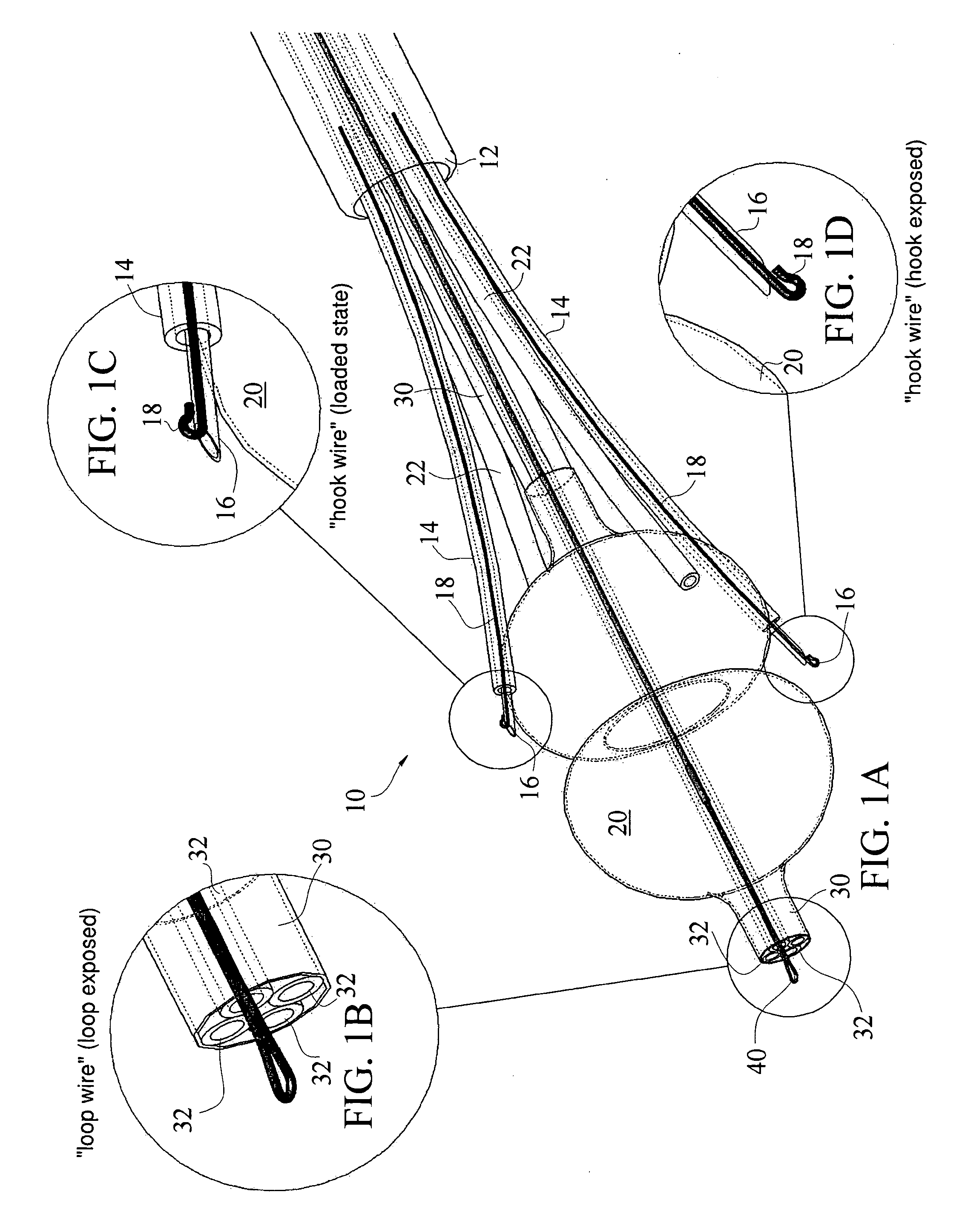
[0088]Non-Invasive Trans-Catheter Septal Defect Repair Device 10

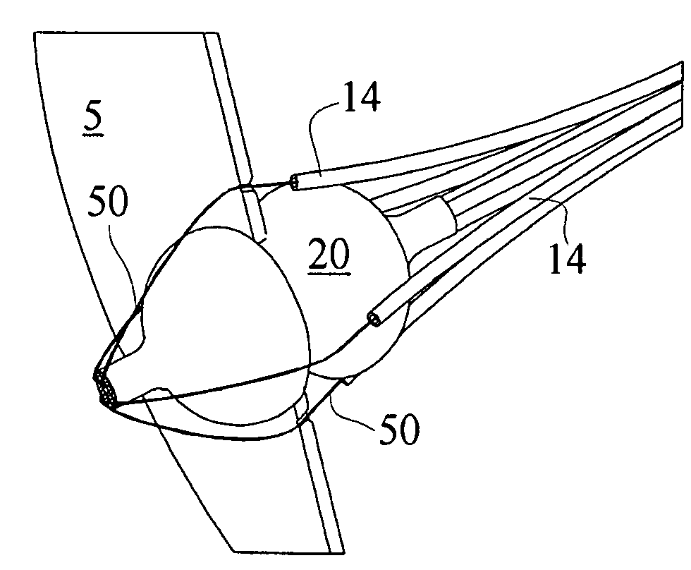
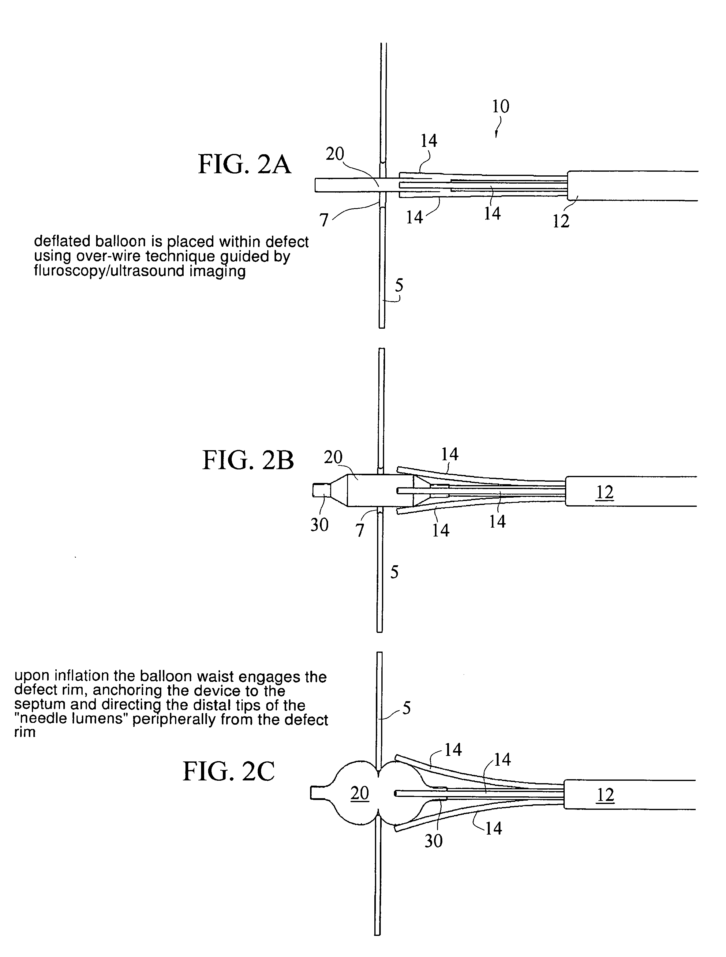
[0089]According to one non-limiting embodiment of the present invention, a summary or overview of a method of non-invasive transcatheter atrial septal defect repair comprises the steps of: advancing a positioning member 20 which is part of device 10 along a catheter 12 into the Atrial Septal Defect 7 of tissue 5, wherein at least one suture deploying lumen 14 is coupled to the positioning member 20 with a piercing member 16 within the suture deploying lumen 14; deploying the positioning member 20 within the Atrial Septal Defect 7 to align each suture deploying lumen 14 within close axial proximity to tissue 5 just adjacent to the Atrial Septal Defect 7; facilitating puncture of the tissue 5 adjacent the Atrial Septal Defect with the piercing member 16 to secure a suture line 50 through the tissue 5. A repair patch 90 may be advanced along suture lines 50 to repair the defect 7 and secured into place using an appropriate...


PUM


Abstract

A noninvasive trans-catheter method and apparatus for remote suture placement can be used in a variety of inner cardiac applications. For example, a method of non-invasive transcatheter atrial septal defect repair comprises the steps of: advancing a positioning member along a catheter into the Atrial Septal Defect, wherein at least one suture deploying lumen is coupled to the positioning member with a piercing member within the suture deploying lumen; deploying the positioning member within the Atrial Septal Defect to align each suture deploying lumen with tissue adjacent the Atrial Septal Defect; and piercing the tissue adjacent the Atrial Septal Defect with the piercing member to secure a suture line through the tissue. A repair patch may be advanced along suture lines to repair the defect and secured into place with the suture lines. The apparatus is also applicable for left atrial appendage closure, mitral valve repair and pacemaker electrode placement.

Description

[0001]The present invention claims priority of U.S. Provisional Patent Application Serial No. [REDACTED] entitled “Noninvasive Trans-Catheter Method and Apparatus for Remote Suture Placement such as for Septal Defect Repair, Left Atrial Appendage Closure, Pacemaker Placement, Mitral Valve Repair, and other Inner-Cardiac and Inner-Arterial Applications” filed [REDACTED].BACKGROUND OF THE INVENTION[0002]1. Field of the Invention[0003]The present invention relates to noninvasive, trans-catheter method and apparatus for remote suture placement, such as non-invasive inner-cardiac suture placement for Atrial Septal Defect (ASD) repair with a suture secured patch.[0004]2. Background Information[0005]Atrial Septal Defect (ASD)[0006]The septum is a wall that separates the heart's left and right sides. Septal defects are sometimes called a “hole” in the heart. A defect between the heart's two upper chambers (the atria) is called an atrial septal defect (ASD). When there is a large defect betw...

Claims


Application Information

Patent Timeline
IPC IPC(8): A61B17/12
CPCA61B17/0057A61B17/0401A61B17/12122A61B17/12131A61B2017/00557A61B2017/00575A61B2017/22069A61B2017/00663A61B2017/0409A61B2017/0417A61B2017/0464A61B2017/0472A61B2017/0061
Inventor FILL, BRIAN J.
Owner ENSION
Learn More