
Methods and Systems for Lesion Localization, Definition and Verification

a technology of lesion localization and definition, applied in the direction of magnetic variable regulation, therapy, instruments, etc., can solve the problems of certain complications, treatment failures, and radiation doses that cannot be delivered to the correct location in the patient's body

Inactive Publication Date: 2009-11-05
FALCO TONY +1
View PDF4 Cites 26 Cited by
  • Summary
  • Abstract
  • Description
  • Claims
  • Application Information

AI Technical Summary

Benefits of technology

[0058]a means for fusing said diagnostic image and said ultrasound image using said first three-dimensional coordinate and said second three-dimensional coordinate so as to obtain an accurate image of said tumour.

Problems solved by technology

However, the delivery of the necessary tumourcidal dose may result in certain complications due to damage of healthy tissue that surround the tumour, or due to damage to other healthy body organs located in the proximity of the tumour.
The basic problem in conformal radiation therapy is knowing the location of the target, or lesion or tumour, or alternatively of the healthy organs with respect to the intended placement of the radiation beam or field (I) prior to the design of a radiation treatment plan and (II) at the time of the radiation treatment.
However, if the position of the target volume is not correctly determined (I) prior to the treatment plan creation or (II) at the time of treatment, treatment failures can occur in a sense that the conformal dose of radiation may not be delivered to the correct location within the patient's body.
This may occur since not all conventional diagnostic imaging devices adequately, completely or fully determine the exact shape, size and orientation of a tumour, resulting in that even with the use of the most up-to-date diagnostic imaging device, some tumours may not be fully diagnosed.
Failures of type (II) can also occur from incorrect positioning of the patient on the treatment table of the radiation treatment unit.
Delivering the maximum radiation dose to a larger volume of healthy tissue or healthy organs may increase the risk of damaging these, and may for example, promote future cancers in the healthy surrounding tissue.
For this reason oncologists using present conformal radiation therapy may decide to deliver a lower radiation dose to the intended treatment volume in order to spare the non-target tissue with the potential disadvantage of compromising the success of the treatment by underdosing some portion of the target organ.
1. The transrectal ultrasound probe may considerably displace the lesion or organ thus providing inaccurate information about the spatial location of the lesion at treatment time if at that time the transrectal probe is not re-inserted. In any event, the insertion and removal of the probe prior to initiating treatment may cause displacement of the lesion, adding further uncertainty to the localization of the tumour. Moreover, inserting the transrectal probe for each treatment session can cause significant patient discomfort, resulting in this method not gaining popularity with physicians.
2. Holupka provides only for two dimensional images, and assumes that the 2D ultrasound image and the image obtained with the conventional diagnostic imaging modality are acquired in the same plane. For this case two identifiable fiducials in both images would be sufficient to register and superimpose the images. However, there is no certainty that the ultrasound image and the image from the conventional diagnostic imaging device are providing images in the same imaging planes and therefore a deviation of one image from the plane of another may considerably compromise the accuracy of the method.
3. The above said method registers and superimposes a two-dimensional ultrasound image onto a 2-dimensional image acquired with a conventional diagnostic imaging modality. Thus the ultrasound definition of the lesion is performed only in a single plane. For the purposes of three-dimensional conformal therapy, a two-dimensional definition of the lesion is incomplete and therefore inadequate since in other imaging planes, the extent of the lesion volume may be larger or smaller.
4. Further, Holupka is of limited application since it may only be used with respect to a very limited number of tumours, such as of the rectum, lower large intestine, and of the prostate. It can not be used for other type of tumours.
However, the following shortcomings may limit the utility of the above said system.
Thus the process of comparing outlines of the outer surfaces of the tumour or lesion or organ as they appear in images obtained with different imaging devices may be inaccurate since these surfaces can be different both in appearance and extent.
In other words, Carol compares apples and oranges, which results in an incomplete assessment of the tumour.
2. Carol also does not address failures of type I whereby the diagnostic images obtained with computed tomography or magnetic resonance imaging devices do not reveal completely the location or the extent of the tumour or lesion or organ, due to the inherent limitation of said devices with respect to certain tumours in certain locations.
Furthermore if the computed tomography or magnetic resonance diagnostic images do not reveal, or completely reveal, the tumour or organ or lesion, Carol will lack the means to outline an outer surface to serve as a reference for the comparison to the outer surface of the tumour or lesion or organ outlined on the one or more ultrasound images.

Method used


Image

  • Methods and Systems for Lesion Localization, Definition and Verification
  • Methods and Systems for Lesion Localization, Definition and Verification
  • Methods and Systems for Lesion Localization, Definition and Verification

Examples


Embodiment Construction

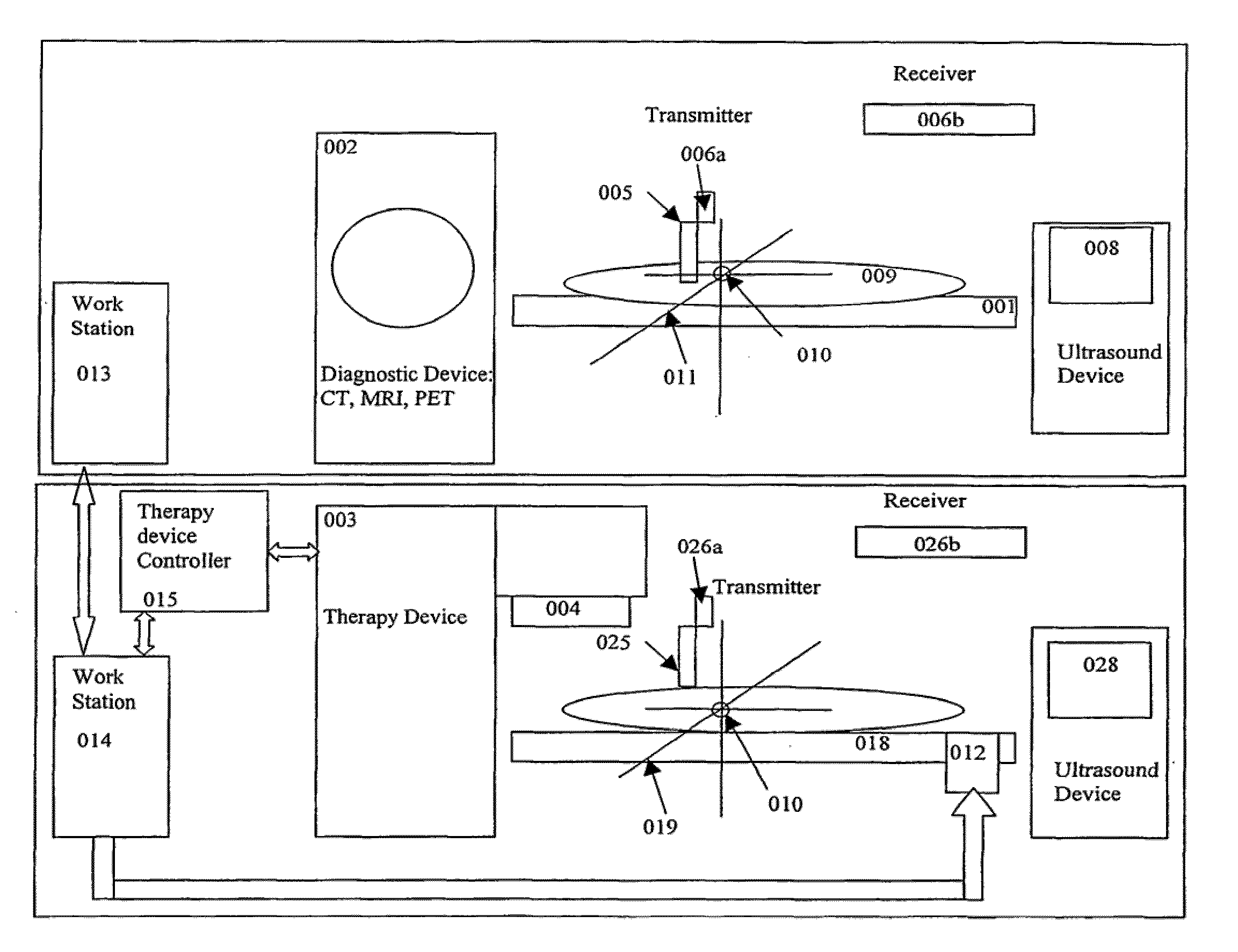
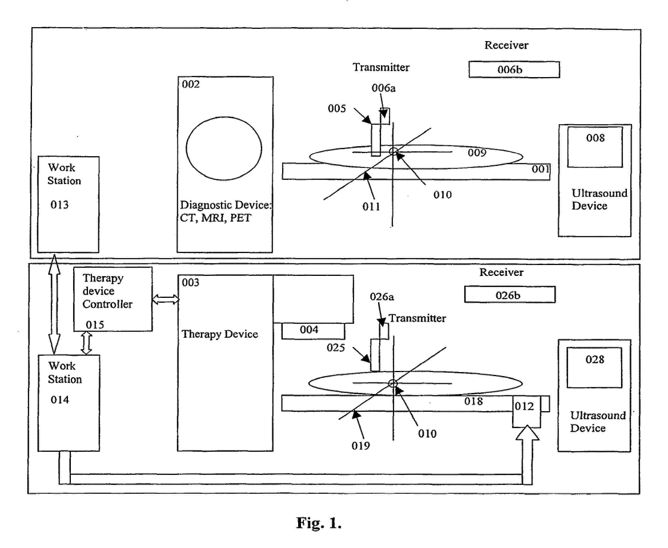
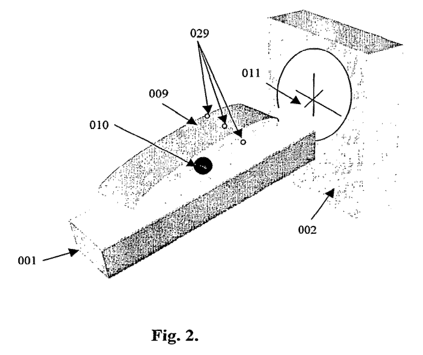
[0080]An illustration of an embodiment of the method and apparatus of the present invention is shown in the components of the apparatus and images derived from the figures. In the schematic diagram of FIG. 1 the embodiment of the invention is generally illustrated. In order to achieve one of the objectives of the present invention, that is, to obtain the most accurate possible definition of the size, location and orientation of a tumour 010, it has been found that the target area of a patient's body 009 believed to comprise a tumour 010 may be scanned or diagnosed using two distinct diagnostic apparatuses, and that the resulting images be compared. This may be achieved by comparing the image of the tumour 010 acquired through the use of a diagnostic device selected from group comprising an MRI, CT or PET with the image of the tumour 010 obtained with an ultrasound apparatus, such as those of Acuson, GE Medical Systems, Siemens, Toshiba and others. The order in which the two images i...


PUM


Abstract

A method and apparatus for lesion or organ definition for the purpose of radiation treatment planning localization and treatment position verification. The apparatus uses a combination of an ultrasound imaging system and a diagnostic imaging system to acquire localization ultrasound images referenced in the coordinate space of the diagnostic imaging system through the use of position sensing system. The method compares the location of the lesion in the localization ultrasound images with the position of the lesion in ultrasound images taken while the patient lies on the treatment table of a therapy treatment unit, suggests corrective measures to place the lesion in its intended treatment position and executes the correction upon confirmation from qualified personnel.

Description

BACKGROUND OF THE INVENTION[0001]1. Field of the Invention[0002]The invention relates to a method wherein the shape, form, position and configuration of a lesion or tumour, to be treated by a radiation therapy device, may be ascertained with greater definition, in order to better design a treatment plan for its eradication. In accordance with a further aspect, the invention also relates to method and apparatus for verification of the position of the lesion with respect to the radiation beam or beams prior to the execution of a radiation treatment. The invention relates to a method wherein the size, location and disposition of a tumour may be determined, updated and tracked prior to and during treatment therefor.[0003]2. Description of Prior Art[0004]The goal of modern day radiation therapy of cancerous tumours or lesions, is to eradicate the tumour while avoiding to the maximum extent possible damage to healthy tissue and organs in the vicinity of the tumour. Since the large majorit...

Claims


Application Information

Patent Timeline
IPC IPC(8): A61B8/13G06K9/00A61B5/055A61B6/03G01R33/28A61B8/00A61B8/08A61N5/10G01T1/161
CPCA61B8/08A61N5/1049A61N2005/1058A61N2005/1051A61N2005/105A61B8/4245
Inventor FALCO, TONYHRISTOV, DIMITRE
Owner FALCO TONY
Learn More