
Apparatus for providing endoscopic high-speed optical coherence tomography

Inactive Publication Date: 2009-09-10
THE GENERAL HOSPITAL CORP
View PDF106 Cites 23 Cited by
  • Summary
  • Abstract
  • Description
  • Claims
  • Application Information

AI Technical Summary

Benefits of technology

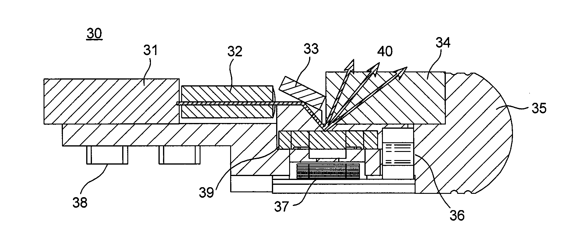
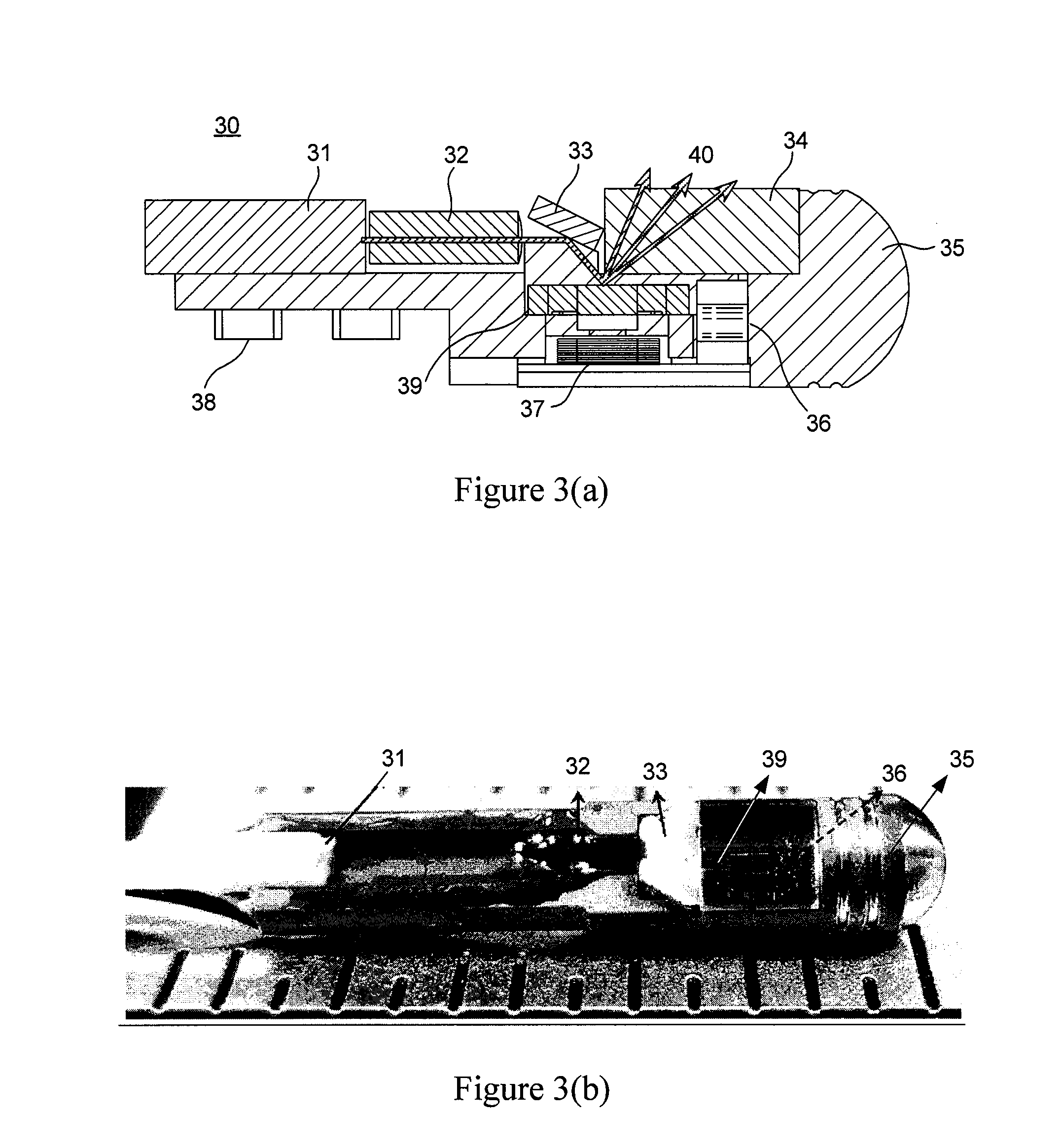
[0021]It is one of the objects of the exemplary embodiments of the present invention to resolve the issue described above with respect to the resonant vibration. Magnetic actuation requires low voltages (1˜3 V), but with somewhat large currents. For example, the power consumed in the catheter can be estimated to be approximately 150 mW by driving both axes. The temperature of the body of the exemplary embodiment of the catheter can be approximately 45° C. due to small body size and thin electrical wires. More beneficial heat sinking may be obtained for larger scanning ranges in the next generation catheter. Additional metal wires can be connected to the catheter body for a heat dissipation, such that certain mechanical rigidity which is preferable for a catheter operation can be obtained. Further, the power consumption can be reduced by moving the coils closer to the mirror.
[0023]According to another exemplary embodiment of the present invention, it may be possible to provide an endoscopic system based on a miniaturized scanning probe. The exemplary probe can have a scanning mirror at the tip, which may be fabricated based on MEMS technology. Such exemplary probe can scan the beam of light in a 2D pattern for imaging or laser treatment. For imaging, the probe may be integrated with a high-speed OCT system, allowing it to be used to image an internal tissue in 3D for diagnosis. The exemplary probe can also be combined with commercial endoscopes in order to navigate to internal organs, and / or used as a light treatment device in addition to 3D imaging. The exemplary embodiment of the system can determine the extent of treatment region based on 3D OCT imaging, so that it can perform a selective and precise treatment. Such treatment can include tissue treatment procedures such as photodynamic therapy using photoactivated sensitizers or drugs, arrangements to kill, ablate or coagulate diseased tissue or blood vessels by photothermal, photochemical or other means, or remove tattoos.
[0031]In a further exemplary embodiment of the present invention, an apparatus can be provided which may include at least one first arrangement which is configured to generate a magnetic field, at least one second arrangement coupled to the at least one first arrangement and configured to receive at least one first electro-magnetic radiation from an energy-generating arrangement to generate at least one second electro-magnetic radiation. The second arrangement(s) can includes at least one surface that may be at least partially reflective, and the magnetic field can control a motion of the at least one surface. The second electro-magnetic radiation(s) can effect a structure of the sample.

Problems solved by technology

However, for imaging non-tubular organs, conventional catheters scanning linearly may be limited in scanning speed to a few frames / s due to various factors including large inertia, friction, and compliance related to proximal actuation.
Furthermore, these translational catheters likely only generate two-dimensional images by scanning along a single axis.
Therefore, the conventional OCT catheters limit the ability to perform high-speed three-dimensional imaging of non-tubular organs.
However, these comb-drive actuators generally use small gaps and high driving voltages (e.g., ˜100V), with their potential failure being a concern for patient safety.
One of the disadvantages of the small size catheters has been that the scanning lengths of such catheters generally decreases with the reduction of outer diameter given a fixed scanning angle.
It still has remained a challenge to provide an OCT system to image asymmetric shaped organs at high speeds.
Further, conventional laser treatment methods are likely not incorporated with OCT systems, and the extent of the lesion or treatment is determined based on wide field imaging.
Further, in the prior art, there is no known cost effective ways to perform the treatment with a precise position control.

Method used


Image

  • Apparatus for providing endoscopic high-speed optical coherence tomography
  • Apparatus for providing endoscopic high-speed optical coherence tomography
  • Apparatus for providing endoscopic high-speed optical coherence tomography

Examples


Embodiment Construction

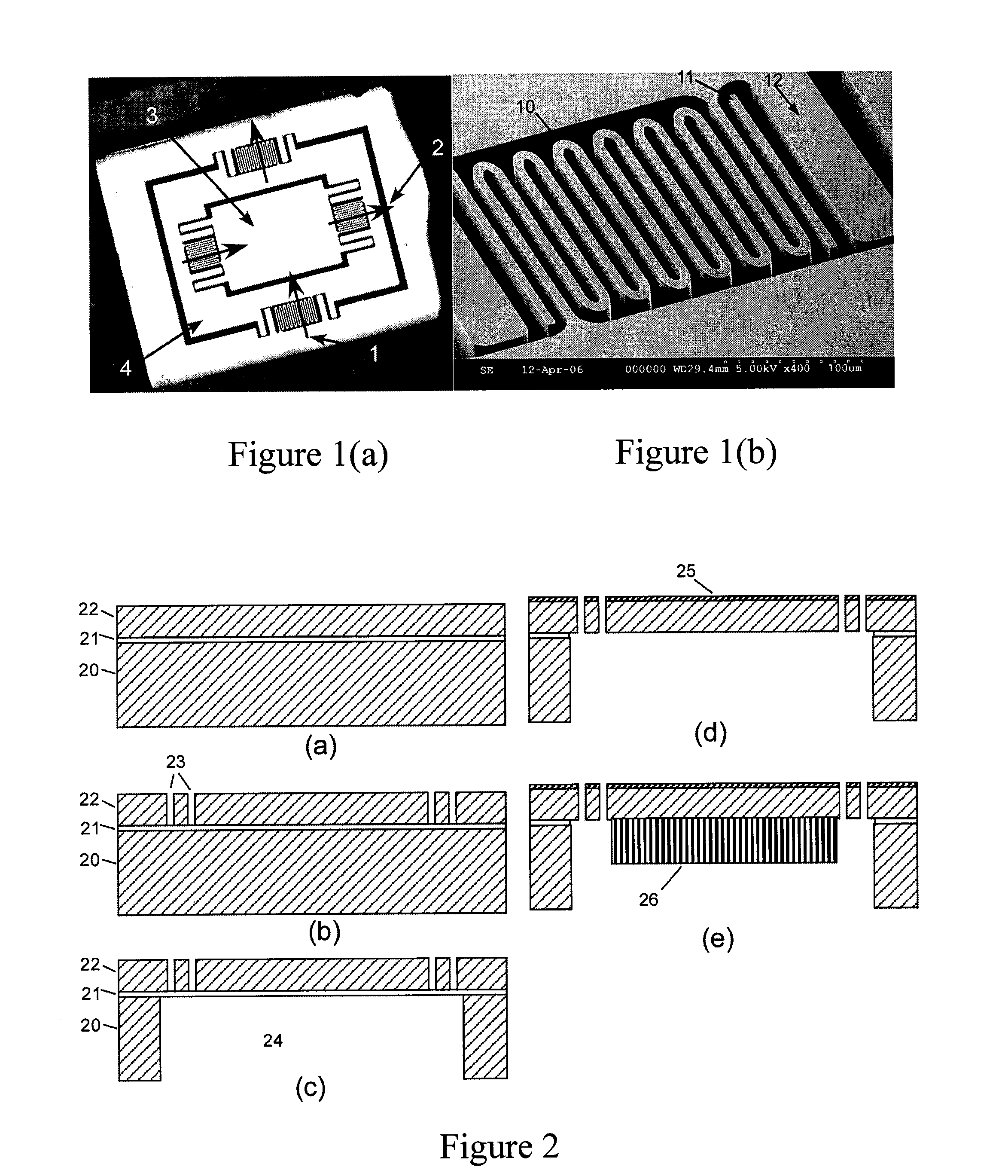
[0018]Accordingly, it may be beneficial to provide an exemplary embodiment of an apparatus for providing endoscopic high-speed optical coherence tomography such as, e.g., a two-axis MEMS micro-mirror scanning catheter according to the present invention which can be actuated by, e.g., a magnetic field for endoscopic SD-OCT imaging. This exemplary embodiment of the MEMS arrangement according to the present invention can be actuated either statically (i.e. below resonance) or at its resonant frequency (e.g., typically between 100 and 1000 Hz). Therefore, the exemplary implementation of high-speed endoscopic OCT imaging procedures may be effectuated using this exemplary catheter.

[0019]For example, the exemplary embodiment of a scanner according to the present invention can have a scanning range of about ±20° in optical angle in both axes with low driving voltages (e.g., 1˜3 V). According to one exemplary embodiment of the present invention, the catheter can have an outer diameter of abo...


PUM


Abstract

Exemplary embodiments of an apparatus can be provided which can include at least one first arrangement which is configured to generate a magnetic field. Further, the exemplary apparatus can include at least one second arrangement coupled to the first arrangement(s) and configured to receive at least one first electro-magnetic radiation from a sample to generate at least one second electro-magnetic radiation. The second arrangement(s) can include at least one surface that is at least partially reflective, and the magnetic field can control a motion of the at least one surface. At least one third interferometric arrangement can also be provided which is configured to receive the second electro-magnetic radiation(s) from the second arrangement(s) and at least one third electro-magnetic radiation from a reference. Further, it is possible for the second electro-magnetic radiation(s) to effect a structure of the sample.

Description

CROSS-REFERENCE TO RELATED APPLICATION(S)[0001]The present invention relates to U.S. Provisional Application No. 61 / 021,829 filed Jan. 17, 2008, the entire disclosure of which is incorporated herein by reference.STATEMENT REGARDING FEDERALLY-SPONSORED RESEARCH[0002]The invention was developed in part with the U.S. Government support from the National Institute of Health under Grant Number NIH-NCRR RO1-RR-019768 and the Department of the Army under Grant Number DAMD17-02-2-0006. Thus, the U.S. Government may have some right to the invention.FIELD OF THE INVENTION[0003]The present invention relates to the field of optical coherence tomography, and more particularly, to an exemplary apparatus for providing endoscopic high-speed optical coherence tomography such as, e.g., a two-axis magnetically-driven microelectro-mechanical systems (MEMS) scanning catheter for endoscopic high-speed optical coherence tomography.BACKGROUND INFORMATION[0004]References are being made below to various publ...

Claims


Application Information

Patent Timeline
IPC IPC(8): G01B9/02
CPCA61B1/018A61B1/04A61B5/0066A61B5/0084A61B5/6852G01B2290/65G01N21/4795G01N2021/4792G01B9/0205G01B9/02091A61B2019/5231A61B2090/373
Inventor BERNSTEIN, JONATHAN J.ROGOMENTICH, FRAN J.KIM, KI HEANLEE, TOM W.DE BOER, JOAHNNES F.BURNS, JAMES A.
Owner THE GENERAL HOSPITAL CORP
Learn More