
Implant composite material

a composite material and composite material technology, applied in the field of composite materials, can solve the problems of not being able to obtain continuous and connecting shifting between cartilage tissues and hard-bone tissues, not being able to use a material as a scaffold for the treatment or reconstruction of a necrotized part of a joint bone head or for the reinforcement, and not being able to develop adherent to a joint, etc., to achieve rapid hydrolysis, high bioceramic-particle proportion, and hydrolysis far more rapid

Inactive Publication Date: 2009-06-18
TAKIRON CO LTD
View PDF8 Cites 31 Cited by
  • Summary
  • Abstract
  • Description
  • Claims
  • Application Information

AI Technical Summary

Benefits of technology

[0045]In the implant composite material of the invention, the porous composite is rapidly hydrolyzed from the surface and inner parts thereof by the action of a body fluid in contact with the surface and of a body fluid which has penetrated into interconnected pores thereof. With this hydrolysis, the inductive growth of bone tissues is triggered by the bioactive bioceramic particles and bone tissues grow up to inner parts of the porous composite. The implant composite material is thus replaced by (cartilage) bone tissues in a relatively short time period. On the other hand, the compact composite is hard and strong and hydrolyzes far more slowly than the porous composite. It retains a sufficient strength until the hydrolysis proceeds to a certain degree and is wholly degraded finally. A living bone conductively grows by the action of the bioactive bioceramic particles and the compact composite is thus replaced by bone tissues. Since the bioceramic particles contained in the porous composite and in the compact composite are bioabsorbable, they neither remain/accumulate in the (cartilage) bone tissues which have replaced and regenerated nor come into soft tissues or blood vessels.
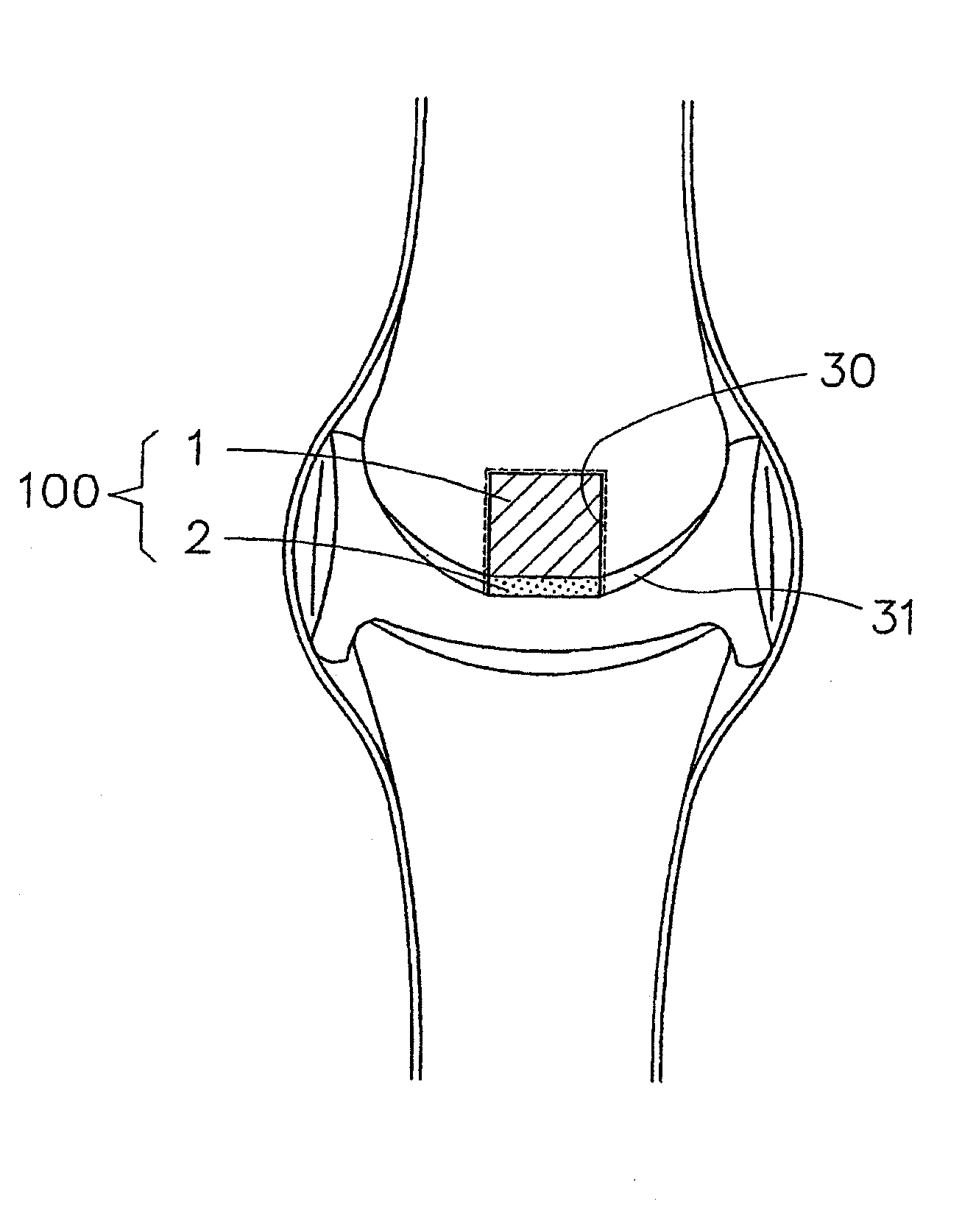
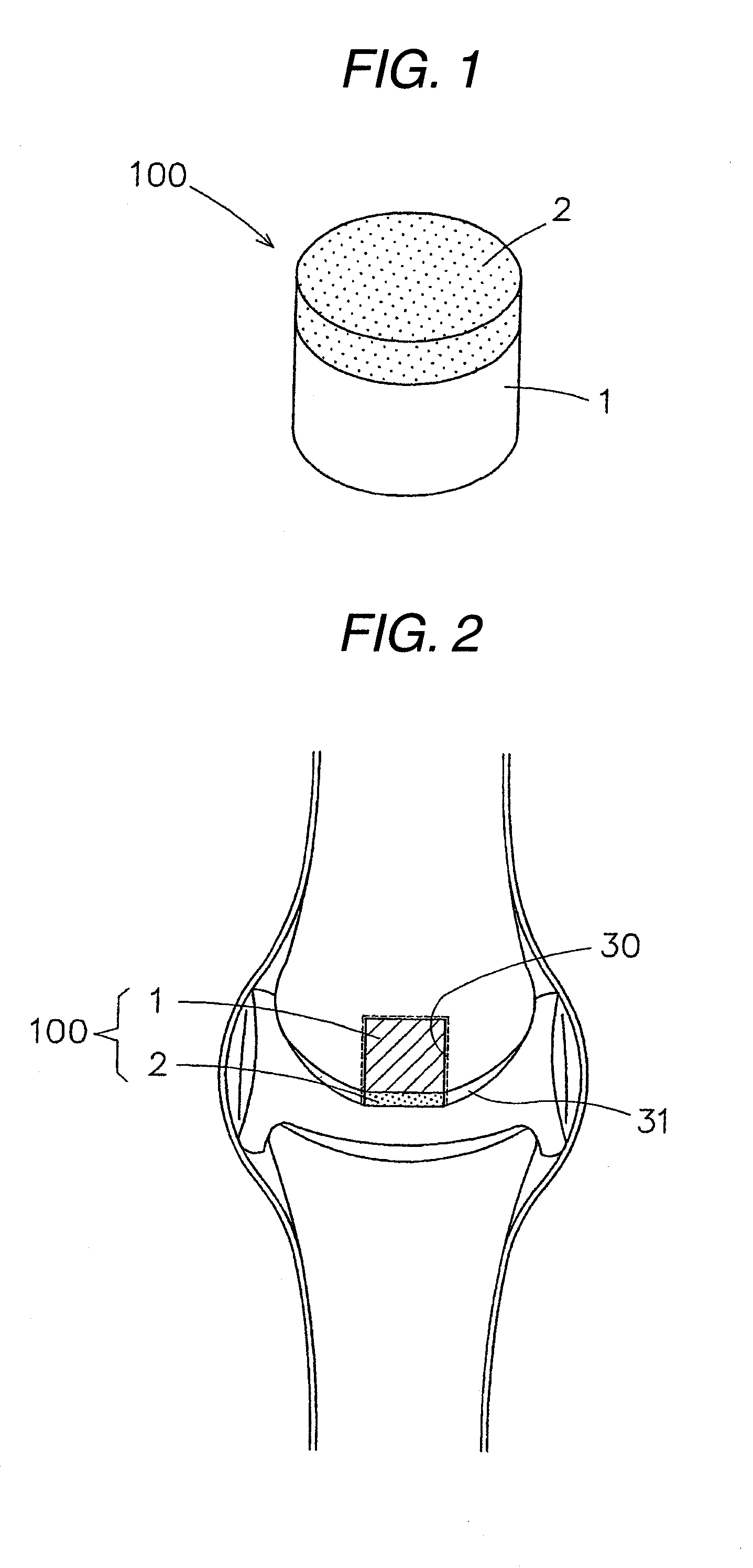
[0046]The implant composite material in which the porous composite has been superposed on and united until one side or all surfaces of the compact composite, like that of the first type of the invention, has the properties or functions required of scaffold materials and the like for use in, e.g., the treatment of articular cartilage disorders as stated above. Because of this, when the implant composite material of the first type in which the porous composite has been superposed on and united with one side of the compact composite is implanted in and fixed to, for example, a part where a necrotized part of an articular bone head has been excised, so that the porous composite is located on the cartilage side of the articular bonehead surface, then it functions by the following mechanism. The porous composite is wholly replaced by cartilage tissues inductively grown in an early stage and disappearance, and the compact composite, which has strength, also is wholly replaced finally by conductively grown hard-bone tissues and disappears. The bioceramic particles also are completely assimilated. Thus, the hard-bone part and cartilage part of the necrotized articular bone head part are regenerated. On the other hand, when the implant composite material of the first type in which the porous composite has been superposed on and united with the all surfaces of the compact composite is implanted in an excised part of an articular bone head, the following effect/advantage is brought about besides those described above. Hard-bone tissues rapidly grow inductively in the porous composite in contact with the hard bone in the excised part, whereby this implant composite material is bonded with and fixed to the excised part of the articular bone head in a short time period.
[0047]The implant composite material of the first type in which the porosity of the porous composite gradually changes to have an inclination so that the porosity increases from an inner-layer part to a surface-layer part of the porous composite in the range of 50-90% has the following advantage. A body fluid and an osteoblast more easily penetrate into the surface side of the high-porosity porous composite having interconnected pores, and hydrolysis and the inductive growth of (cartilage) bone tissues proceed rapidly. Consequently, this implant composite material bonds with a living (cartilage) bone in an earlier stage to complete regeneration. The content of the bioceramic particles in the porous composite may be even throughout the porous composite. However, the porous composite in which the content thereof gradually changes to have an inclination so that it increases from an inner-layer part to a surface-layer part of the porous composite in the range of 30-80% by mass has the following advantage. Since the surface side of the porous composite has a high bioceramic-particle proportion and hence has higher bioactivity, the inductive growth of an osteoblast and bone tissues on the surface side is especially enhanced. As a result, replacement by (cartilage) bone tissues is further accelerated. The porous composite containing at least one biological bone growth factor selected from a BMP, TGF-β, EP4, b-FGF, and PRP and/or an osteoblast derived from a living organism has the following advantage. Osteoblast multiplication/growth is greatly accelerated and, hence, (cartilage) bone tissues grow vigorously. Thus, regeneration proceeds more rapidly.
[0048]The implant composite material of the second type of the in

Problems solved by technology

However, no material usable as a scaffold for the treatment or reconstruction of a necrotized part of a joint bone head or for the reinforcement of a ligament part adherent to a joint has been developed because of difficulties in material science.
However, in the case where the two substitutes are not in a united form but a combination of separate members, continuous and connecting shifting is not obtained between cartilage tissues and hard-bone tissues.
In addition, a problem that the two substitutes separate from each other upon joint movements arises.
In this case, when the target prosthetic material is one not assimilable in the living body, such as a metal, ceramic, or polymer, it is not replaced by living tissues with the lapse of time and the long-term holding of the implanted material continuously has a fear concerning problems such as infection and mechanical troubles.
However, these fixing techniques generally have a drawback that the part where the ligament or tendon has been fixed becomes loose with the lapse of time.
In the fixing (1), although metallic screws have conventionally been mainly employed, this fixing arouses troubles in extreme knee bends, e.g., sitting on the heels.
However, such screw fixing has a drawback that the screw does not directly bond with the bone in the implantation part.
The screw receives a load caused by bends over a prolonged time period and this is a cause of getting loose.
However, this fixing technique unavoidably has a possibility that metallic cross pins, when present over long in a joint part involved in heavy movements, might shift their positions to cause stimulation and this might sometimes produce a serious harmful effect.
Furthermore, assimilable ones have poor reliability with respect to flexural strength and deformation by flex relaxation.
However, these screws have a high modulus of elasticity and, in particular, the metallic interference screws may adversely influence the living body due to metal ion dissolution.
There is hence a problem that a reoperative surgery should be performed for taking the screws out of the body in an early stage after the treatment.
However, since these bone-uniting materials have a far higher modulus of elasticity than living bones, there are problems, for example, that dependence on their strength reduces rather than increases the strength of the bones surrounding the uniting materials.
In particular, in the case of metallic screws, there is a fear that metal ions gradually released therefrom may adversely influence the living body in a prolonged time period exceeding 10 years after implantation.
There is hence a fear that a reoperative surgery for taking the screws out of the body must be performed in an early stage.

Method used


Image

  • Implant composite material
  • Implant composite material
  • Implant composite material

Examples


Embodiment Construction

[0114]Specific embodiments of the invention will be described below in detail by reference to drawings.

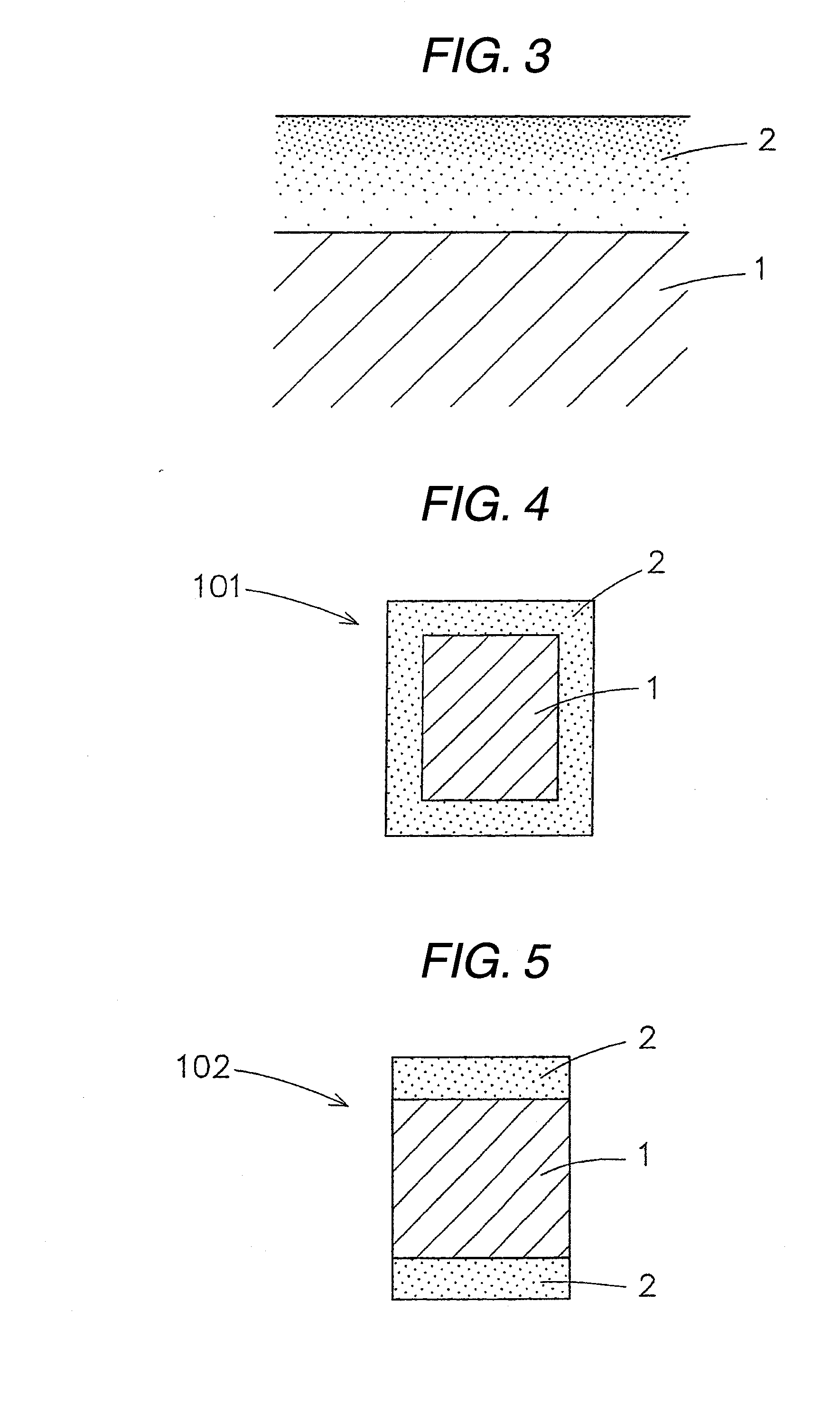
[0115]FIG. 1 is a slant view of an implant composite material of the first type as one embodiment of the invention; FIG. 2 is a view illustrating an example in which this implant composite material is used; and FIG. 3 is an enlarged sectional view illustrating part of the implant composite material.

[0116]The implant composite material 100 shown in FIG. 1 is an implant composite material of the first type which comprises a compact composite 1 and a porous composite 2 superposed on and united with one side (upper side in this embodiment) of a surface-layer part of the compact composite 1.

[0117]The compact composite 1 is a compact block composite comprising a biodegradable and bioabsorbable polymer containing bioabsorbable and bioactive bioceramic particles. Although the compact composite 1 in this embodiment is in the form of a solid cylinder, it can have a quadrangular solid prism, ...


PUM

PropertyMeasurementUnit
Pore sizeaaaaaaaaaa
Percent by massaaaaaaaaaa

Abstract

An implant composite material is provided which is for use in the treatment of articular cartilage disorders such as hip joint femur head necrosis and knee joint bone head necrosis, the reconstruction / fixing of a bio-derived or artificial ligament or tendon, the uniting / fixing of a bone, etc. Part of the implant composite material is replaced by bone tissues in an early stage to enable the material to stably bond with a living bone, while the other part retains a necessary strength over a necessary time period. Finally, the implant composite material is wholly replaced by the living bone and disappears.It is an implant composite material having a constitution which comprises a compact composite of a biodegradable and bioabsorbable polymer containing bioabsorbable and bioactive bioceramic particles and a porous composite of a biodegradable and bioabsorbable polymer containing bioabsorbable and bioactive bioceramic particles, the porous composite being united with the compact composite. The porous composite is replaced by bone tissues in an early stage to enable the material to stably bond with a living bone, while the compact composite retains a necessary strength over a necessary time period. Finally, the material is wholly replaced by the living bone and disappears. Consequently, this implant composite material can sufficiently meet desires in this medical field.

Description

TECHNICAL FIELD[0001]The present invention relates to an implant composite material which is for use in the treatment of articular cartilage disorders such as hip joint femur head necrosis and knee joint bone head necrosis, the reconstruction / fixing of a bio-derived or artificial ligament or tendon, the uniting / fixing of a bone, etc.BACKGROUND ART[0002]Various regenerative medical techniques have hitherto been investigated in order to reconstruct, regenerate, or reinforce hard-bone or cartilage parts which have been destroyed or damaged considerably. It is widely understood that the reconstruction of a damaged part having a given shape essentially necessitates a scaffold which serves to help completion of the reconstruction by avoiding an external mechanical load or a cytological or physiological attack and forming / maintaining the desired shape until the regeneration of tissues is completed.[0003]At present, various ideas have been proposed on scaffold materials for use in the case ...

Claims


Application Information

Patent Timeline
IPC IPC(8): A61F2/02
CPCA61B17/8625A61L27/58A61B17/866A61B17/8685A61B2017/00004A61F2/08A61F2/0811A61F2/28A61F2/30756A61F2/30767A61F2/38A61F2/44A61F2/4455A61F2002/0841A61F2002/0858A61F2002/087A61F2002/2828A61F2002/30004A61F2002/30013A61F2002/30062A61F2002/30113A61F2002/30125A61F2002/30128A61F2002/30153A61F2002/30224A61F2002/30904A61F2002/448A61F2210/0004A61F2230/0006A61F2230/0008A61F2230/0019A61F2230/0069A61F2250/0014A61F2250/0024A61F2310/00179A61L27/446A61L27/56A61B17/864A61F2002/30011
Inventor SHIKINAMI, YASUO
Owner TAKIRON CO LTD
Learn More