
Nanocells for Diagnosis and Treatment of Diseases and Disorders

a technology of cancer cells and tumors, applied in the direction of immunological disorders, drug compositions, therapy, etc., can solve the problems of inability to deliver specific imaging agents, inability to precisely deliver imaging agents, and inability to achieve tumor-specific imaging. to achieve the effect of fine tuning the delivery of specific drugs

Inactive Publication Date: 2009-04-30
MASSACHUSETTS INST OF TECH
View PDF43 Cites 14 Cited by
  • Summary
  • Abstract
  • Description
  • Claims
  • Application Information

AI Technical Summary

Benefits of technology

[0014]In another embodiment, a targeting ligand is attached to the outer surface of the nanocell (i.e. on the PEG or lipid nanoshell) to further enhance and target delivery of the imaging agent to particular organs, tissue, or cells.
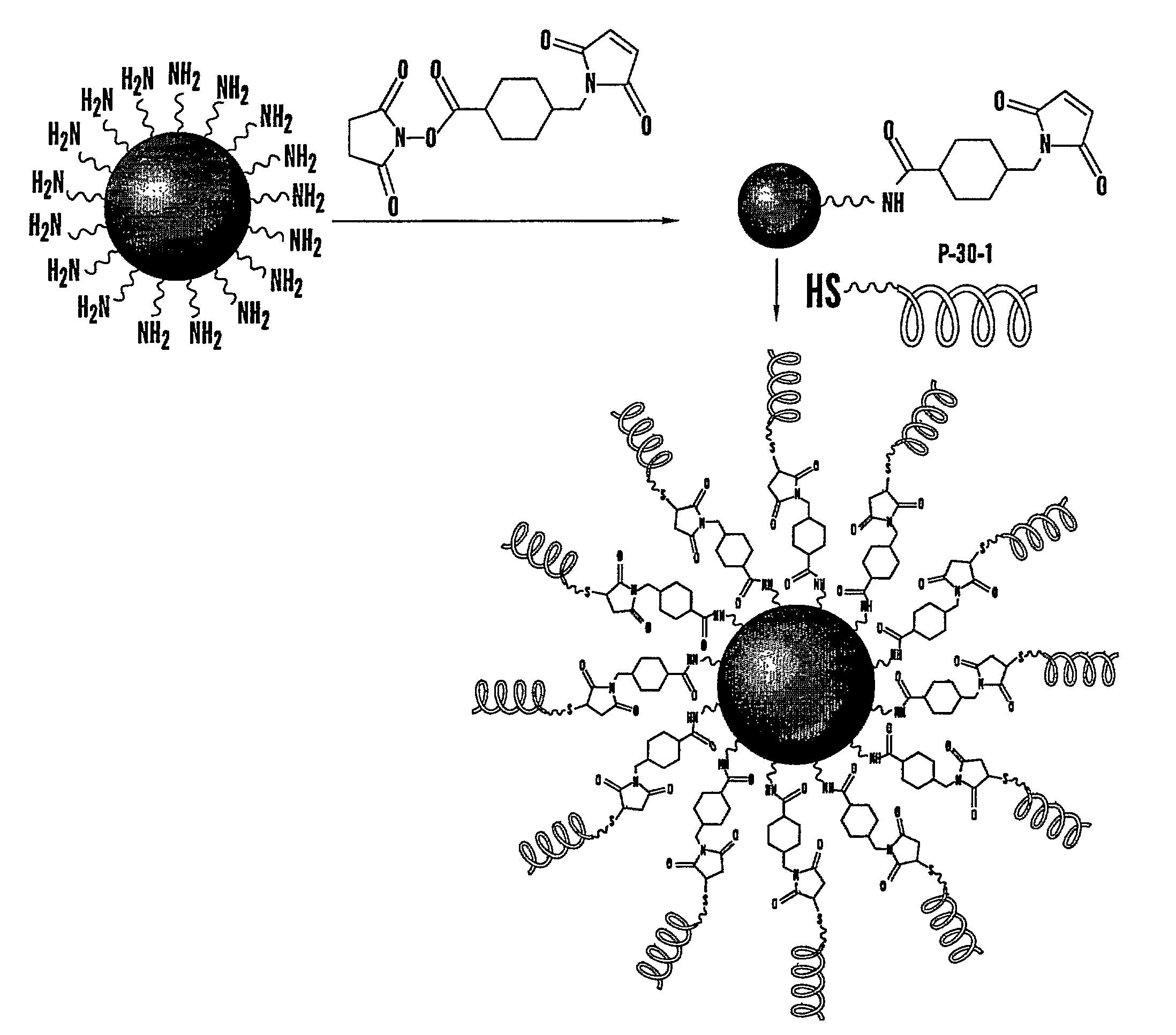
[0023]In another embodiment, a composition and method for the treatment of asthma is disclosed. In this embodiment, the tailored nanocell composition comprises a nanocore with a first therapeutic consisting of a corticosteroid and a nanoshell with a second therapeutic consisting of a bronchodilator. One can also add additional layers around the nanocell to further fine tune delivery of specific drugs. The corticosteroid may be selected from the group consisting of cortisol, cortisone, hydrocortisone, fludrocortisone, prednisone, methylprednisonlone, or prednisolone etc. The bronchodilator may be selected from the group consisting of an anticholinergic, such as ipratropium or a beta-agonist such as albuterol, metaproterenol, salmeterol, pirbuterol, or levalbuteral. The composition for the treatment of asthma allows for an individual to be administered a smaller dose of corticosteroid than is normally available because the bronchodilator in the nanoshell acts first to make available the biological sites of action for the corticosteroid. In one embodiment, the nanocore may comprise a biodegradable polymer such as PLGA and the nanoshell may comprise a water soluble carrier such as lactose. The size may be about 102 to about 104 nm.

Problems solved by technology

A range of imaging agents, for example radioimaging agents, have been developed, but have suffered from problems such as cost, complexity, and the need to identify specific ligands that target desired tissues.
A limitation of current diagnostic imaging methods is that it is often not possible to deliver the imaging agent specifically to the tissue or cell type that one wishes to image.
In the area of diagnostic imaging of cancer, current methods for tumor-specific imaging are hindered by imaging agents that also accumulate in normal tissues.
Despite increased understanding of many aspects of cancer, the methods available for its detection continue to have limited success.
The specific coordination requirements of particular radionuclides place constraints on the ligands that can be used, which in turn place limits on what are viable targets.
Although numerous therapies currently exist for cancers, diabetes, asthma, cystic fibrosis, and other diseases and disorders, the actual results are not entirely satisfactory.
One problem may be the presently available modes, dosage, and timing of delivery.
For example, while anti-inflammatory therapy is a vital treatment for alleviating asthmatic attack, delivering an anti-inflammatory during an acute attack can be ineffective due to its inability to reach its target site.
However, current therapies provide for a large dose corticosteroid and bronchodilator administered concurrently, which results in ineffective treatment and unwanted side effects due to unnecessarily large doses of pharmaceutical compounds.

Method used


Image

  • Nanocells for Diagnosis and Treatment of Diseases and Disorders
  • Nanocells for Diagnosis and Treatment of Diseases and Disorders
  • Nanocells for Diagnosis and Treatment of Diseases and Disorders

Examples


example 1

[0192]The present invention overcomes several limitations of using nanoparticles for imaging, including their insolubility and tendency to aggregate and the general distribution when injected into systemic circulation, which would prevent the discrimination between normal and diseased tissues. Various approaches have been made to keep them stable in suspension including the attachment of pegylated groups, or coating them with various functional groups and peptides for targeted delivery. However, such approaches still fail to overcome the potential for uptake by the reticuloendothelial system (RES) or uptake by normal tissues because of their nanoscale size.

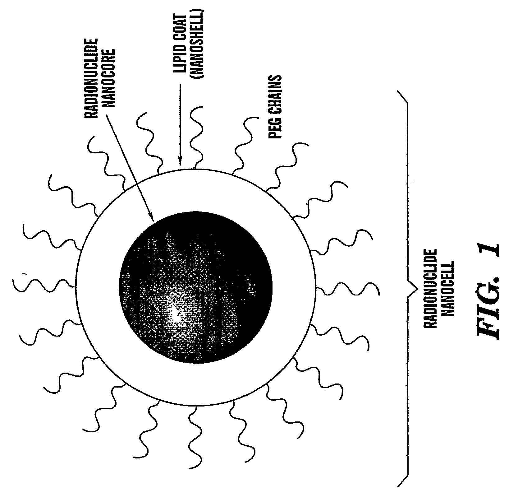
[0193]The present invention describes a modified, nuclear nanocell, where the nuclear nanocore is a quantum dot or a nanoparticle that emits a radiation following excitation (FIG. 1). The encapsulation of the nuclear nanocore inside the lipid bilayer, and the presence of the PEG on the surface of the bilayer prevents the RES from ...

example 2

Preparation of Nanocells for Treatment of Asthma

[0211]Nanoparticles with dexamethasone were synthesized from PLGA using PVA as a stabilizer using an emulsion-solvent evaporation technique. The nanoparticles were then coated with a shell of lactose using a spray drying technique. The bronchodilator, salbutamol, was dissolved in the lactose solution prior to spray drying. The nanocell formed was then lyophilized overnight before being administered in vivo. For SEM, dehydrated nanoparticles were gold-coated on a carbon grid. They were analyzed using a Jeol EM (magnification, 3700×).

[0212]As shown in FIG. 5, electron micrograph revealed that the nanoparticles formed were spherical and were of a diverse size range from 5×101-20×103 nm. The nanoparticles were then coated with a lactose layer, which made the size of the particles in the 103 to 105 nm range.

[0213]Release Kinetics Characterization

[0214]Drug-loaded nanocells were suspended in 1 ml of PBS buffer or hypoxic-cell lysate and seal...

example 3

[0227]Despite major advances in the development in anticancer drugs and imaging agents, a major disadvantage is their lack of selectivity for malignant tissue. Currently, most common drug delivery systems and imaging agents target proteins that are overexpressed on the surface of cancer cells. Alterations to the normal function of the glycosylation machinery have been increasingly recognized as a consistent indication of malignant transformations and tumorigenesis. In many cases, these alterations result in the overexpression of specific cancer-associated carbohydrates, specifically on the malignant tissue. Due to the complexity of molecular interactions with carbohydrates, very few systems have been designed to specifically target carbohydrates for imaging and drug delivery purposes. Despite the use of lectins for detection of carbohydrates in different tissues, their low affinity, high molecular weight, the stability of their active structures and their complexity for selective ch...


PUM


Abstract

The present invention relates to novel nanocell compositions and their use in imaging, diagnostic and treatment methods. In one embodiment, nanocells tailored for imaging methods comprise a nanocore surrounded by a lipid matrix, and are modified to contain a radionuclide core or a nanocore with an emission spectra. The nanocells may be size restricted such as being greater than about 60 nm so that they selectively extravasate at sites of angiogenesis (e.g. tumor) and do not pass through normal vasculature or enter non-tumor bearing tissue. In this way, angiogenic sites can be both detected and treated. In another embodiment, nanocells are tailored for various treatment methods, including the treatment of brain cancer, asthma, Grave's Disease, Cystic Fibrosis, and Pulmonary Fibrosis.

Description

CROSS REFERENCE TO RELATED APPLICATIONS[0001]This application claims the benefit under 35 U.S.C. 119(e) of U.S. Provisional Patent Application Ser. No. 60 / 661,627, filed Mar. 14, 2005 and U.S. Provisional Patent Application Ser. No. 60 / 708,012, filed Aug. 12, 2005, the contents of which are herein incorporated by reference in their entirety.FIELD OF THE INVENTION[0002]The present invention relates to novel diagnostic agents, method for their use in imaging, such as identification of malignant cells, preferably solid tumor detection, and kits for preparing and using such diagnostic agents. Also encompassed are novel nanocell platforms for targeting cells, method for their use in treatment of diseases or disorders, and kits for preparing and using the same.BACKGROUND OF THE INVENTION[0003]The ability to obtain in vivo images has assisted in treatment, diagnosis and prognosis of a variety of diseases and disorders. A range of imaging agents, for example radioimaging agents, have been d...

Claims


Application Information

Patent Timeline
Patent Type & Authority Applications(United States)
IPC IPC(8): A61K51/00
CPCA61K9/1271A61K47/48861A61K47/48884A61K51/1244A61K51/1241B82Y10/00A61K9/14A61K45/06B82Y5/00A61K47/6923A61K47/6929A61P1/00A61P1/04A61P11/00A61P11/06A61P19/02A61P27/00A61P27/02A61P27/06A61P29/00A61P35/00A61P35/04A61P37/06A61P43/00A61P9/00
Inventor SENGUPTA, SHILADITYASASISEKHARAN, RAMBOSQUES, CARLOS J.EAVARONE, DAVID A.SHUM, POCHI
Owner MASSACHUSETTS INST OF TECH
Learn More