
Systems and methods for quantification and classification of fluids in human cavities in ultrasound images

a technology of ultrasound images and fluids, applied in the field of ultrasound imaging of bodily tissues, bodily fluids, and fluidfilled cavities, can solve the problems of unnecessary catheterization, image generation that fails to properly distinguish blood from other bodily fluids, and serious infection possibilities

Inactive Publication Date: 2008-06-12
MCMORROW GERALD +6
View PDF2 Cites 53 Cited by
  • Summary
  • Abstract
  • Description
  • Claims
  • Application Information

AI Technical Summary

Problems solved by technology

Moreover, when blood within the selected portion and other bodily fluids are imaged using B-mode ultrasound, images may be generated that similarly fail to properly distinguish the blood from the other bodily fluids.
However, serious disadvantages to unnecessary catheterization range from. the uncomfortable situation for the patient to serious possibilities of infection.
Non-invasive procedures for bladder volume estimation are known, but are either unreliable or expensive or have some other significant disadvantages.
Palpation and auscultatory percussion are known to be unreliable, while radiography and dye-excretion techniques are known to be similarly inaccurate.
However, it is invasive, painful and might produce traumas or infections.
His method is not adjusted to specific, filling dependent, measuring configurations.
Since the bladder changes in shape when filling, a single distance measurement is not precise enough to predict the entire bladder volume.
The instruments are costly and require a skilled operator.
This further increases complexity, acquisition time of the data, and costs of the instrument.
The used apparatus is complex and is different from the method described in this application.
The acquisition procedure is time consuming and thus no instantaneous volume measurement results.
This nevertheless still requires an acquisition time equivalent to full acquisition procedure and thus does not yield an instantaneous display of volume.
No instantaneous feedback on optimal positioning is thus available.
The three dimensional scanning requirement makes this instrument complex.
This explains the large error that several studies obtained when a single geometric model was used.
Non-linear distortion increases with distance, insonifying ultrasound energy and frequency.

Method used


Image

  • Systems and methods for quantification and classification of fluids in human cavities in ultrasound images
  • Systems and methods for quantification and classification of fluids in human cavities in ultrasound images
  • Systems and methods for quantification and classification of fluids in human cavities in ultrasound images

Examples


Embodiment Construction

[0062]The present invention relates to the ultrasound imaging of tissues and / or fluid-filled cavities having linear or non-linear acoustic properties. Many specific details of certain embodiments of the invention are set forth in the following description and in FIGS. 1 through 21 to provide a thorough understanding of such embodiments. One skilled in the art, however, will understand that the present invention may have additional embodiments, or that the present invention may be practiced without several of the details described in the following description.

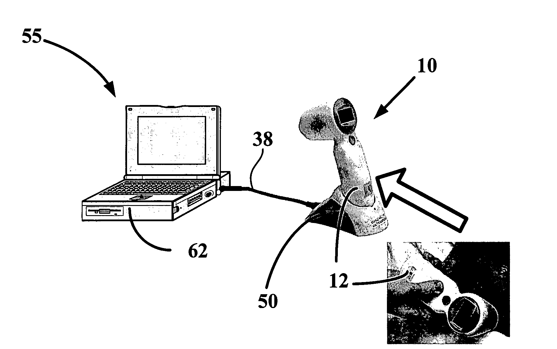
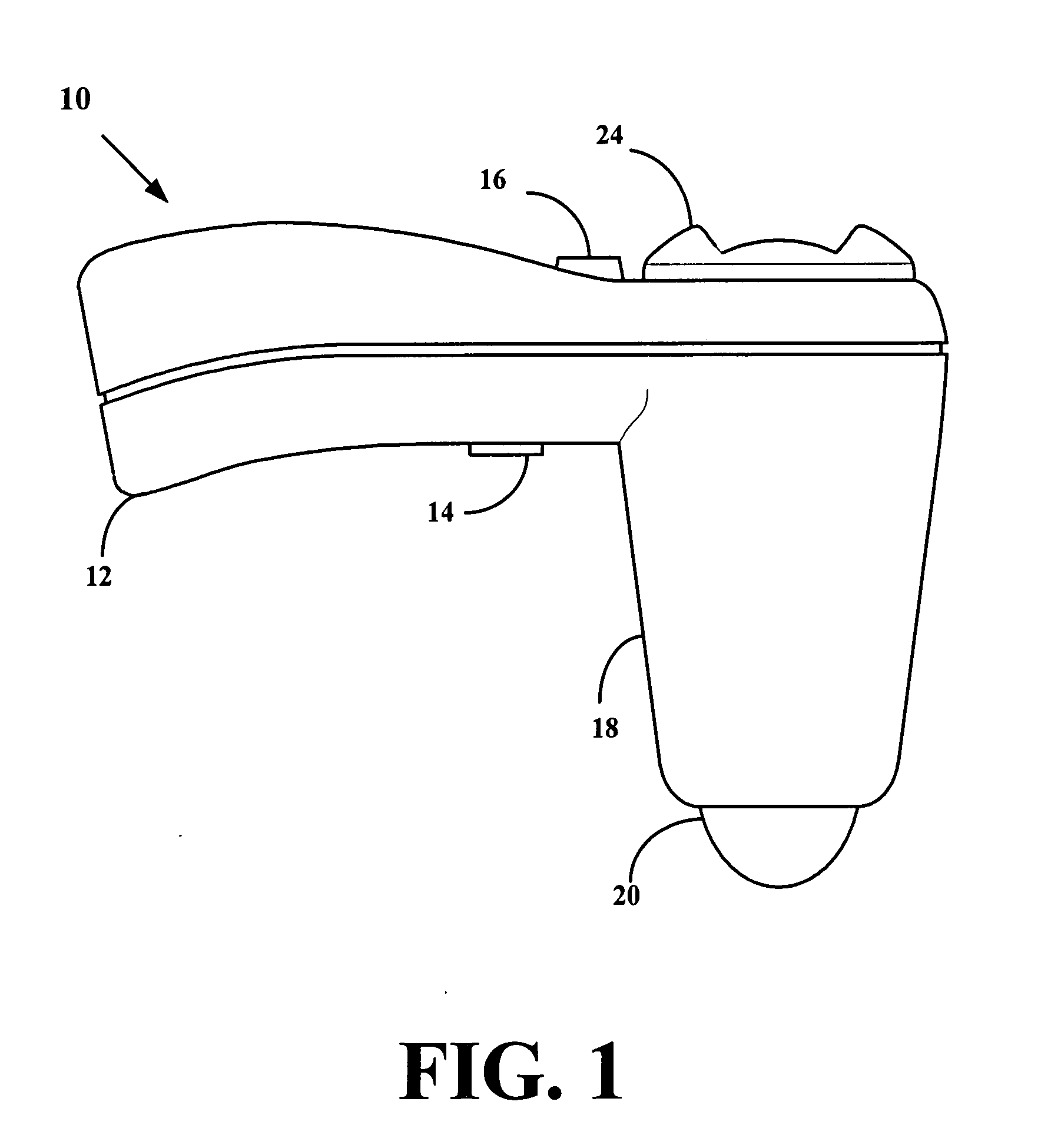
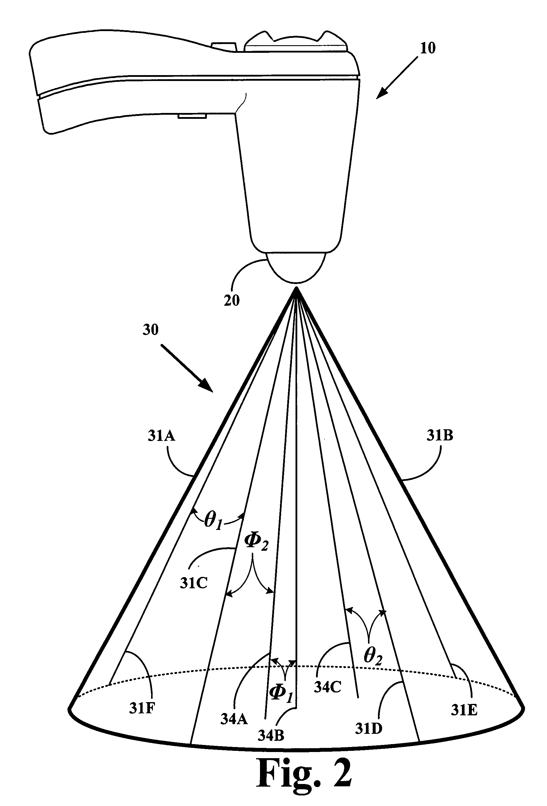
[0063]FIG. 1 is a side elevational view of an ultrasound transceiver 10 according to an embodiment of the invention. The transceiver 10 includes a transceiver housing 18 having an outwardly extending handle 12 that is suitably configured to allow a user to manually manipulate the transceiver 10. The handle 12 includes a trigger 14 that allows the user to initiate an ultrasound scan of a selected anatomical portion, and a cavity ...


PUM


Abstract

Ultrasound imaging systems and methods are disclosed. In one embodiment, an ultrasonography method includes creating a database that is representative of a tissue, a fluid, or a cavity of a body, and transmitting ultrasound pulses into a region-of-interest in a patient. Echoes are received from the region of interest, and based upon the received echoes, compiling an ultrasonic pattern of the region-of-interest is compiled. The pattern is processed by comparing the region-of-interest patterns to the pattern information stored in the database. A composition within the region-of-interest of the patient is then determined.

Description

FIELD OF INVENTION[0001]This invention relates to ultrasound imaging of bodily tissues, bodily fluids, and fluid-filled cavities.BACKGROUND OF THE INVENTION[0002]The following applications are incorporated by reference as if fully set forth herein: U.S. application Ser. Nos. 11 / 213,284 filed on Aug. 26, 2005; 11 / 010,539 filed on Dec. 13, 2004; and 10 / 523,681 filed on Feb. 3, 2005.[0003]Ultrasound imaging is accomplished by placing an ultrasound transducer on a selected location of a body and projecting ultrasound energy into the body. Acoustic waves reflecting from internal structures in the body are then received by the transducer and are processed to form an image of the internal structures. In a particular ultrasound method, amplitudes of selected harmonics of the returned signal are processed to form the ultrasound image. Briefly and in general terms, harmonic generation by the internal structures in the body is at least partially determined by the properties of the tissue that ...

Claims


Application Information

Patent Timeline
Patent Type & Authority Applications(United States)
IPC IPC(8): A61B8/00A61B5/20A61B8/08A61B8/12A61B8/14
CPCA61B5/204A61B8/08A61B8/0858A61B8/4472A61B8/483G06T2207/30004G01S15/102G01S15/8909G06T7/0012G06T7/602G06T2207/10132G01S7/52036G06T7/62
Inventor MCMORROW, GERALDCHALANA, VIKRAMYUK, JONGTAEBAARTMANS, HENRIBOM, NICOLAASLANCEE, CHARLES THEODOORMERKS, EGON J.W.
Owner MCMORROW GERALD
Learn More