
Diagnostic and therapeutic methods based on the l1 adhesion molecule for ovarian and endometrial tumors

Inactive Publication Date: 2008-06-12
DEUTES KREBSFORSCHUNGSZENT STIFTUNG DES OFFENTLICHEN RECHTS +1
View PDF4 Cites 2 Cited by
  • Summary
  • Abstract
  • Description
  • Claims
  • Application Information

AI Technical Summary

Benefits of technology

[0008]It has surprisingly been found that the L1 adhesion molecule represents a highly specific marker in body fluids (e.g. in the blood serum, ascites and fluid from Douglas coule de sac) and tissue from ovarian and endometrial tumors, above all for highly aggressive forms of these tumors. L1 is a neuronal adhesion molecule of 200-230 kDa, which as regards its structure belongs to the Ig superfamily. L1 is a protein associated with cell migration, which in the brain is involved in the neuronal migration in the cerebellum and in the fasciculation of neurites. L1 has several binding partners among them L1 itself, the proteoglycan neurocan and various integrins (Kadmon et al., Differentiation 61: 143-150, 1997). Soluble L1 can stimulate the migration of tumor cells and hence promote metastasis (Mechtersheimer et al., JBC 2001, in press). In the screening experiments resulting in the present invention it turned out that L1 is not expressed in normal tissues with the exception of peripheral nerves and ganglions. In benignant ovarian and endometrial tumors no L1 expression could be detected, and the screening of other malignant tumors (breast, prostate, cervix) was also negative. It showed that L1 is expressed on highly aggressive serous papilloma carcinomas of the ovary and the endometrium. Soluble L1 was detectable in the serum and ascitic or pleural fluids of tumor patients but not in normal sera and body fluids or in patients having benignant tumors. Preoperative samples which were obtained by gynecologic curettage were positive in serous papilloma carcinomas of the endometrium and with the involvement of the ovary. Thus, the expression of L1 as cell surface molecule in the serum and in body fluids is highly significant as regards the early detection of aggressive ovarian and endometrial tumors and also as regards the prognostication. Therefore, the L1 detection permits the early detection and accurate diagnosis or prognosis of aggressive ovarian and endometrial tumors.

Problems solved by technology

A further technical problem is to provide a therapeutic approach by using L1 as therapeutic target.

Method used


Image

  • Diagnostic and therapeutic methods based on the l1 adhesion molecule for ovarian and endometrial tumors
  • Diagnostic and therapeutic methods based on the l1 adhesion molecule for ovarian and endometrial tumors
  • Diagnostic and therapeutic methods based on the l1 adhesion molecule for ovarian and endometrial tumors

Examples


example 1

The Examination of Curettage Tissue Shows a High Correlation Between the Expression of L1 and the Clinical Stage and the Pathological Degree of the Tumors, Respectively

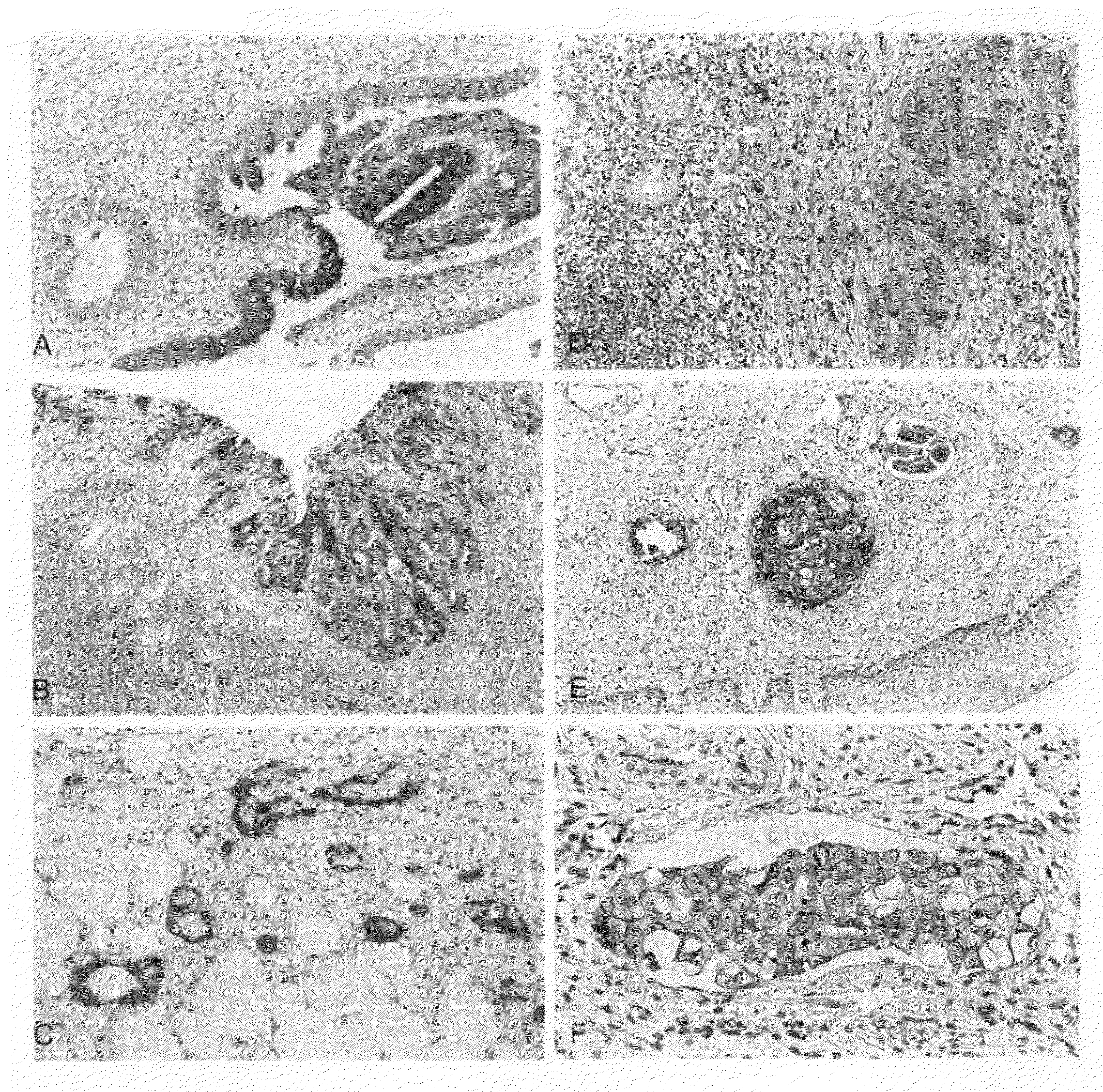
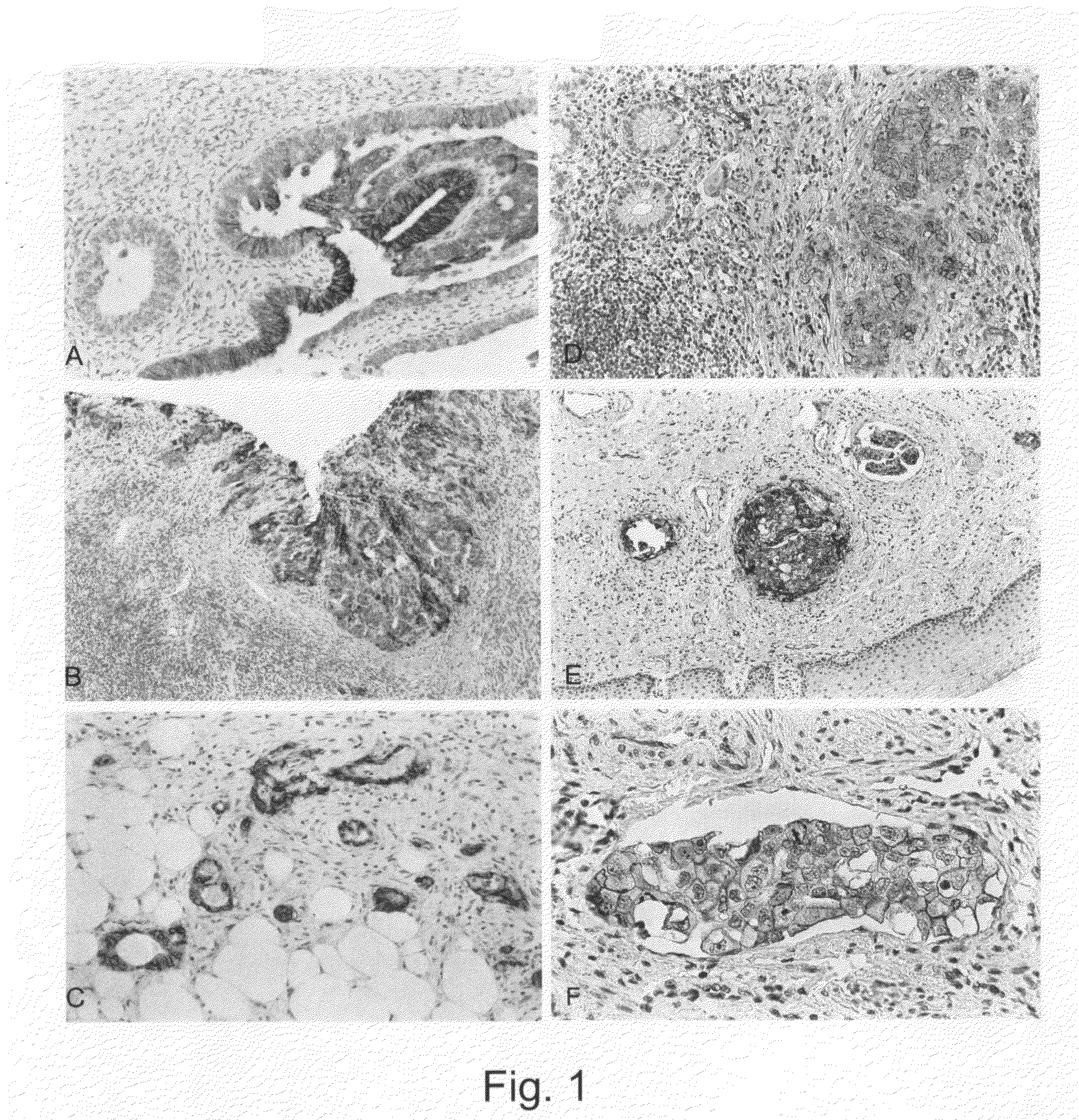
[0044]Tumor tissue is embedded in paraffin according to standard methods and serial sections are made. Following the treatment of the sections in the microwave oven (10 min., 92° C.) in the presence of 1 mmol EDTA (pH 8.0) immunostaining of the tissue is carried out by means of the MCA1753 antibody (Biozol Diagnostica Vertiebs GmbH). The bound first antibody is detected by means of an enzyme-coupled second antibody (Vector ABC Kit; www.vectorlabs.com). Some exemplary stainings are shown in FIG. 1. The clinical data are listed in Tables 1 and 2.

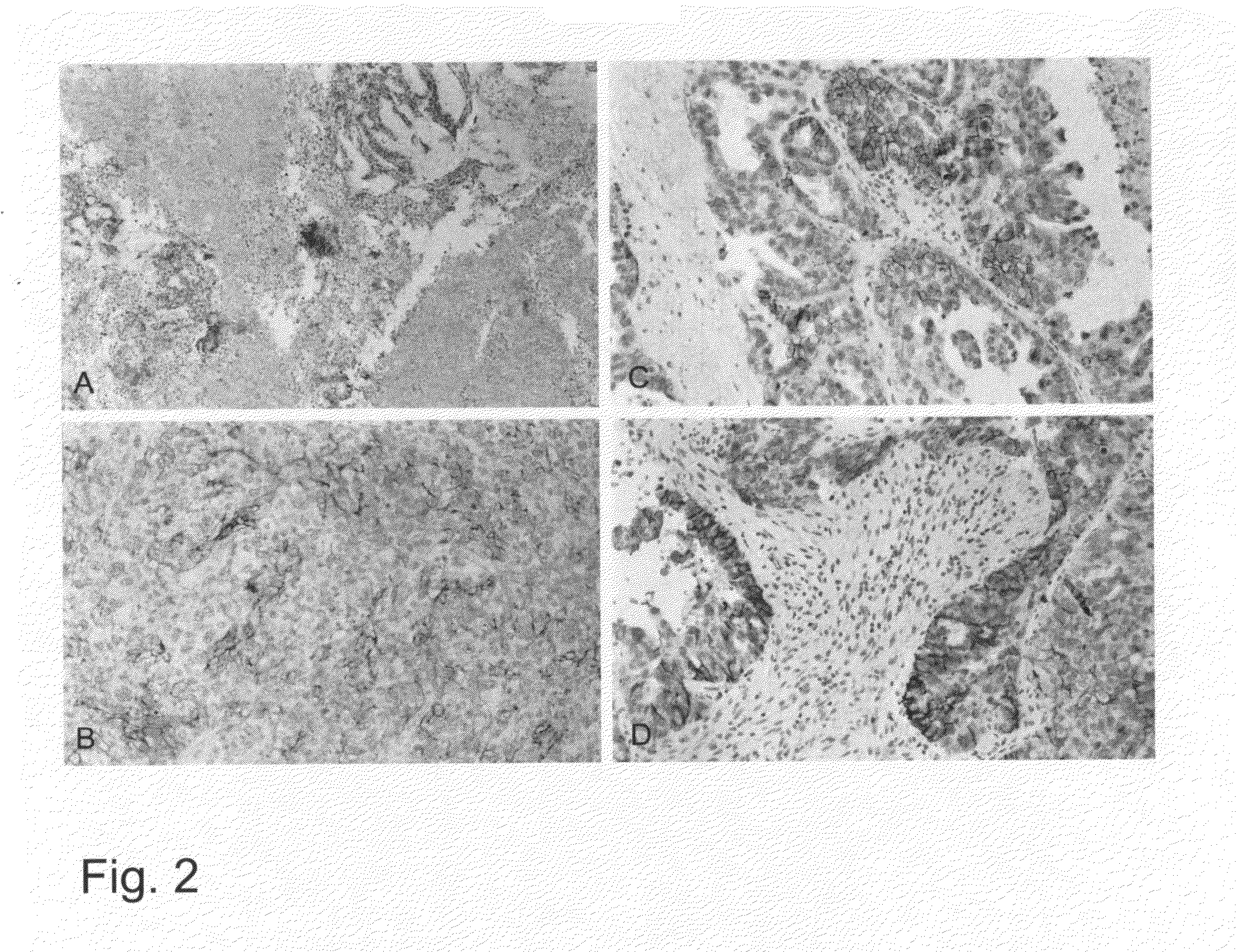
[0045]FIG. 2 shows that L1 can also be detected by means of curettage tissue. Using the above described procedure, a similar staining pattern as on the tumor sections results. The determination of L1 by means of curettage material permits an early preoperative classification of ...

example 2

ELISA Shows the Presence of Soluble L1 in Sera and Ascitic Fluid of Tumor Patients, a Correlation Between the Presence of L1 and the Tumor Type (Ovarian and Endometrial Tumor) Being Observed

[0046]Samples of body fluids (ascites, serum) were tested for the presence of soluble L1 using a “capture” ELISA. For this purpose, microtitration plates were coated with the human anti L1 antibody (concentration: 1 μg / ml) described in Example 1 and then a blocking step was carried out with 3% BSA in PBS (45 min., room temperature) to eliminate the non-specific bonding to the plate. The body fluid was added at differing concentrations (1:2 and 1:10 of the fluid in 3% BSA in PBS), and incubation was carried out at room temperature for 1 hour. Therefore, four wash steps followed in Tris-buffered common salt solution (TBS, pH 8.0, in the presence of 0.02% Tween-20). Bound soluble L1 was determined by the addition of human biotin-conjugated anti L1 antibody. For this purpose, the biotinylated antibod...

example 3

A New ELISA Format for the Detection of Soluble L1

[0048]The ELISA described above in Example 2 used the coating of the microtiter plate with L1 mAb 1 (capturing monoclonal antibody) and the detection of soluble L1 with a biotinylated (or otherwise labelled) L1 mAb 2 (detecting monoclonal antibody). This type of ELISA is termed G / K format. We have now developed another format in which we use both for capturing and detection the same mAb to L1 (K / K format). In FIGS. 5 A and B a comparison of both types of ELISA is shown using a positive serum (CA526, ovarian tumor patient) and seval control sera from unrelated tumors. It is obvious that the new ELISA format in FIG. 5B gives a better signal to noise ratio (appr. 5-8 fold) than the previous format shown in FIG. 5A. As the K / K format can only work when the antibody epitope for detection is not blocked by the antibody used for capturing, these results implicate that in the serum sample the soluble L1 is dimeric or multimeric. The control ...


PUM

PropertyMeasurementUnit
pHaaaaaaaaaa
concentrationaaaaaaaaaa
adhesionaaaaaaaaaa

Abstract

A method is described for the diagnosis of an ovarian or endometrial tumor or for a prognosis, characterized in that the L1 level is determined in a patient sample, preferably via an anti L1 antibody, the presence of L1 being an indication of the presence of an ovarian or endometrial tumor or a predisposition for such a tumor. Furthermore, methods for treating of ovarian or endometrial tumors are provided.

Description

CROSS-REFERENCE TO RELATED APPLICATIONS[0001]This application is a divisional of and claims priority to U.S. patent application Ser. No. 10 / 332,876, filed May 19, 2003, which claims priority of International Patent Application No. PCT / EP01 / 07924 filed Jul. 10, 2001, which in turn claims priority of European Patent Application No. 00 114 788.3, filed Jul. 10, 2000, the entire contents of which are incorporated by reference herein in their entirety.BACKGROUND OF THE INVENTION[0002]1. Field of the Invention[0003]The present invention relates to a method for the diagnosis of an ovarian or endometrial tumor or for a prognosis, characterized in that the L1 level is determined in a patient sample, the presence of L1 being an indication of the presence of an ovarian or endometrial tumor or a possible predisposition for such a tumor. This detection is made preferably via a monoclonal anti L1 antibody or a fragment thereof. The invention also relates to pharmaceutical preparations for use in ...

Claims


Application Information

Patent Timeline
Patent Type & Authority Applications(United States)
IPC IPC(8): A61K39/395A61P37/00G01N33/53A61K31/7088A61K38/00A61K39/00A61K45/00A61K48/00A61P35/00C12N15/09C12Q1/68G01N33/566G01N33/574
CPCG01N33/57442G01N2333/70596G01N2333/70503G01N33/57449A61P35/00A61P37/00
Inventor ALTEVOGT, PETERFOGEL, MINA
Owner DEUTES KREBSFORSCHUNGSZENT STIFTUNG DES OFFENTLICHEN RECHTS
Learn More