Ultrasonic Myocardial Tagging

a myocardial wall and ultrasonic technology, applied in the field of ultrasonic myocardial wall tagging, can solve the problems of infrequent use of myocardial tagging, affecting the accuracy of myocardial tagging, so as to achieve the effect of increasing spatial resolution

Inactive Publication Date: 2008-04-24
KONINKLIJKE PHILIPS ELECTRONICS NV
View PDF2 Cites 7 Cited by
  • Summary
  • Abstract
  • Description
  • Claims
  • Application Information

AI Technical Summary

Benefits of technology

[0009]Thus, the present invention provides the ultrasonic tagging equivalent to MRI tagging, in the sense that the additional clinical information available to the clinician in respect of the visualisation of the motion of the heart muscle is provided without the associated disadvantages, i.e. the ultrasonic tagging system of the present invention provides the advantages of MRI tagging but the imaging technology (i.e. ultrasound) is relatively inexpensive and an increased spatial resolution is achievable.

Problems solved by technology

Historically, the use of myocardial tagging has been infrequent due to the limited availability of easy-to-use software and an inexpensive imaging modality.
In recent years, the use of magnetic resonance imaging to perform tagged imaging has become more widespread, but although tagging allows the fall 3D displacement field to be calculated, this method is time-consuming and relatively expensive.
However, such two-dimensional analysis is still hampered by the expense, and also by limited spatial resolution.

Method used


Image

  • Ultrasonic Myocardial Tagging
  • Ultrasonic Myocardial Tagging
  • Ultrasonic Myocardial Tagging

Examples


Embodiment Construction

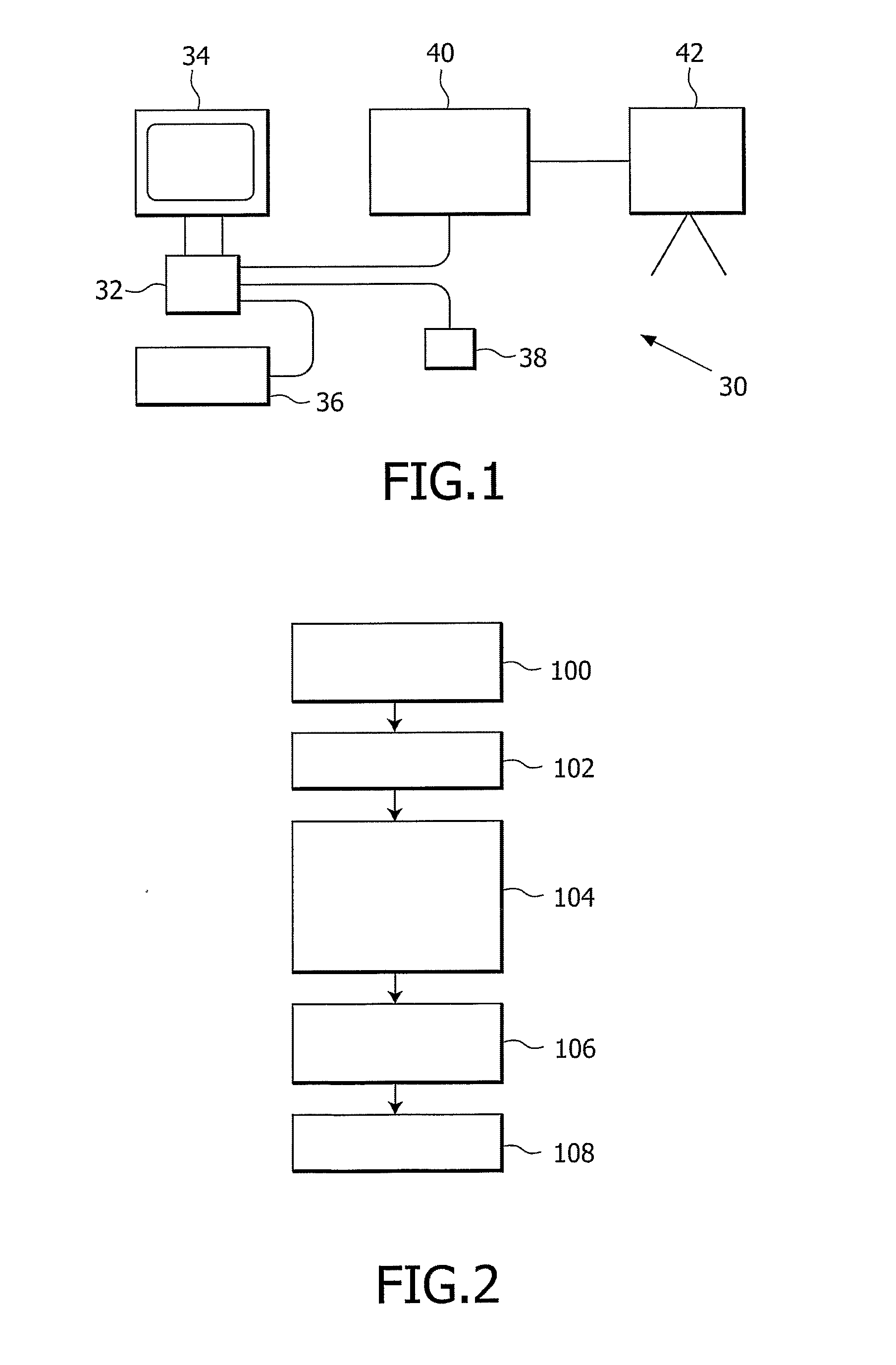
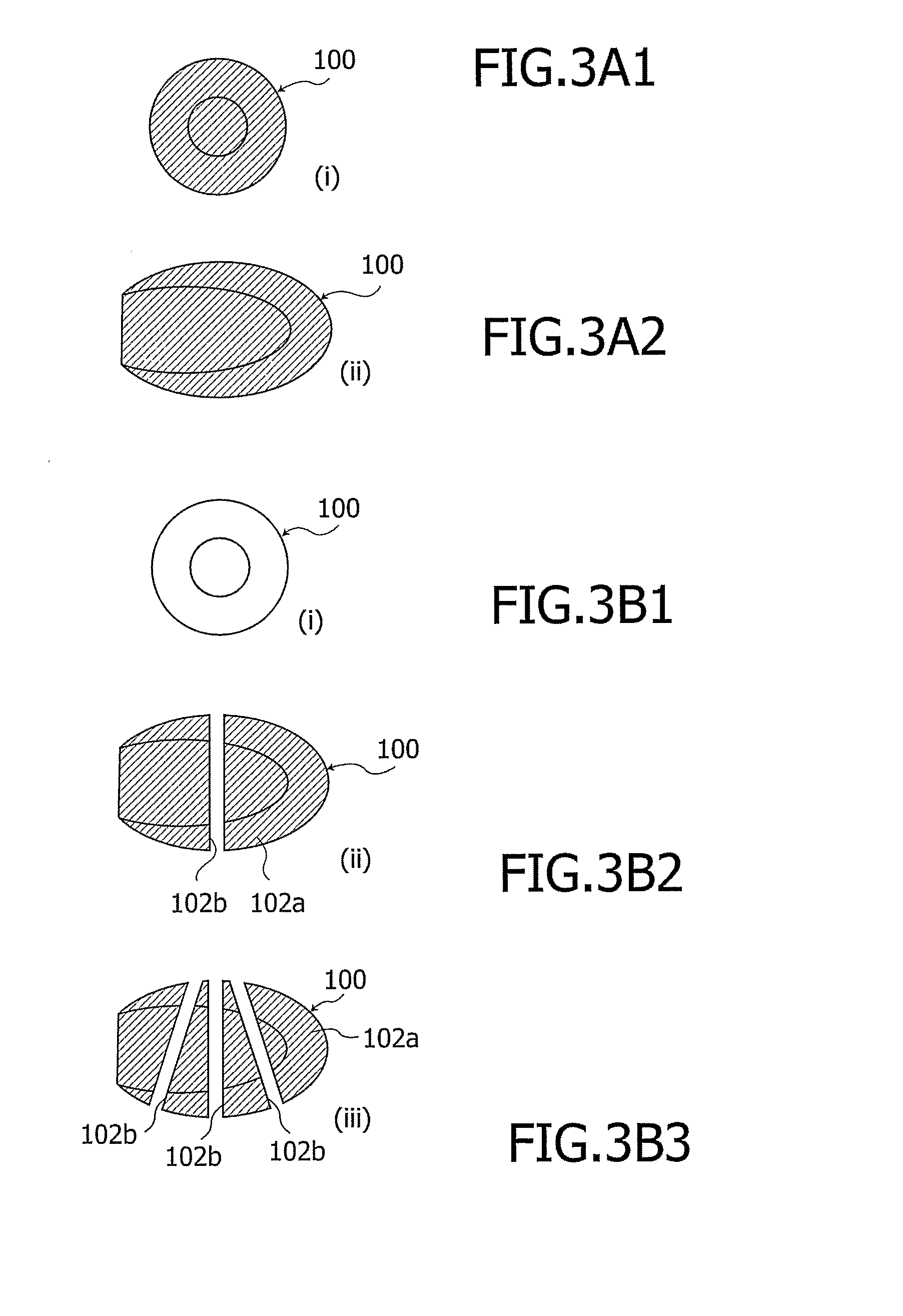
[0025]Thus, as explained above, myocardial tagging provides a clinician with information about the ability of a heart to contract, by labelling specific segments of the heart muscle and following them throughout the heart cycle. The present invention involves the use of an ultrasound imaging system in conjunction with an ultrasonic, microbubble contrast agent to follow motion of the heart muscle, with the same effective clinical utility as current MRI tagging methods, without the associated limitations in relation to spatial resolution, data acquisition time and cost.

[0026]Ultrasonic imaging systems are known which make use of contrast agents in circulation to enhance ultrasound returns, and such contrast agents are currently primarily used for blood pool measurements of left ventricular opacification to visualise the myocardium and flow patterns through valves. Another desired use for ultrasonic contrast agents is in the assessment of perfusion of the heart muscle, and several clin...


PUM


Abstract

The present invention relates to an ultrasonic myocardial tagging and imaging system (30), in which an ultrasonic contrast agent, e.g. microbubbles, is introduced to the myocardial area and allowed to circulate. a high-MI pressure field is applied to selectively destroy regions of the contrast agent in a known geometry so as to create a “tag” (102) comprising a predetermined pattern of non-echogenic regions (102b) and echogenic regions (102a). Movement of the tag (102) relative to the myocardium (100) is tracked so as to monitor movement and/or de formation of the wall tissue and assess myocardial viability.

Description

FIELD OF THE INVENTION[0001]This invention relates generally to myocardial tagging techniques and, more particularly, a system and method for tracking myocardial wall motion through the use of ultrasonic contrast agents and an ultrasonic imaging system.BACKGROUND OF THE INVENTION[0002]Heart performance can be assessed by measuring the detailed strain patterns of the myocardium. Strain patterns can reflect the normal and abnormal myocardial motion, which can be used to correlate the myocardial motion abnormalities with coronary artery disease. Regional wall-motion abnormalities are an excellent indicator of coronary stenosis, and actually precede both ECG abnormalities and chest pain as an indicator of myocardial ischemia. In recent years, several techniques have been developed for fast and accurate tracking of cardiac wall motion.[0003]MRI (magnetic resonance imaging) myocardial tagging is a well-developed method for the evaluation of regional myocardial motion abnormalities, and th...

Claims


Application Information

Patent Timeline
Patent Type & Authority Applications(United States)
IPC IPC(8): G06K9/00
CPCA61B8/0883A61B8/483A61B8/481
Inventor HALL, CHRISTOPHER
Owner KONINKLIJKE PHILIPS ELECTRONICS NV
Why Eureka
Try Eureka