
Texture analysis for mammography computer aided diagnosis

a technology of mammography and computer assisted diagnosis, applied in image analysis, image enhancement, instruments, etc., can solve the problems of limiting the effectiveness of conventional image processing techniques employed by cad systems with some types of mass shapes, and unable to meet the needs of patients, so as to achieve the effect of minimizing over- or under-sampling

Inactive Publication Date: 2008-02-07
CARESTREAM HEALTH INC
View PDF5 Cites 41 Cited by
  • Summary
  • Abstract
  • Description
  • Claims
  • Application Information

AI Technical Summary

Benefits of technology

[0012]An advantage of the present invention is that it can offer a method for assessment of spiculated masses that is relatively straightforward and minimizes over- or under-sampling.

Problems solved by technology

Accurate detection and classification of spiculated masses, including differentiating suspicious spiculate structures from normal structures having some of the same shape characteristics, presents a challenge to Computer-Aided Diagnostic (CAD) systems.
However, it has been observed that conventional image processing techniques employed by CAD systems can fail to detect masses that are small, but have considerable spiculation and are, therefore, indicative of malignancy in early stages.
As is described in more detail subsequently, the method given in the Sahiner et al. paper may provide some improvement in classification accuracy; however, this method has some drawbacks that constrain its effectiveness with some types of mass shapes.

Method used


Image

  • Texture analysis for mammography computer aided diagnosis
  • Texture analysis for mammography computer aided diagnosis
  • Texture analysis for mammography computer aided diagnosis

Examples


Embodiment Construction

[0026]The present description is directed in particular to elements forming part of, or cooperating more directly with, apparatus in accordance with the invention. It is to be understood that elements not specifically shown or described may take various forms well known to those skilled in the art.

[0027]The method of the present invention uses hardware and software components, but is independent of any particular component characteristics such as architecture, operating system, or programming language, for example. In general, the type of system equipment that is conventionally employed for scanning, processing, and classification of mammography image data, or of other types of medical image data, is well known and includes at least some type of computer or computer workstation, having a logic processor which may be dedicated solely to the assessment and maintenance of medical images or may be used for other data processing functions in addition to image processing. Typically, resul...


PUM


Abstract

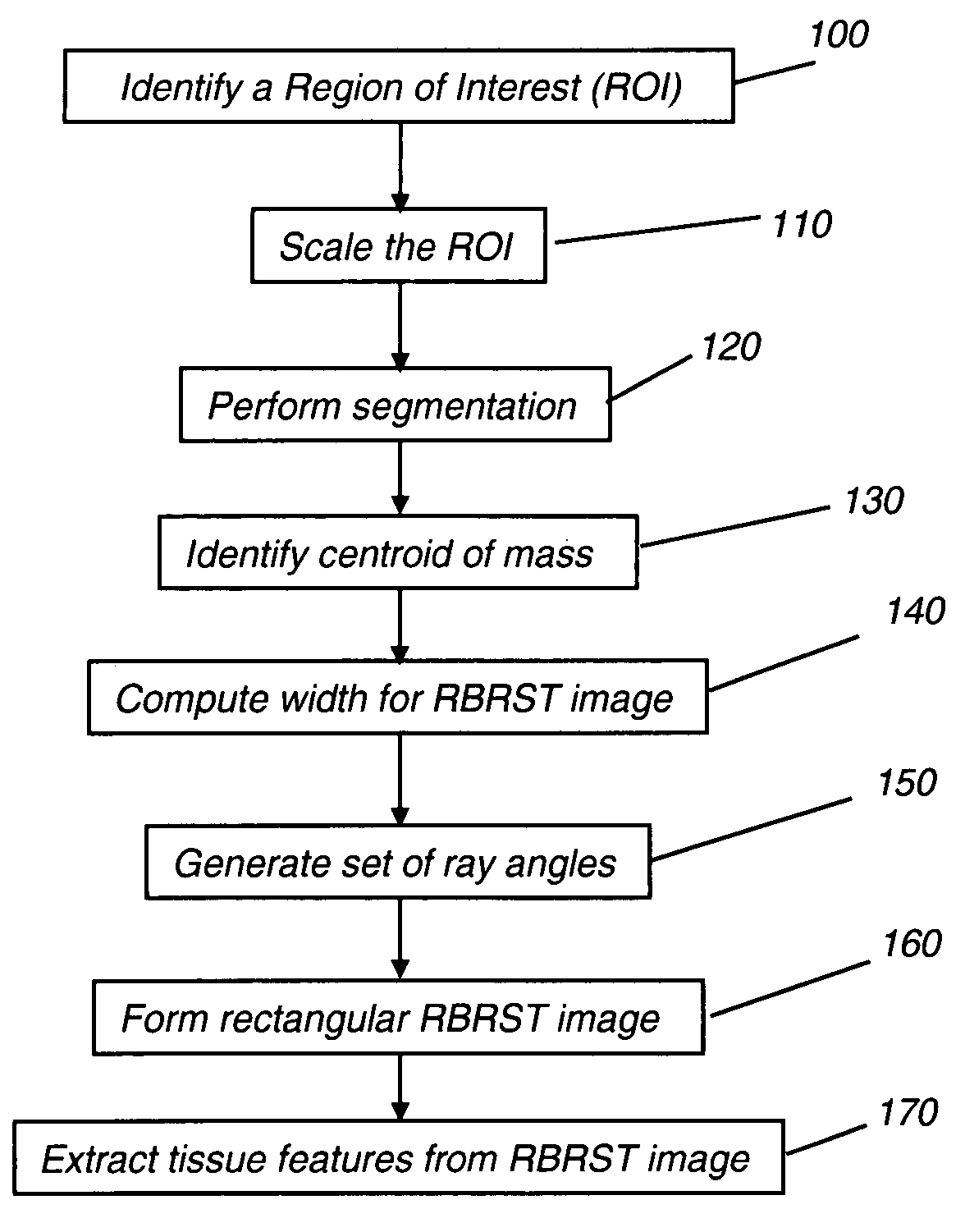
A method of characterizing a mass within a digital mammogram. A region of interest is identified that includes the mass and surrounding tissue. A border outline of the mass is identified. A rectangular image is formed wherein each column of the image is formed by repetition of steps. A vector is employed for each of a set of ray angles, wherein the vector extends from a central point of the mass and intersects the border outline at an intersection point. A starting pixel along the vector is identified, between the intersection point and the central point, at a first distance before the intersection point. An ending pixel along the vector is identified at a second distance beyond the intersection point. Pixels along the vector, from the starting pixel to the ending pixel, are remapped as the respective column in the rectangular image. Texture features are extracted from the formed rectangular image.

Description

FIELD OF THE INVENTION[0001]This invention generally relates to medical image analysis and more particularly relates to an automated method for obtaining texture information for imaged tissue.BACKGROUND OF THE INVENTION[0002]The benefits of computer-aided diagnosis in radiology in general, and particularly in mammography, have been recognized. To date, there has been considerable effort directed toward computer-aided methods that assist the diagnostician to correctly and efficiently identify problem areas detected in a mammography image and to improve the accuracy with which diagnoses are made using this information.[0003]Functions of computer-aided diagnosis include detection of mass structures within imaged tissue and characterization of their features. In general, it has been observed that sharply defined masses that have somewhat “regular” shapes are rarely malignant, while more irregularly shaped masses are of higher concern. Salient examples of irregularly shaped masses includ...

Claims


Application Information

Patent Timeline
IPC IPC(8): G06K9/00
CPCG06T7/0012G06T2207/30068G06T2207/10116
Inventor AGATHEESWARAN, ANURADHAZHANG, DAOXIAN H.ZHENG, YANG
Owner CARESTREAM HEALTH INC
Learn More