
Method and apparatus for automated detection of target structures from medical images using a 3d morphological matching algorithm

a morphological matching and target structure technology, applied in image analysis, image enhancement, instruments, etc., can solve the problem of difficult differentiation of perivascular nodules, and achieve the effect of saving computing power and simplifying detection

Inactive Publication Date: 2007-06-21
BAE KYONGTAE T +1
View PDF25 Cites 35 Cited by
  • Summary
  • Abstract
  • Description
  • Claims
  • Application Information

AI Technical Summary

Benefits of technology

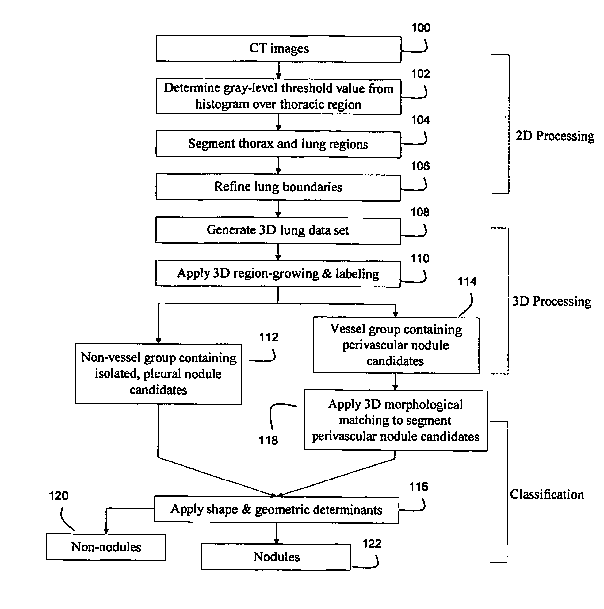
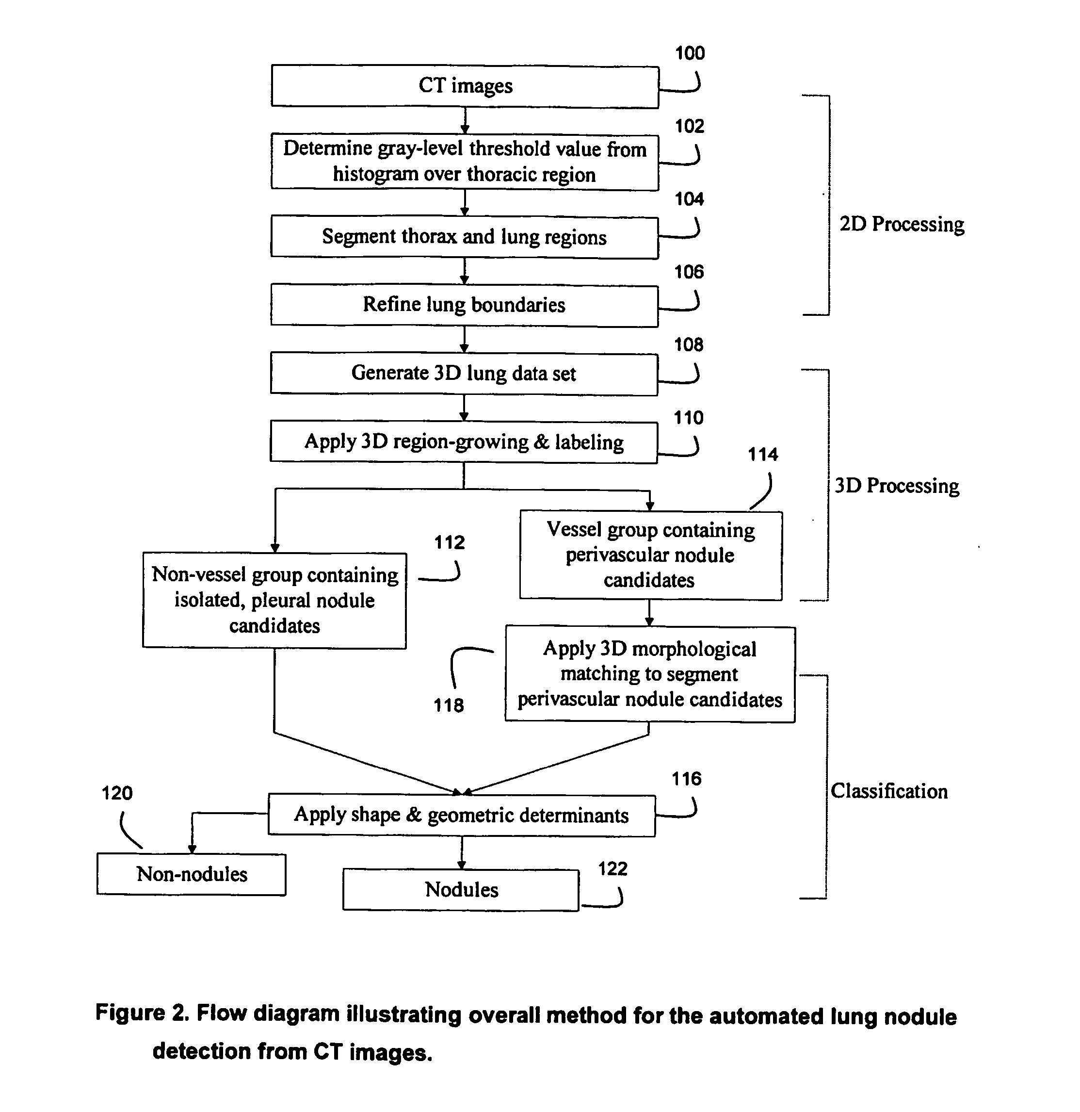
[0014] By classifying 3D structures found in the image data into different groups, the present invention allows for the particularized detection techniques to be applied to each group, wherein each detection technique is optimized to the specifics of its group. For example, because accurate detection of perivascular nodules in a patient's lung region requires a more strenuous analysis than does detection of isolated nodules or pleural nodules, the present invention allows application of the more rigorous detection algorithm to the perivascular nodule candidates while providing application of the simpler detection algorithm to the other nodule candidates. Such a bifurcated approach preserves computing power and represents an efficient approach to CAD.
[0015] Also, by application of 3D morphological filters to candidate structures that may include perivascular nodules, the present invention allows for a level of automated detection of perivascular nodules that, to the inventors' knowledge, has previously been unattainable.

Problems solved by technology

However, perivascular nodules are not easily differentiated from the nearby blood vessels because they are closely associated with adjacent blood vessels and because some blood vessels that are perpendicular to the imaging plane exhibit a similar appearance as perivascular nodules in 2D slices.

Method used


Image

  • Method and apparatus for automated detection of target structures from medical images using a 3d morphological matching algorithm
  • Method and apparatus for automated detection of target structures from medical images using a 3d morphological matching algorithm
  • Method and apparatus for automated detection of target structures from medical images using a 3d morphological matching algorithm

Examples


Embodiment Construction

[0026] The lung nodule detection algorithm of the preferred embodiment is a combination of 2D and 3D operations. While the 3D operation is more general and more powerful, in some image processing steps the 2D operation is preferred because it is more efficient and easier to implement. The combined 2D / 3D approach of the preferred embodiment represents an efficient technique for pulmonary nodule detection and may be used by radiologists when the CT images are reviewed for the presence of lung nodules.

[0027] The CT images may be generated by a CT scanner as is well known in the art. The generated CT images are preferably electronically transferred from the scanner to a computer such as a PC workstation. The automated detection algorithm of the preferred embodiment is then executed on the CT image data by software.

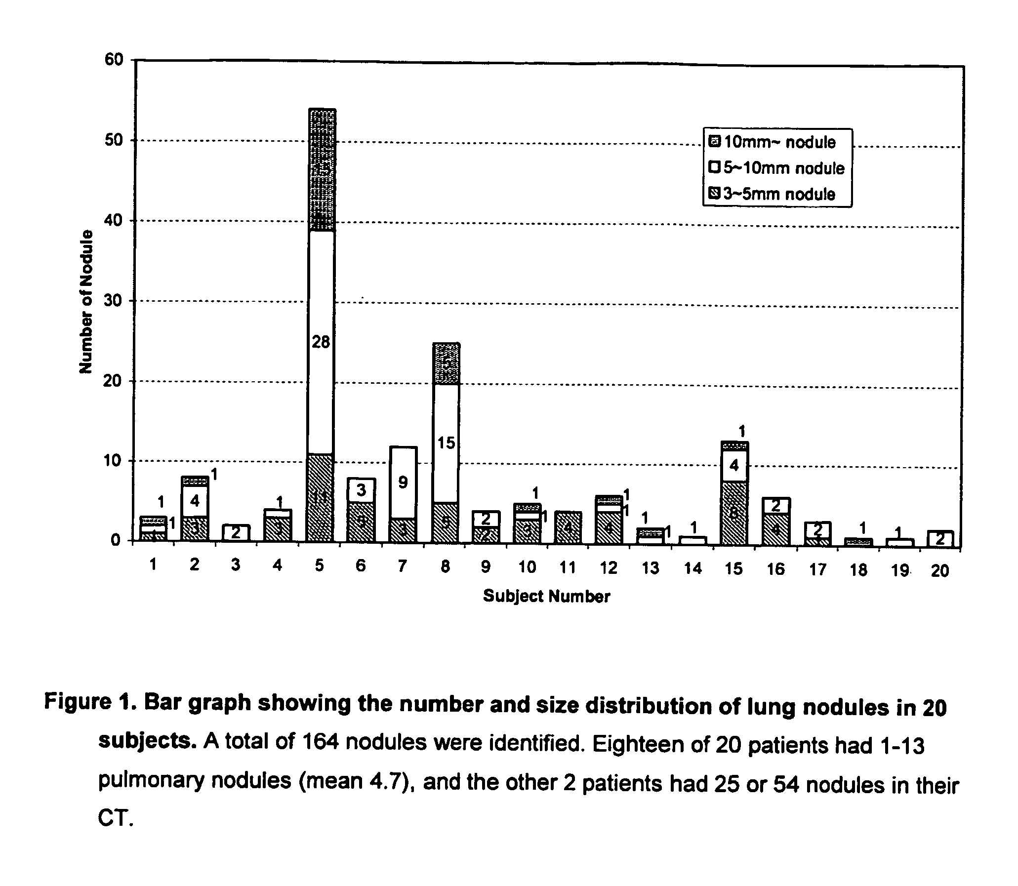
[0028] In testing the preferred embodiment, a database of 20 diagnostic thoracic CT scans from 20 patients (13 men, 7 women; age range of 40-75 years with a mean age of 57 y...


PUM


Abstract

A method for the detection of target structures shown in digital medical images, the method of comprising: (1) generating a three dimensional (3D) volumetric data set of a patient region within which the target structure resides from a plurality of segmented medical image slices; (2) grouping contiguous structures that are depicted in the 3D volumetric data set to create corresponding grouped structure data sets; (3) assigning each grouped structure data set to one of a plurality of detection algorithms, each detection algorithm being configured to detect a different type of target structure; and (4) processing each grouped structure data set according to its assigned detection algorithm to thereby detect whether any target structures are present in the medical images. Preferably, the target structures are pulmonary nodules, and a specialized detection algorithm is applied to image data classified as a candidate for depicting perivascular nodules. To segment perivascular nodule candidates from surrounding vessels, the image data is preferably correlated with a plurality of 3D morphological filters.

Description

CROSS REFERENCE AND PRIORITY CLAIM TO RELATED APPLICATION [0001] This application claims the benefit under 35 U.S.C. § 119(e) of provisional patent application Ser. No. 60 / 430,954 entitled “Method and Apparatus for Automated Detection of Pulmonary Nodules from Computed Tomography (CT) Images Using A 3D Morphological Matching Algorithm”, filed Dec. ______, 2002, the disclosure of which is incorporated by reference herein.FIELD OF THE INVENTION [0002] The present invention relates to a system for computer-aided diagnosis (CAD) of target structures using digital medical images. In particular, the present invention relates to the application of CAD techniques to CT images to thereby automate the detection of target structures such as pulmonary nodules. BACKGROUND OF THE INVENTION [0003] Lung cancer is the second most commonly diagnosed cancer in the United States, and other cancers frequently metastasize to the lung parenchyma as pulmonary nodules. Chest CT is the most sensitive diagnos...

Claims


Application Information

Patent Timeline
IPC IPC(8): G06K9/00G06T7/00
CPCG06T7/0012G06T2207/30061G06T2207/30101
Inventor BAE, KYONGTAE T.KIM, JINSUNG
Owner BAE KYONGTAE T
Learn More