
Method and apparatus for obtaining aliquot from liquid-based cytological sample

a liquid-based cytological sample and aliquot technology, applied in diagnostic recording/measuring, laboratory glassware, instruments, etc., can solve the problems of molecular contamination, molecular contamination problems, and laboratory weary of molecular contamination of hpv dna tests

Inactive Publication Date: 2007-06-14
CYTYC CORP
View PDF13 Cites 26 Cited by
  • Summary
  • Abstract
  • Description
  • Claims
  • Application Information

AI Technical Summary

Benefits of technology

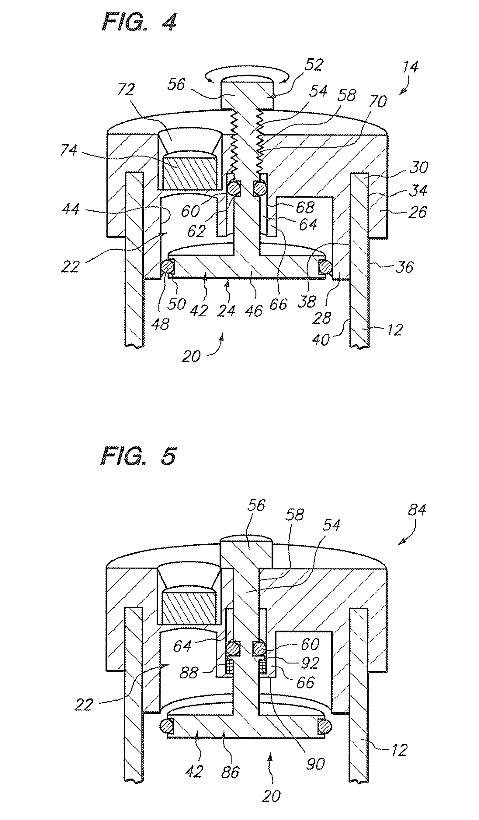
[0021] In one embodiment, the valve mechanism includes a valve configured to be selectively located within the aliquot chamber to prevent fluid communication between the aliquot chamber and the collection chamber, and displaced from the aliquot chamber to allow fluid communication between the aliquot chamber and the collection chamber. In another embodiment, the valve mechanism includes a valve configured to be selectively located at an interface between the aliquot chamber and the collection chamber to prevent fluid communication between the aliquot chamber and the collection chamber, and displaced from the interface to allow fluid communication between the aliquot chamber and the collection chamber.

Problems solved by technology

However, because it is currently not commercially practical to perform the HPV DNA test in conjunction with every Pap smear, the HPV DNA test is typically performed in response to a ASC-US Pap smear result.
Thus, in the case of liquid-based Pap smears, the aliquot required for HPV DNA testing would have to be taken from the sample after being processed for slide preparation, which may potentially lead to molecular contamination issues, and in the case of automated Pap smear processing, aerosol cross-contamination resulting from the use of air pressure to manipulate the sample or liquid contamination resulting from the transfer of filtered solution found in the plumbing of the automated processor.
Significantly, laboratories that perform HPV DNA tests are weary of molecular contamination—a well-known problem in molecular diagnostic laboratories, which typically employ special engineering and design features, laboratory practices, and monitoring activities to minimize molecular contamination in all of their activities.
Because automated systems for making Pap smear slides are designed to safely and effectively handle cells—not nucleic acids, the sterilization protocols followed by technicians when operating these systems do not satisfy the strict molecular contamination control safeguards and procedures required by molecular diagnostic laboratories.
Thus, due to the risk of cross-contamination, molecular diagnostic laboratories may not accept aliquots that have been taken from an already processed liquid-based Pap smear for fear of unnecessarily generating false HPV positives.
However, false positives are a special problem when testing for Chlamydia trachomatis and Neisseria gonorrhoeae, because they could have enormous family and social repercussions.
Thus, molecular diagnostic laboratories are even more reluctant to accept aliquots from already processed liquid-based Pap smear samples.
However, this step may still not satisfy the strict contamination prevention requirements imposed by molecular diagnostic laboratories.
Besides contamination issues, the pipetting of an aliquot from a fluid-based Pap smear sample, whether done before or after the sample is processed, and whether done for HPV testing or testing of any other sexually transmitted disease, increases cost in the form of manual labor (which involves not only pipetting the aliquot into an extra vial, but also labeling the vial), material costs for the pipettes and extra vials, and storage space, which when multiplied by the thousands of samples processed, becomes significant.

Method used


Image

  • Method and apparatus for obtaining aliquot from liquid-based cytological sample
  • Method and apparatus for obtaining aliquot from liquid-based cytological sample
  • Method and apparatus for obtaining aliquot from liquid-based cytological sample

Examples


Embodiment Construction

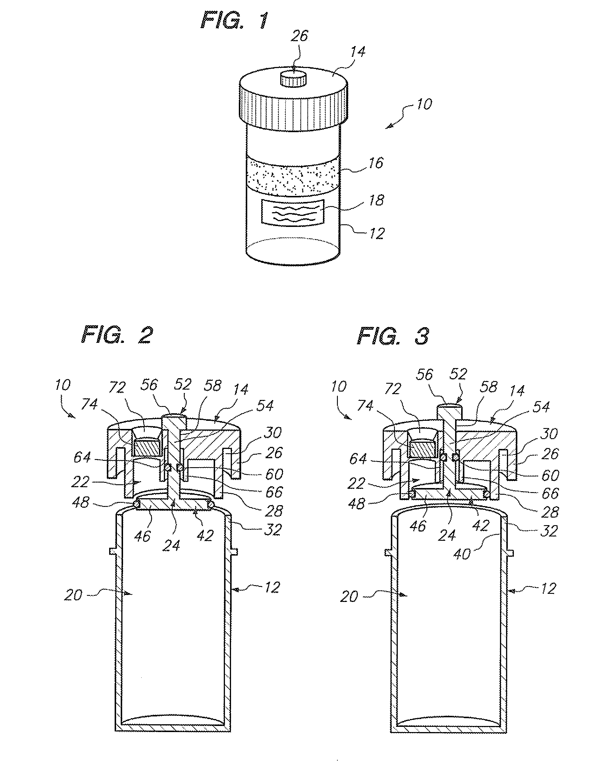
[0037] Referring to FIG. 1, a sample vial 10 constructed in accordance with one embodiment of the present invention will be described. The vial 10 may be used to contain a fluid-based sample, such as a cervical-vaginal sample collected from a patient at a physician's office. The fluid-based sample typically comprises cytological material suspended in an aqueous preservative fluid.

[0038] To this end, the vial 10 comprises a hollow vial container 12 and a vial cap 14 that can be placed onto the vial container 12 to enclose a sample contained within the vial container 12. As depicted, the vial container 12 and vial cap 14 are generally cylindrical in shape. The selected size of the vial container 12 and vial cap 14 may vary, but preferably is large enough to contain the minimum amount of sample necessary to perform the intended diagnostic test. In the illustrated embodiment, the vial container 12 is capable of containing at least 20 ml of fluid, which is the minimum amount of sample r...


PUM

PropertyMeasurementUnit
deoxynucleic acidaaaaaaaaaa
structureaaaaaaaaaa
areaaaaaaaaaaa

Abstract

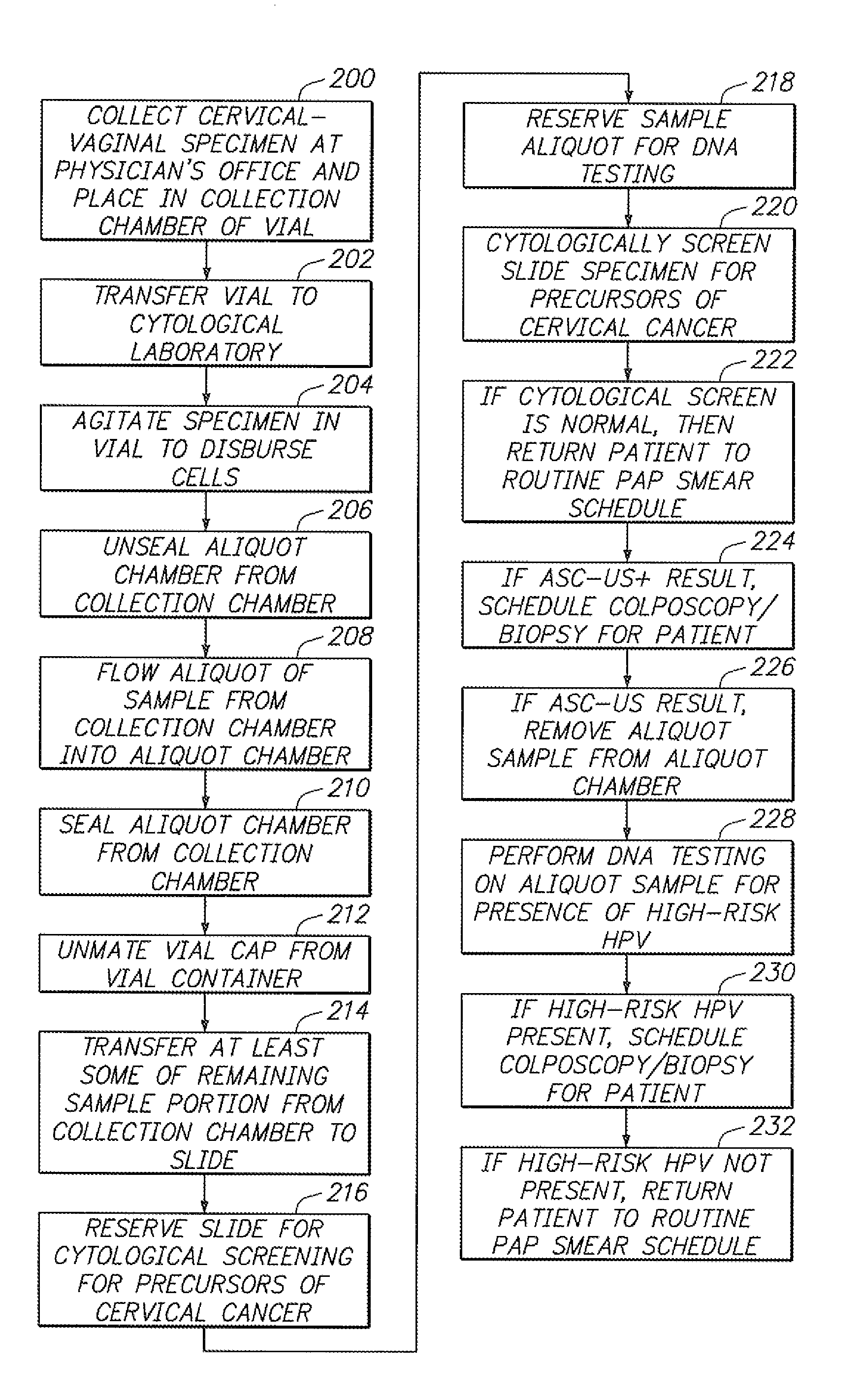
Sample vials and methods of processing the sample vials are provided. The sample vial comprises a vial container, a sample collection chamber within the vial container, a vial cap configured to be mated with the vial container to enclose the collection chamber, an aliquot chamber, which may be carried by the vial cap or the vial container, and a valve mechanism for selectively sealing and unsealing the aliquot chamber from the collection chamber. The method may comprise flowing an aliquot of the sample from the collection chamber while the sample within the collection chamber is isolated from an environment exterior to the vial, sealing the aliquot chamber from the collection chamber to isolate the aliquot sample from the remaining sample portion, and transferring at least some of the remaining sample portion from the collection chamber to a microscope slide while the aliquot chamber is sealed from the collection chamber. The method further comprises reserving the slide for cytological screening of the sample, and reserving the aliquot sample for deoxynucleic acid (DNA) testing.

Description

FIELD OF THE INVENTION [0001] The invention pertains to the preparation of cytological samples, and more specifically, to a method and apparatus for obtaining aliquots from cytological samples, such as fluid-based Papanicolaou (“Pap”) smears. BACKGROUND [0002] Cytology is a branch of biology dealing with the study of the formation, structure, and function of cells. As applied in a laboratory setting, cytologists, cytotechnologists, and other medical professionals make medical diagnoses of a patient's condition based on visual examination of a sample of the patient's cells. Cytological techniques lend themselves well to the detection of abnormal cells and disease in the human body. [0003] Cytological techniques are widely employed, because collection of cell samples for analysis is generally less invasive than traditional surgical pathological procedures such as biopsies, whereby a tissue sample is excised from the patient using specialized biopsy needles having spring loaded transla...

Claims


Application Information

Patent Timeline
Patent Type & Authority Applications(United States)
IPC IPC(8): B01L3/00C12Q1/68C12M1/00
CPCA61B10/0096B01L3/0272B01L3/50825B01L2200/0605B01L2200/141B01L2300/042B01L2300/047B01L2300/049B01L2400/0633
Inventor WATTS, HALSAKAL, ROBERT
Owner CYTYC CORP
Learn More