
Method for projecting radiological image data into a neuroanatomical coordinate system

a radiological image and coordinate system technology, applied in image enhancement, image analysis, instruments, etc., can solve the problems of requiring a great time outlay for users of image data, affecting the efficiency of radiological image evaluation, and affecting the statistical evaluation of radiological image data of a plurality of peopl

Inactive Publication Date: 2007-04-12
SIEMENS AG
View PDF6 Cites 2 Cited by
  • Summary
  • Abstract
  • Description
  • Claims
  • Application Information

AI Technical Summary

Benefits of technology

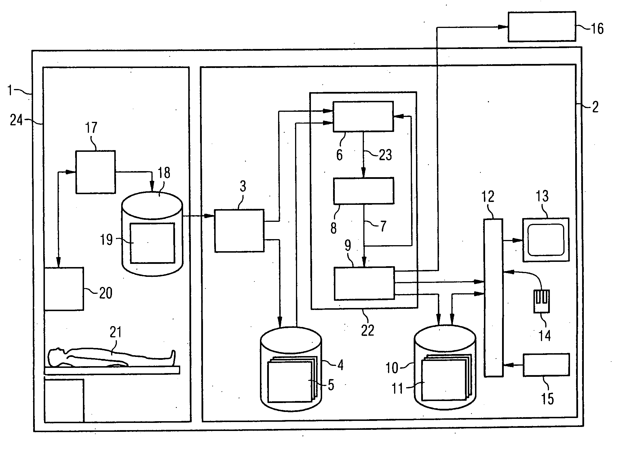
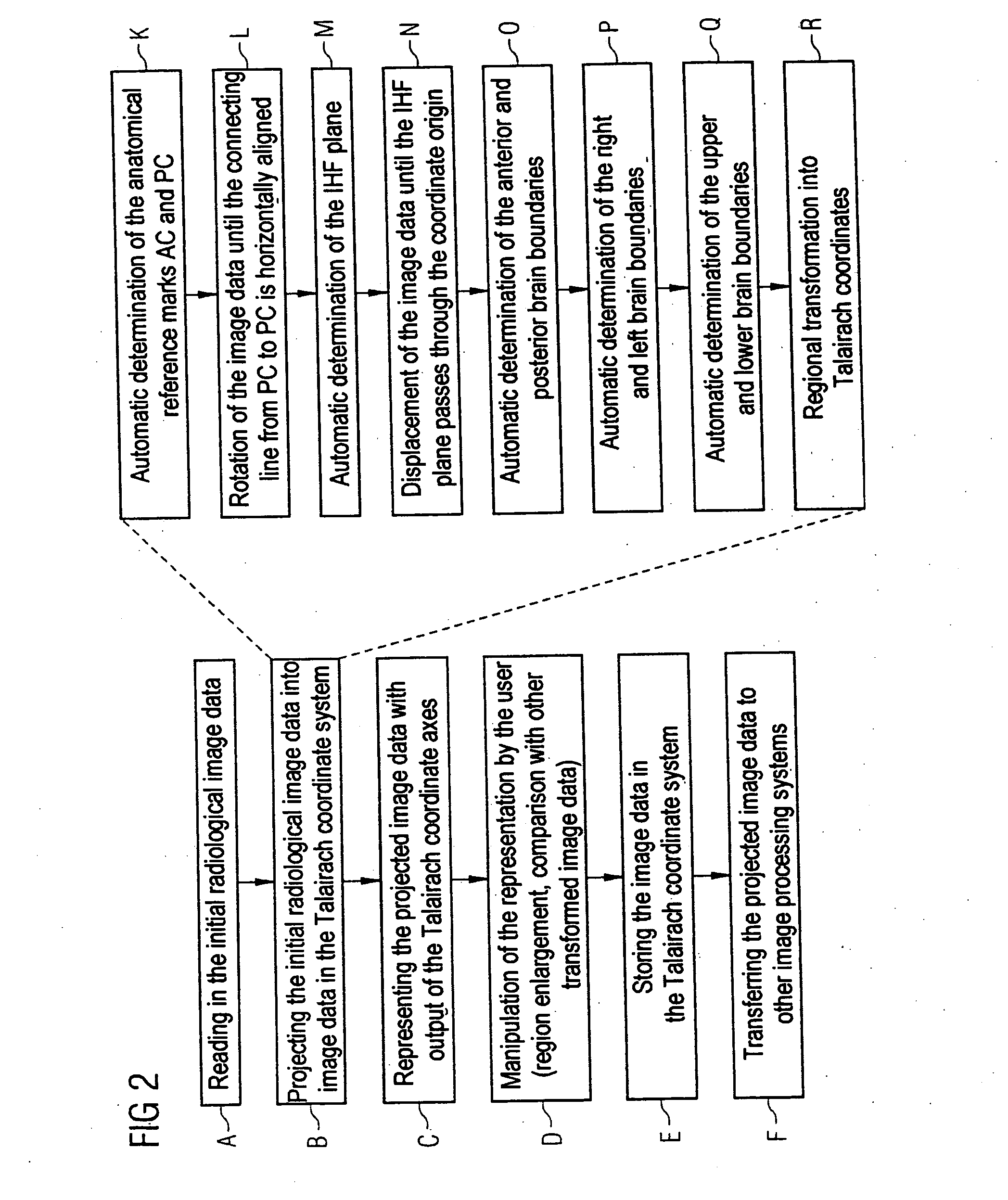
[0008] In at least one embodiment of the present invention, a method is provided for projecting initial radiological image data of at least a part of a brain into a neuroanatomical coordinate system, which can be carried out in a substantially automated fashion so that the image data are available for the user promptly and in a uniform form.
[0014] With the aid of the method according to at least one embodiment of the invention, radiological image data of the brain can be preprocessed in a substantially automated fashion and therefore relatively quickly so that they can be compared easily with image data of other brains, even when the initial radiological image data differ owing to the respective individual anatomical properties of the brain and a special situation possibly existing during the image data recording. In particular, the “standardization” of the determination of the anatomical reference marks makes it possible to generate anatomical or functional average-value images for a multiplicity of patients or subjects efficiently by the method according to the invention, which facilitates especially the conduct of clinical studies. Divergences, which could result if the anatomical reference marks are identified with the involvement of a user, are precluded.
[0020] In an example embodiment of the method according to the invention, the transformation of the rotated and / or displaced image data also involves scaling. Such scaling may inter alia be used to modify the radiological image data of different persons so that the extents of the brains, i.e. for example the distance between the anterior and posterior brain boundaries, are projected onto uniform values. This improves the comparability of the radiological image data.
[0026] In a further example embodiment of the representation of radiological image data, which have been projected into a neuroanatomical coordinate system, the transformed image data of different brains are represented simultaneously. This makes it relatively easy for the observer to study common features and differences between the image data.
[0029] Such an implementation of the method according to at least one embodiment of the invention is recommendable particularly when a multiplicity of image data are to be projected into a neuroanatomical coordinate system, for example in the scope of clinical studies. The image data processing device may alternatively be a component of a control device (also referred to as a scanner console) of a modality, that is to say it is in spatial proximity to the recording instrument. This makes it possible for the image data to be projected into a neuroanatomical coordinate system while the patient or subject is still inside the recording instrument. In particular, this offers the advantage that the transformed image data are made available rapidly and sometimes even allow immediate decisions to be made about the further procedure of the image data recording.

Problems solved by technology

It is clear that the individual anatomical properties of the brain, for example differences in the respective geometrical extent and shape of a brain, hinder efficient evaluation of the radiological image data.
Yet since the patient image data remain substantially unchanged in this method, the individual properties of a brain make it more difficult to statistically evaluate the radiological image data of a plurality of persons, for example in order to generate anatomical or functional average-value images.
However, identification of the anatomical landmarks and projection of the radiological image data by the user of the image data require a great time outlay.
Such a process is furthermore susceptible to error since, for example, correct identification of the anatomical landmarks depends on the respective experience of the user.
It is therefore also clear that the comparison of image data which have been projected by different users onto a neuroanatomical coordinate system is only limitedly possible owing to the individual influence of the user during the projection.

Method used


Image

  • Method for projecting radiological image data into a neuroanatomical coordinate system
  • Method for projecting radiological image data into a neuroanatomical coordinate system
  • Method for projecting radiological image data into a neuroanatomical coordinate system

Examples


Embodiment Construction

[0035] The terminology used herein is for the purpose of describing particular embodiments only and is not intended to be limiting of the present invention. As used herein, the singular forms “a”, “an” and “the” are intended to include the plural forms as well, unless the context clearly indicates otherwise. It will be further understood that the terms “includes” and / or “including”, when used in this specification, specify the presence of stated features, integers, steps, operations, elements, and / or components, but do not preclude the presence or addition of one or more other features, integers, steps, operations, elements, components, and / or groups thereof.

[0036] In describing example embodiments illustrated in the drawings, specific terminology is employed for the sake of clarity. However, the disclosure of this patent specification is not intended to be limited to the specific terminology so selected and it is to be understood that each specific element includes all technical e...


PUM


Abstract

A method is described for projecting radiological image data of at least a part of a brain into a neuroanatomical coordinate system. To this end, anatomical reference marks in the initial radiological image data are determined in an automated fashion and a rotation and / or displacement of the initial radiological image data is carried out, with at least some of the anatomical reference marks in the rotated and / or displaced image data match corresponding anatomical reference marks in the neuroanatomical coordinate system. The rotated and / or displaced image data are then converted into the neuroanatomical coordinate system by a transformation. An image data processing device for projecting initial radiological image data according to the method and a radiological system having a corresponding image data processing device are also described.

Description

PRIORITY STATEMENT [0001] The present application hereby claims priority under 35 U.S.C. §119 on German patent application number DE 10 2005 045 908.0 filed Sep. 26, 2005, the entire contents of which is hereby incorporated herein by reference. FIELD [0002] The invention generally relates to a method for projecting initial radiological image data of at least a part of a brain into a neuroanatomical coordinate system. The invention also generally relates to a corresponding image data processing device which converts initial radiological image data into a neuroanatomical coordinate system according to such a method, as well as to a radiological system having such an image data processing device. Background [0003] Imaging systems in medical technology (often also referred to as “modalities”) such as X-ray systems, computer tomographs or magnetic resonance tomographs are nowadays regularly used for studying the brain. The radiological image data generated by the imaging systems are used...

Claims


Application Information

Patent Timeline
Patent Type & Authority Applications(United States)
IPC IPC(8): G06K9/00
CPCG06T19/00G06T3/0075G06T7/0028G06T2207/10072G06T2207/30016G06T7/33G06T3/147
Inventor HEMPEL, ECKHARD
Owner SIEMENS AG
Learn More