Deposit contrast agents and related methods thereof
a contrast agent and contrast technology, applied in the field of depositing contrast agents, can solve the problems of insufficient spatial resolution of the clinical system to image fine structures in people, limitations of contrast perfusion imaging using high frequency ultrasound with available microbubble contrast agents and protocols, and inability to destroy microbubbles, etc., to achieve the effect of improving the quality of ultrasound images
- Summary
- Abstract
- Description
- Claims
- Application Information
AI Technical Summary
Benefits of technology
Problems solved by technology
Method used
Image
Examples
example 1
[0127] Microbubbles were prepared with a lipid monolayer shell and perfluorocarbon gas core. Particles for ultrasound contrast enhancement were prepared by sonication of an aqueous suspension of either dimyrstyl- or distearylphosphatidycholine, and PEG-sterate in a saturated atmosphere of decafluorobutane gas. This process resulted in the production of decafluorobutane microbubbles with a lipid monolayer shell. Electrozone counting of the particles with a Coulter Multisizer revealed a broad range in microbubble size with a mean diameter of just under 2 μm and a small fraction of microbubbles with a diameter greater than 5 μm. By flotation separation, a population of relatively large microbubbles (5-15 microns) were separated from smaller bubbles. These microbubbles were of sufficient size that they pass through pulmonary arteriovenous shunts (accounting for up to 3-5% of total pulmonary flow in a small animal), and yet lodge in the coronary or other tissue microcirculation or microv...
example 2
Methods
Microbubble Preparation
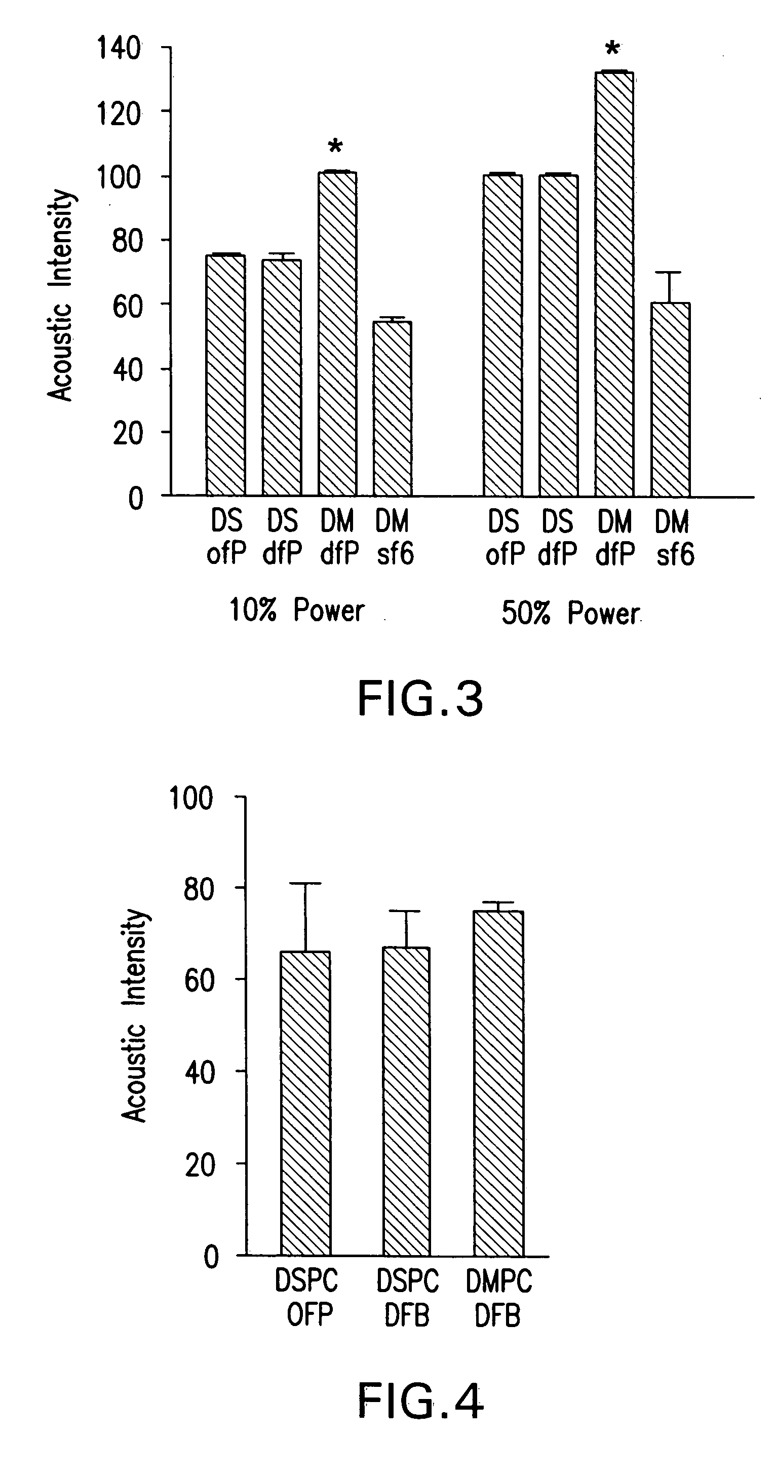
[0130] Lipid-shelled perfluorocarbon gas microbubble agents were prepared by sonication of a gas-saturated aqueous lipid dispersion. Four separate agents were prepared: distearyl phosphatidylcholine (1.6 mg·mL−1) and PEG-5000 with ocafluoropropane gas (DSPC-OFP); distearyl phosphatidylcholine (1.6 mg·mL−1) and PEG-5000 with decafluorobutane gas (DSPC-DFB); distearyl phosphatidylcholine (1.6 mg·mL−1) and PEG-5000 with sulphur hexafluoride gas (DSPC-SF6); and dimyrystylphosphatidylcholine (1.6 mg·mL−1) and PEG-5000 with decafluorobutane gas (DMPC-DFB). All microbubbles were washed by flotation centrifugation in phosphate-buffered saline (PBS) and their concentration and size distribution were determined by electrozone sensing with a Coulter Multisizer IIe (Beckman-Coulter, Fullerton, Calif.). Separation of microbubbles into small and large size fraction was performed by flotation-centrifugation at 400 g for 15 seconds and separation of the turbid subn...
example 3
Contrast Agent Preparation
[0146] Lipid-shelled microbubbles were prepared by sonication of an aqueous lipid dispersion of PEG-5000-stearate and distearoyl phosphatidylcholine saturated with decafluorobutane gas. For intravital microscopy studies, the microbubble shell was fluorescently labeled by adding a trace amount of DiI to the suspension prior to sonication. Microbubble concentration and size distribution were determined by electrozone sensing (Coulter Multisizer IIe, Beckman-Coulter, Fullerton, Calif.).
Animal Preparation
[0147] Twenty-seven male wild-type C57B1 / 6 mice 8-12 weeks of age were used. Mice were anesthetized with an intraperitoneal injection (12.5 μL·g−1) of a solution containing ketamine hydrochloride (10 mg·mL−1), xylazine (1 mg·mL−1) and atropine (0.02 mg·mL−1). Body temperature was maintained at 37° C. with a heating platform. A jugular vein was cannulated for administration of microbubbles.
Intravital Micrososcopy
[0148] In 4 anesthetized mice, the cremast...
PUM
 Abstract
Description
Claims
Application Information
 - R&D
- Intellectual Property
- Life Sciences
- Materials
- Tech Scout
- Unparalleled Data Quality
- Higher Quality Content
- 60% Fewer Hallucinations
Browse by: Latest US Patents, China's latest patents, Technical Efficacy Thesaurus, Application Domain, Technology Topic, Popular Technical Reports.
© 2025 PatSnap. All rights reserved.Legal|Privacy policy|Modern Slavery Act Transparency Statement|Sitemap|About US| Contact US: help@patsnap.com


