Oncoprotein protein kinase
a technology of oncoprotein and kinase, which is applied in the direction of transferases, peptide/protein ingredients, instruments, etc., can solve the problems of overexpression of a particular gene, affecting healthy cells as well as neoplastic cells, and prone to act indiscriminately,
- Summary
- Abstract
- Description
- Claims
- Application Information
AI Technical Summary
Benefits of technology
Problems solved by technology
Method used
Image
Examples
example 1
Plasmids and Expression of GST Fusion Proteins
[0097] The glutathione-S-transferase (GST)-cJun expression vector, pGEX2T-cJun(wt), was constructed by inserting a filled-in BspHI-PstI fragment (encoding AA 1-223) from RSV-cJun(BspHI) into the SmaI site of pGEX2T (Pharmacia). RSV-cJun(BspHI) was constructed by changing the translation initiation sequence CTATGA of RSV-cJun to TCATGA by site-directed mutagenesis. The GSTcJun(Ala63 / 67)(BspHI) expression vector was derived in the same manner from RSV-cJun(Ala63 / 73) (Smeal, et al., supra, 1991) and was used to construct pGEX2T-cJun(Ala 63 / 67). The various GSTcJun truncation mutants were constructed using the polymerase chain reaction (PCR) to amplify various portions of c-Jun coding region. The sequences of the primers are indicated below:
N-terminal primers:(SEQ ID NO: 2)TCTGCAGGATCCCCATGACTGCAAAGATGGAAACG (underlinedcodon: amino acid 1);(SEQ ID NO: 3)TCTGCAGGATCCCCGACGATGCCCTCAACGCCTC (a.a.11);(SEQ ID NO: 4)TCTGCAGGATCCCCGAGAGCGGACCTTA...
example 2
Kinase Assays
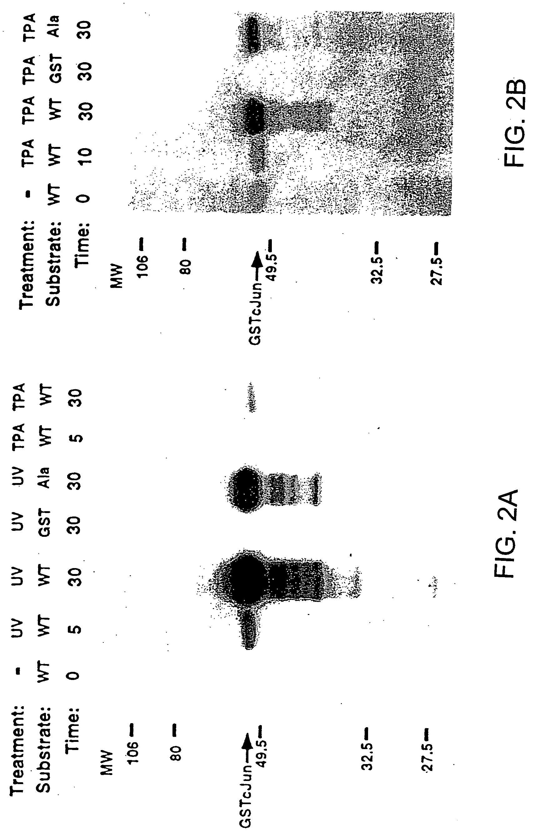
[0104] Cell extracts were diluted so that the final composition of the WCE buffer was 20 mM HEPES pH 7.7, 75 mM NaCl, 2.5 mM MgCl2, 0.1 mM EDTA, 0.05% Triton X-100, 0.5 mM DTT, 20 mM β-glycerolphosphate, 0.1 mM Na3VO4, 2 μg / ml leupeptin, 100 μg / ml PMSF. The extracts were mixed with 10 μl of GSH-agarose suspension (Sigma) to which 10 μg of either GST or GST-Jun fusion proteins were bound. The mixture was rotated at 4° C. for 3 hours in a microfuge tube and pelleted by centrifugation at 10,000×g for 20 sec. After 4×1 ml washes in HEPES binding buffer (20 mM HEPES pH 7.7, 50 mM NaCl. 2.5 mM MgCl2, 0.1 mM EDTA, 0.05% Triton X-100), the pelleted beads were resuspended in 30 μl of kinase buffer (20 mM HEPES pH 7.6, 20 mM MgCl2, 20 mM β-glycerolphosphate, 20 μM p-nitrophenyl phosphate, 0.1 mM Na3VO4, 2 mM DTT) containing 20 μM ATP and 5 μCl γ-32P-ATP. After 20 minutes at 30° C. the reaction was terminated by washing with HEPES binding buffer. Phospho...
example 3
Binding of a Protein Kinase to GST-cJun-GSH-Agarose Beads
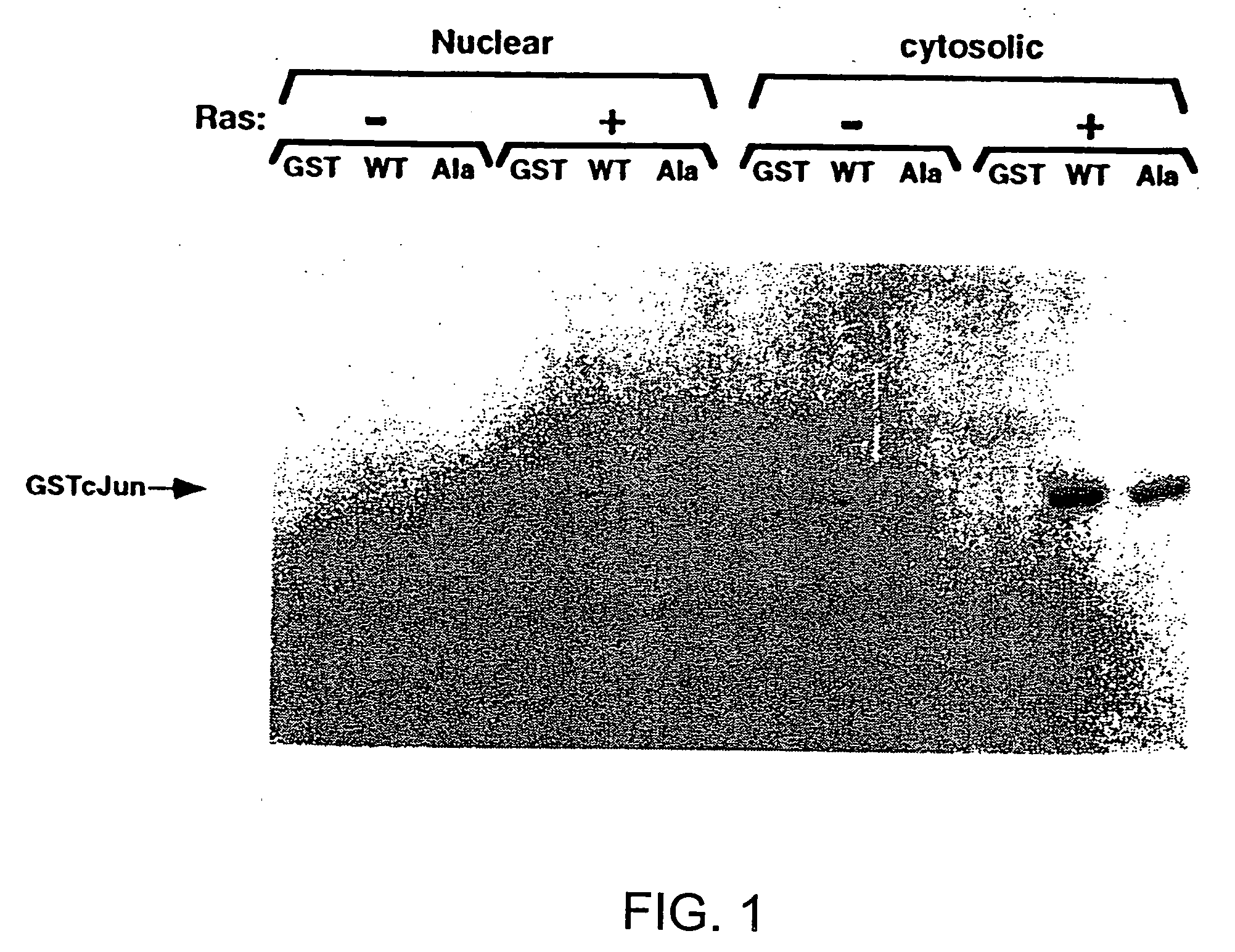
[0106] The fusion protein, GSTcJun(wt), can bind through its GST moiety to glutathione (GSH)-agarose beads to generate an affinity matrix for identification of c-Jun binding proteins, which may include protein kinases. Ha-ras transformation of FR3T3 cells results in increased phosphorylation of c-Jun on Ser 63 and 73 (Binetruy, et al., supra, 1991; Smeal. et al., supra, 1991). Preliminary experiments indicated that transformed cells contained higher levels of c-Jun N-terminal kinase activity, while the levels of c-Jun C-terminal kinase activity remaned unchanged. To develop a more convenient assay for characterizing the c-Jun N-terminal kinase activity, nuclear and cytoplasmic extracts of untransformed and transformed FR3T3 cells were mixed with GSTcJun(wt)-GSH-agarose beads. FRT3T3(−) and Ha-ras-transformed FR3T3(+) cells were kept in 0.5% FCS for 24 hours and harvested to prepare nuclear and cytosolic extracts. These extrac...
PUM
 Abstract
Description
Claims
Application Information
 - R&D
- Intellectual Property
- Life Sciences
- Materials
- Tech Scout
- Unparalleled Data Quality
- Higher Quality Content
- 60% Fewer Hallucinations
Browse by: Latest US Patents, China's latest patents, Technical Efficacy Thesaurus, Application Domain, Technology Topic, Popular Technical Reports.
© 2025 PatSnap. All rights reserved.Legal|Privacy policy|Modern Slavery Act Transparency Statement|Sitemap|About US| Contact US: help@patsnap.com


