
Instantaneous ultrasonic measurement of bladder volume

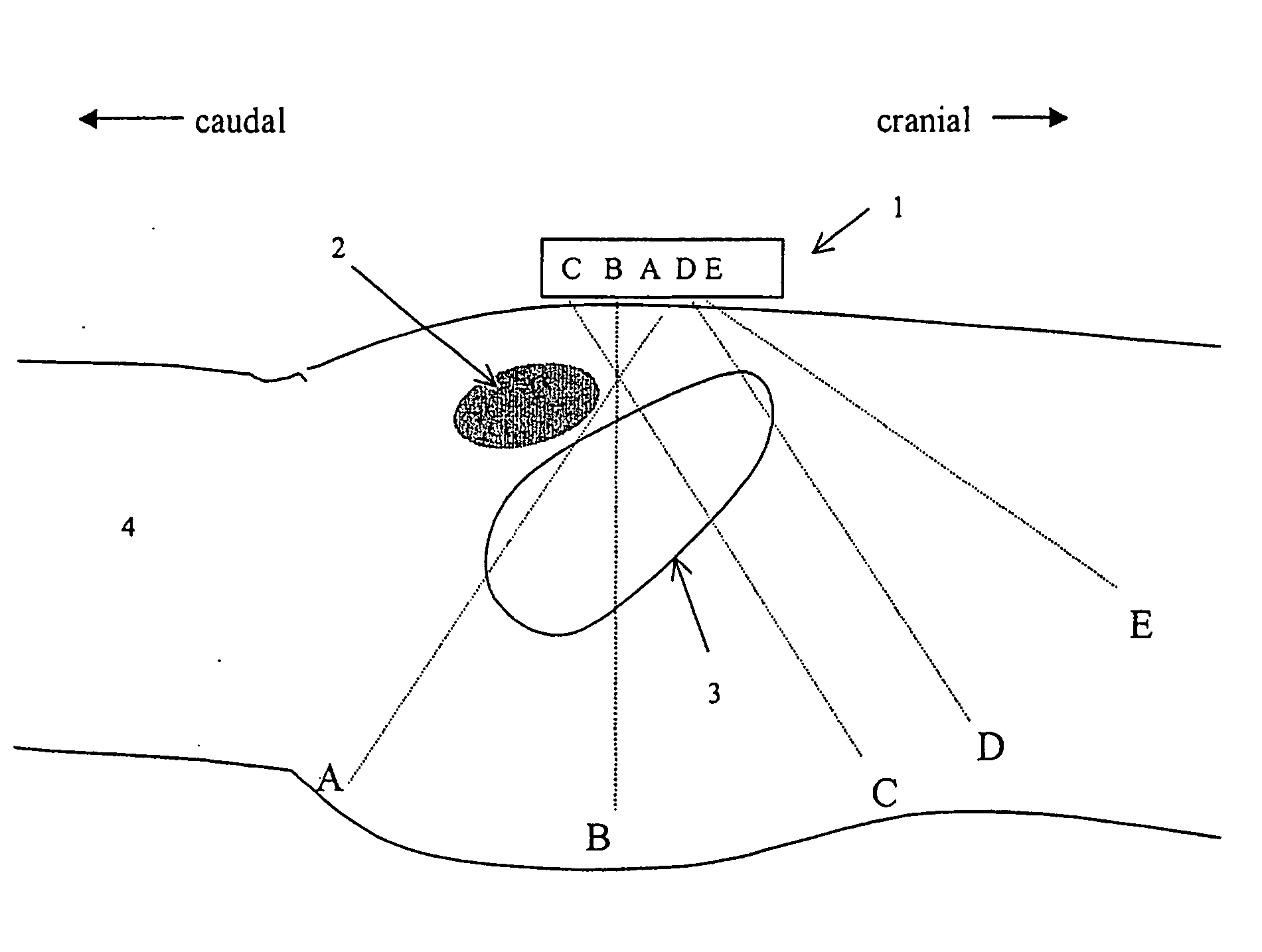
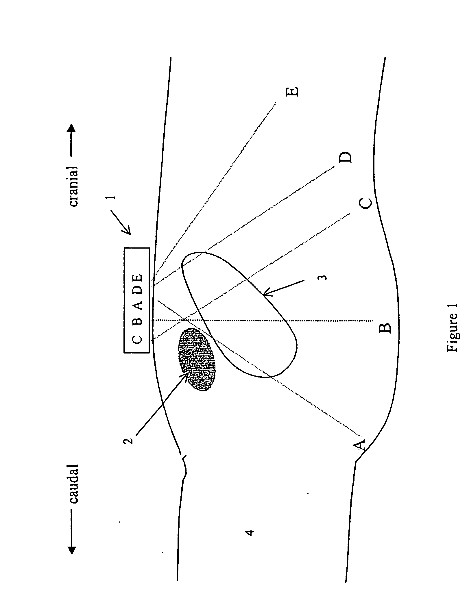
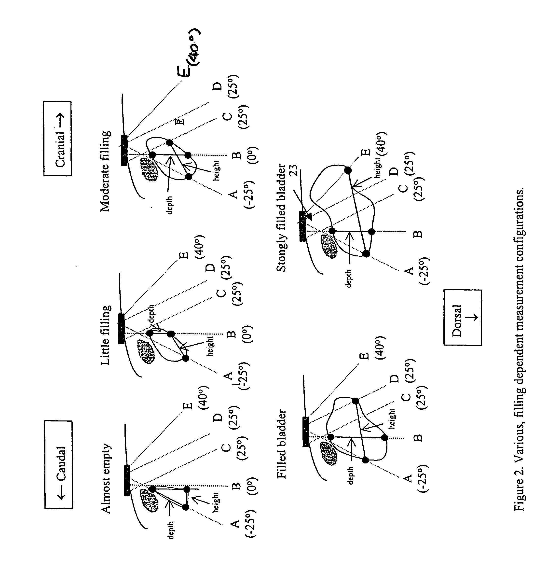
a technology of ultrasonic measurement and bladder volume, which is applied in the direction of instruments, catheters, image enhancement, etc., can solve the problems of unnecessary catheterization, serious infection, uncomfortable patient situation,

Active Publication Date: 2006-05-25
VERATHON
View PDF20 Cites 44 Cited by
  • Summary
  • Abstract
  • Description
  • Claims
  • Application Information

AI Technical Summary

Problems solved by technology

However, serious disadvantages to unnecessary catheterization range from the uncomfortable situation for the patient to serious possibilities of infection.
Non-invasive procedures for bladder volume estimation are known, but are either unreliable or expensive or have some other significant disadvantages.
Palpation and auscultatory percussion are known to be unreliable, while radiography and dye-excretion techniques are known to be similarly inaccurate.
However, it is invasive, painful and might produce traumas or infections.
His method is not adjusted to specific, filling dependent, measuring configurations.
Since the bladder changes in shape when filling, a single distance measurement is not precise enough to predict the entire bladder volume.
The instruments are costly and require a skilled operator.
This further increases complexity, acquisition time of the data, and costs of the instrument.
The used apparatus is complex and is different from the method described in this application.
The acquisition procedure is time consuming and thus no instantaneous volume measurement results.
This nevertheless still requires an acquisition time equivalent to full acquisition procedure and thus does not yield an instantaneous display of volume.
No instantaneous feedback on optimal positioning is thus available.
The three dimensional scanning requirement makes this instrument complex.
This explains the large error that several studies obtained when a single geometric model was used.
Non-linear distortion increases with distance, insonifying ultrasound energy and frequency.

Method used


Image

  • Instantaneous ultrasonic measurement of bladder volume
  • Instantaneous ultrasonic measurement of bladder volume
  • Instantaneous ultrasonic measurement of bladder volume

Examples


Embodiment Construction

[0062] The second version of the device is based on a different principle. The approach consists of using a single acoustic beam with a very wide width such that it encloses approximately the entire volume of the bladder when it is filled up. Such a wide beam width can be obtained using a single element transducer with a defocusing lens as drawn in FIG. 7 or a curved single element transducer.

[0063] The schematic principle of transducer positioning is illustrated in FIG. 7. The sagittal cross section through the bladder is shown. The cone like shape of the acoustic beam allows to encompass approximately the full bladder volume, and therefore any harmonic distortion detected in the echo signal returning from a region beyond the posterior wall of the bladder around depth W, would correlate to the amount of fluid contained in the bladder.

[0064] It has been demonstrated that the propagation of ultrasound waves is a nonlinear process. The nonlinear effects, which increase with higher i...


PUM


Abstract

An apparatus and methods to quantify the volume of urine in a human bladder with a limited number of acoustic beams is disclosed. In a first version the apparatus is composed of a transducers assembly that transmits a plurality of narrow ultrasound beams in different directions towards the bladder and receives the returning ultrasound signals; a receiver detector for processing the returned signals; an analog-to-digital converter; a memory to store the digitized data and a volume display allowing to define the optimal position of the transducer assembly. The apparatus also includes a signal processing software that automatically determines the bladder Depth D and Height H and computes the volume of urine using an empirical formula corrected by specific, empirically measured, filling dependant correction factors. In a second version a single wide angle ultrasound beam transducer transmitting ultrasound signals at fundamental frequency is used to quantify the urine volume. Return signals originating from a depth beyond the usual position of the posterior wall depth of a filled bladder are analyzed for presence of higher harmonic signals which in turn are related to presence or absence of urine. Both methods or a combination thereof can be used us a simple warning device for presence of residual urine after voiding or indicate the presence of a critical bladder urine filling level.

Description

[0001] The present invention relates to methods and apparatus for the measurement of volume of a fluid filled cavity in a human or animal body, such as a bladder, using ultrasound techniques. TECHNICAL FIELD [0002] This invention concerns an apparatus which, in a first version, with a limited number of fixed ultrasound transducers with narrow sound beams oriented in well defined directions, automatically determines the volume of the human bladder without assumption of any geometrical bladder shape, where volume is calculated by (Height×Depth×K) and the empirically measured K factor varies with bladder filling degree, which in turn is indicated by the number of ultrasonic beams that intercept the filled bladder. In this first version, standard echographic technique is used where short ultrasound pulses are transmitted at fundamental frequency and the echo travel time is used to calculate distance. [0003] In a second version, with a wide ultrasound beam, pulses are transmitted at fund...

Claims


Application Information

Patent Timeline
Patent Type & Authority Applications(United States)
IPC IPC(8): A61B8/00A61B8/06A61B5/20A61B8/08A61B8/12A61B8/14
CPCA61B5/204A61B8/08A61B8/0858A61B8/4472A61B8/483G01S7/52036G01S15/102G01S15/8909G06T7/0012G06T7/602G06T2207/10132G06T2207/30004G06T7/62
Inventor MCMORROW, GERALDBAARTMANS, HENRIBOM, NICOLAASLANCEE, CHARLES THEODOOR
Owner VERATHON
Learn More