
Endoscopic mucosal resection method and associated instrument

a mucosal resection and endoscopic technology, applied in the field of endoscopic medical procedures, can solve the problems of muscularis layer, poor surgical candidates for this difficult surgery, significant complications and lifestyle problems of patients, etc., and achieve the effect of reducing the likelihood of organ perforation and accurate tissue removal

Inactive Publication Date: 2006-03-23
GRANIT MEDICAL INNOVATION
View PDF14 Cites 95 Cited by
  • Summary
  • Abstract
  • Description
  • Claims
  • Application Information

AI Technical Summary

Benefits of technology

[0011] It is a more particular object of the present invention to provide an instrument that will enable accurate removal of tissue that lies flatly on the mucosal wall of the gastrointestinal tract.
[0012] It is another more particular object of the present invention to provide such a method and / or instrument that reduces the likelihood of organ perforation.
[0014] It is even a more particular object of the present invention to provide an instrument and accompanying method that enables control of the depth and breadth of resection.

Problems solved by technology

This is an effective but drastic treatment and presents significant complications and lifestyle problems for the patient.
Many patients are poor surgical candidates for this difficult surgery.
As discussed above, if the cut is too deep, injuring the muscularis layer, a perforation may occur.
Conversely, a cut too shallow may not remove enough of the affected tissue and therefore may require additional procedures, or worse, result in the development of metastatic cancer.
Similar complications may occur during the removal of sessile colonic polyps.
A perforation as a result of cutting into the muscularis layer will cause a colonic perforation, while a lesion that is not completely removed, either due to insufficient depth or breath, will result in recurrence of the dysplastic tissue.
The complications resulting from EMR as performed with today's devices and methods include perforation, bleeding, and strictures that occur from scar formation resulting from EMR procedures.
Furthermore, all ablative techniques destroy the tissue and prevent adequate pathologic examination of the specimen.

Method used


Image

  • Endoscopic mucosal resection method and associated instrument
  • Endoscopic mucosal resection method and associated instrument
  • Endoscopic mucosal resection method and associated instrument

Examples


Embodiment Construction

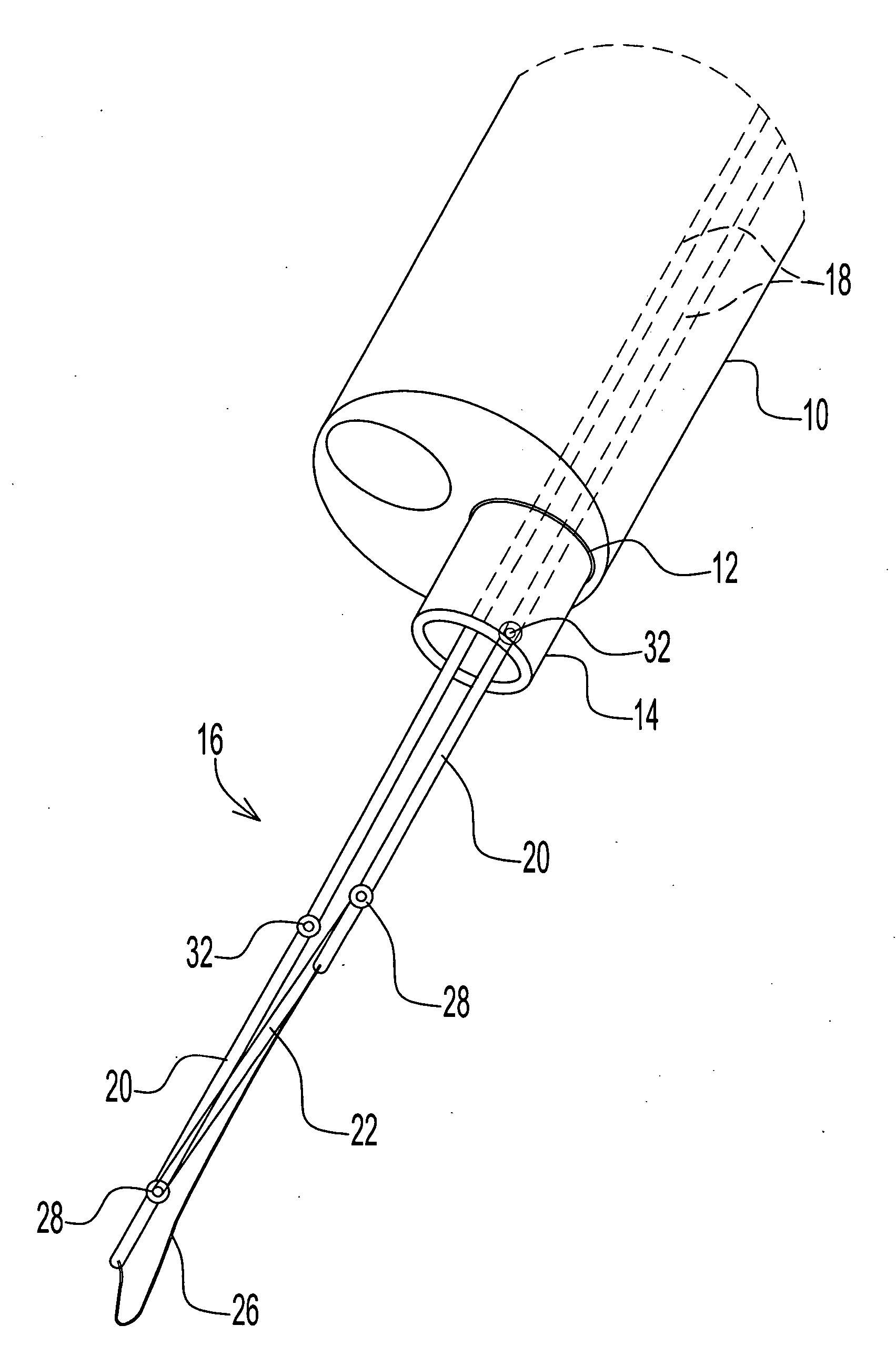
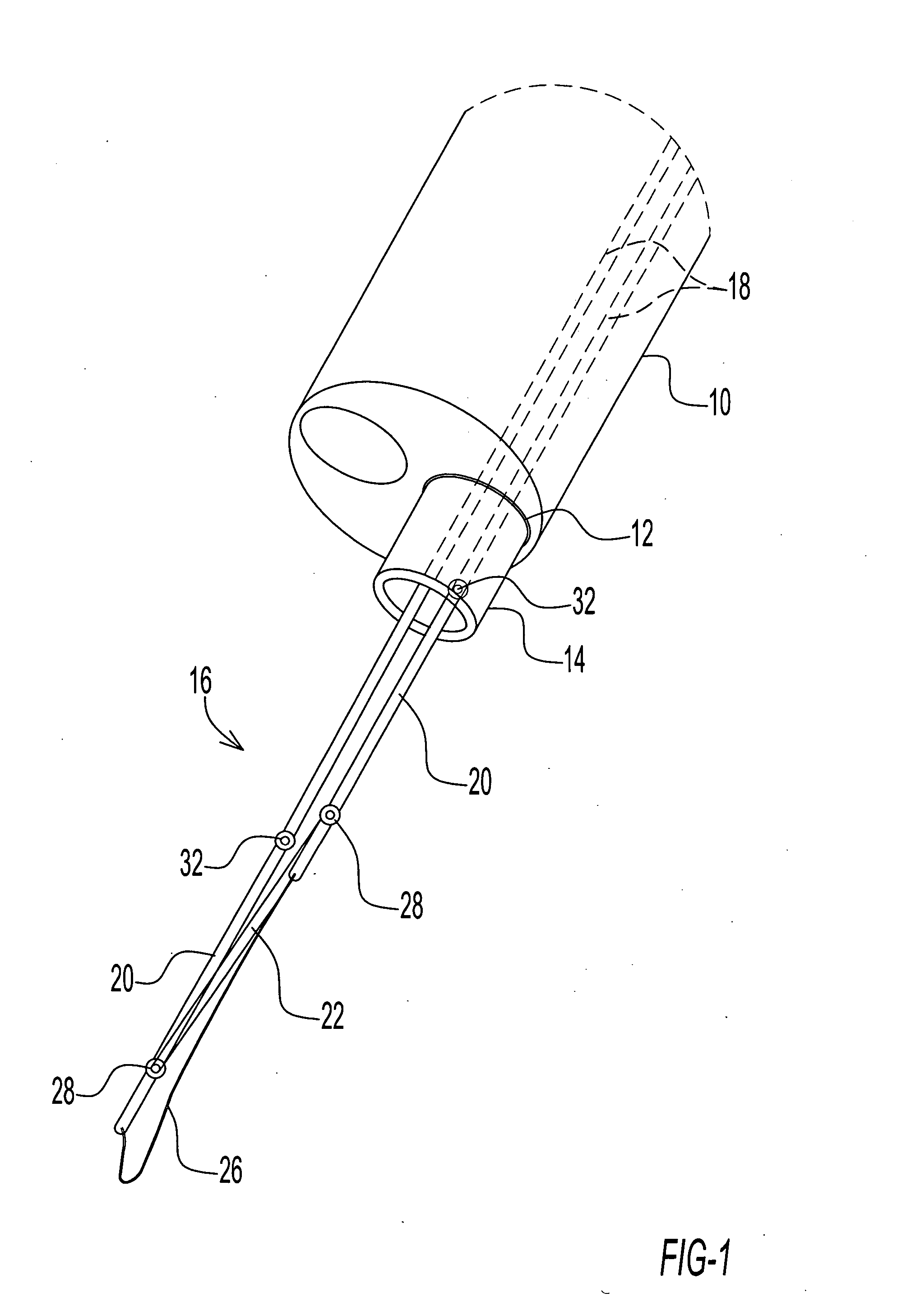
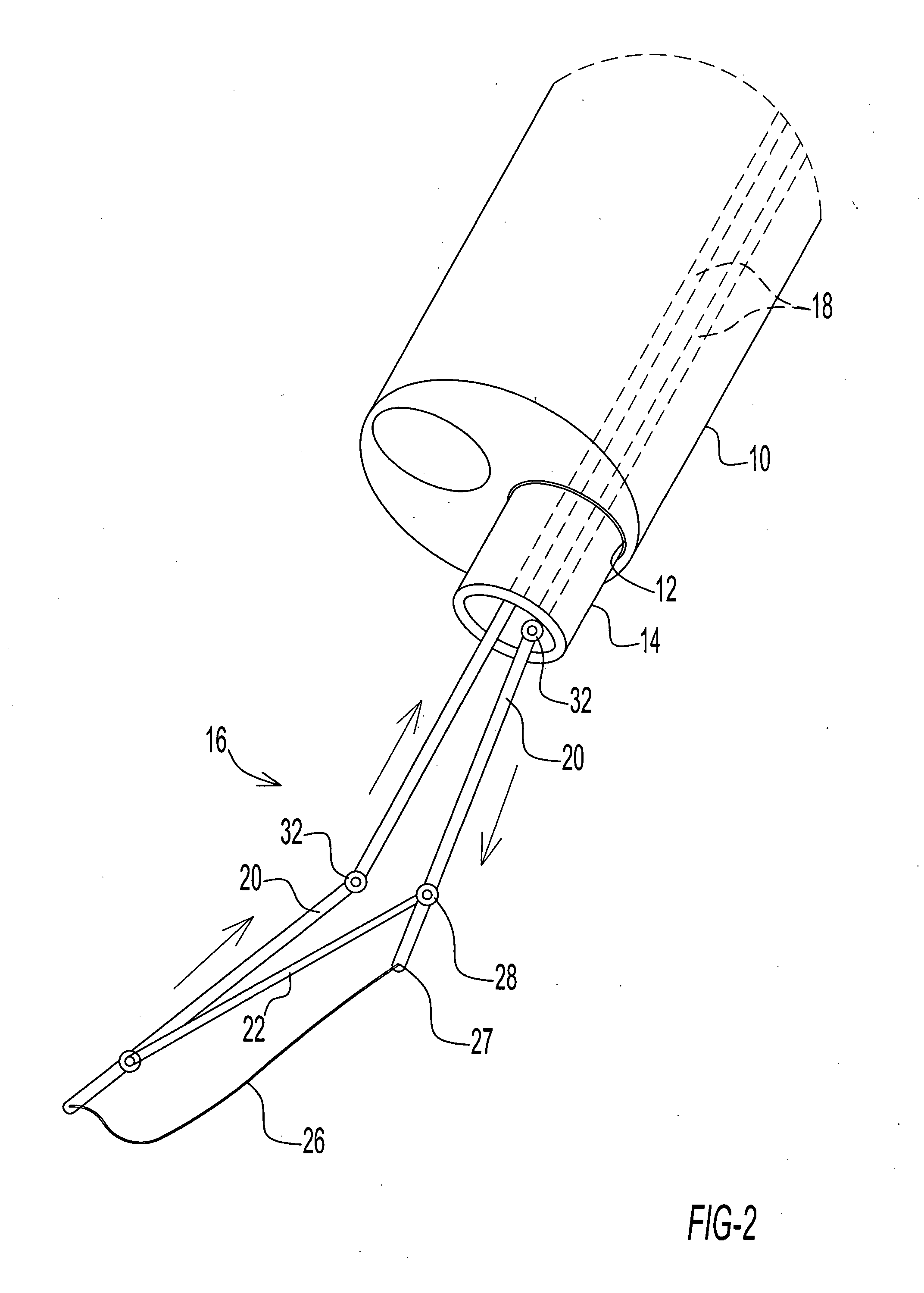
[0075]FIGS. 1-5 illustrate one embodiment of the invention that includes an electrocautery device capable of being passed through the working channel 12 of an endoscope 10. Referring to FIG. 1, an elongated tube 14 might be positioned in the channel 12 to emerge from the distal end of endoscope 10. The cutting device or assembly 16 is moved through the elongate tube 14 to emerge therefrom. The device 16 includes one or more push bars or wires 18 that are generally parallel to one another. The push bars or wires 18 are coupled to a corresponding pair of rigid or semi rigid electrically conductive rod elements or rods 20 and a crossbar element 22 that spans between the rod elements 20. Rod elements 20 and crossbar 22 sever as a holder for a cutting wire 26. Together, rod elements 20, crossbar 22 and wire 26 are an operative tip of the electrocautery device.

[0076] As further illustrated in FIG. 2, cutting wire 26 spans between the distal ends 27 of the rod elements 20. FIG. 1 shows th...


PUM


Abstract

An endoscopic tissue resection device and related method is used in conjunction with a flexible or rigid endoscope. Tissue is resected by shaving thin layers of tissue for diagnostic and therapeutic purposes.

Description

CROSS-REFERENCE TO RELATED APPLICATION [0001] This application claims the benefit of U.S. Provisional Patent Application No. 60 / 611,260 filed Sep. 17, 2004.BACKGROUND OF THE INVENTION [0002] This invention relates to endoscopic medical procedures and more particularly to endoscopic mucosal resection procedures. This invention also relates to an endoscopic instrument or assembly utilizable in performing an endoscopic mucosal resection procedure. [0003] The precancerous nature of high-grade dysplasia and the difficulty in detection of invasive carcinoma by endoscopy make esophagectomy and ablative therapy important considerations to treating those patients with this serious condition. The gold standard treatment for early esophageal cancer and high grade dysplasia is esophagectomy, the surgical removal of the diseased segment of the esophagus. This is an effective but drastic treatment and presents significant complications and lifestyle problems for the patient. Many patients are poo...

Claims


Application Information

Patent Timeline
IPC IPC(8): A61B17/26
CPCA61B10/06A61B17/320016A61B17/32056A61B2018/144A61B2017/00269A61B2018/00482A61B18/1492
Inventor NAKAO, NAOMI L.
Owner GRANIT MEDICAL INNOVATION
Learn More