
Crystal structure of factor Vai and method for identifying blood factor Va modulators

a technology of factor vai and crystal structure, which is applied in the field of crystal structure of factor vai, an inactivated form of factor v, can solve the problems of lacked sufficient detail to determine binding surfaces or inhibitors, no information was provided that would allow the construction of factor vai or the method of producing vai crystals

Inactive Publication Date: 2006-01-19
EVERSE STEPHEN J +3
View PDF1 Cites 1 Cited by
  • Summary
  • Abstract
  • Description
  • Claims
  • Application Information

AI Technical Summary

Benefits of technology

[0012] The present invention provides a crystalline isoform of bovine factor Vai, which is an inactivation product of factor Va. The crystal structure of factor Vai differs in structural arrangement from the known crystal structure of factor Va and now provides a tool f

Problems solved by technology

In developed countries, the majority of deaths can be directly or indirectly attributed to an imbalance in hemostasis, leading to thrombosis.
Unfortunately, only 2-D drawings were presented for the erroneously identified structural model, which lacked sufficient detail to determine binding surfaces or to design inhibitors.
Likewise, no information was provided that would allow construction of factor Vai or methods to produce Vai crystals.
In fact the authors acknowledged that this model could not be correct as shown because “several loop regions overlap and many of the others are lying outside of the density”.
Furthermore the model did not adequately reflect data showing that the R factor remained at 38%.
All the disclosed models were in the form of 2D ribbon drawings which cannot be used to design inhibitors because they lack the detail required to adequately determine binding surfaces.

Method used


Image

  • Crystal structure of factor Vai and method for identifying blood factor Va modulators
  • Crystal structure of factor Vai and method for identifying blood factor Va modulators
  • Crystal structure of factor Vai and method for identifying blood factor Va modulators

Examples


example 1

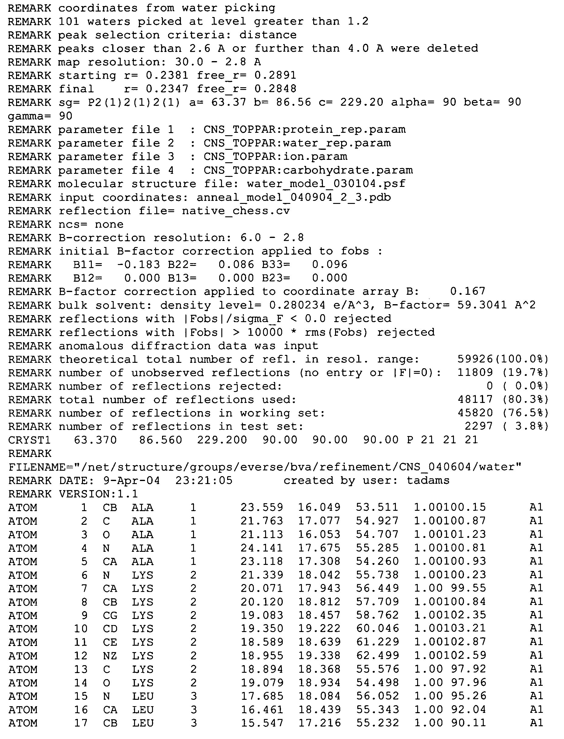
[0073] The search results yielded two unique A domain solutions with correlation coefficients of 0.198 and 0.186 as well as two unique C domain solutions with correlation coefficients of 0.249 and 0.217. Model phases combined with experimental phases produced interpretable density allowing for manual model fitting and rebuilding of the molecular replacement solution. The structure was refined with alternating rounds of refinement including simulated annealing using CNS (Brunger, et al. (1998) Acta Crystallographica D Biological Crystallography. 54, 905-21) and model rebuilding in 0 (Jones, et al. (1991) Acta Crystallographica. A47, 110-119) (Table 1).

example 2

Inactivation of Bovine Factor Va by Bovine APC

[0074] Bovine factor Va (40 μM) was extensively dialyzed against 20 mM HEPES, 150 mM NaCl, 2 mM CaCl2, pH 7.4 (HBSCa). Factor Va was incubated with 100 μM phospholipid vesicles (75% phosphatidylcholine: 25% phosphatidylserine) at 37° C. for 1 hour. Bovine APC was added (250 nM) and the sample was incubated at 37° C. for 3 hours. Factor V activity was monitored by single-stage clotting assays. The sample was loaded onto a Poros HQ20 (4.6×100 mm) equilibrated in 20 mM HEPES, 2 mM CaCl2 and eluted with a gradient elution of 0 to 500 mM NaCl in equilibration buffer over 10 minutes. Fractions identified by SDS-PAGE as containing A2-domainless factor Va., were pooled and analyzed for residual factor Va activity. Purified protein was stored in HBS-Ca at −20° C.

example 3

Crystallization and Data Collection

[0075] Purified bovine factor Va., in 20 mM HEPES, 150 mM NaCl, 2 mM CaCl2 (pH 7.4) was crystallized at ˜6.5 mg / mL by the vapor diffusion sitting-drop method at 12° C. against 200 mM MgCl2, 16% PEG 3350 (pH 5.0). After 521 days, diffraction quality crystals appeared (Table 1). Three isomorphous heavy atom derivative crystals were identified from native crystals soaked in mother liquor containing either 10 mM tetrakismercuroxymethane (TAMM), 10 mM ethylmercury (EtHg) or 2.5 mM lead acetate (PbAc) Supporting data are shown in Table 2.

TABLE 1Data Collection and Refinement StatisticsNativebresolution limits (Å)30-2.8Space groupP212121Cell dimensions (Å)a = 63.37b = 86.56 c = 229.20Reflections30822completeness (%)a97.4 (94.9)Redundancy3.6I / oa16.9 (4.1) Rsyn t (%)a 6.9 (29.4)model details7012no. protein atomsno. solvent molecules390additional ligands5 NAG, 1Cu2+,1Ca2+Average B-factor (A2)protein main-chain46.2protein side-chain47.3solvent molecules50....


PUM

PropertyMeasurementUnit
Temperatureaaaaaaaaaa
Timeaaaaaaaaaa
Fractionaaaaaaaaaa

Abstract

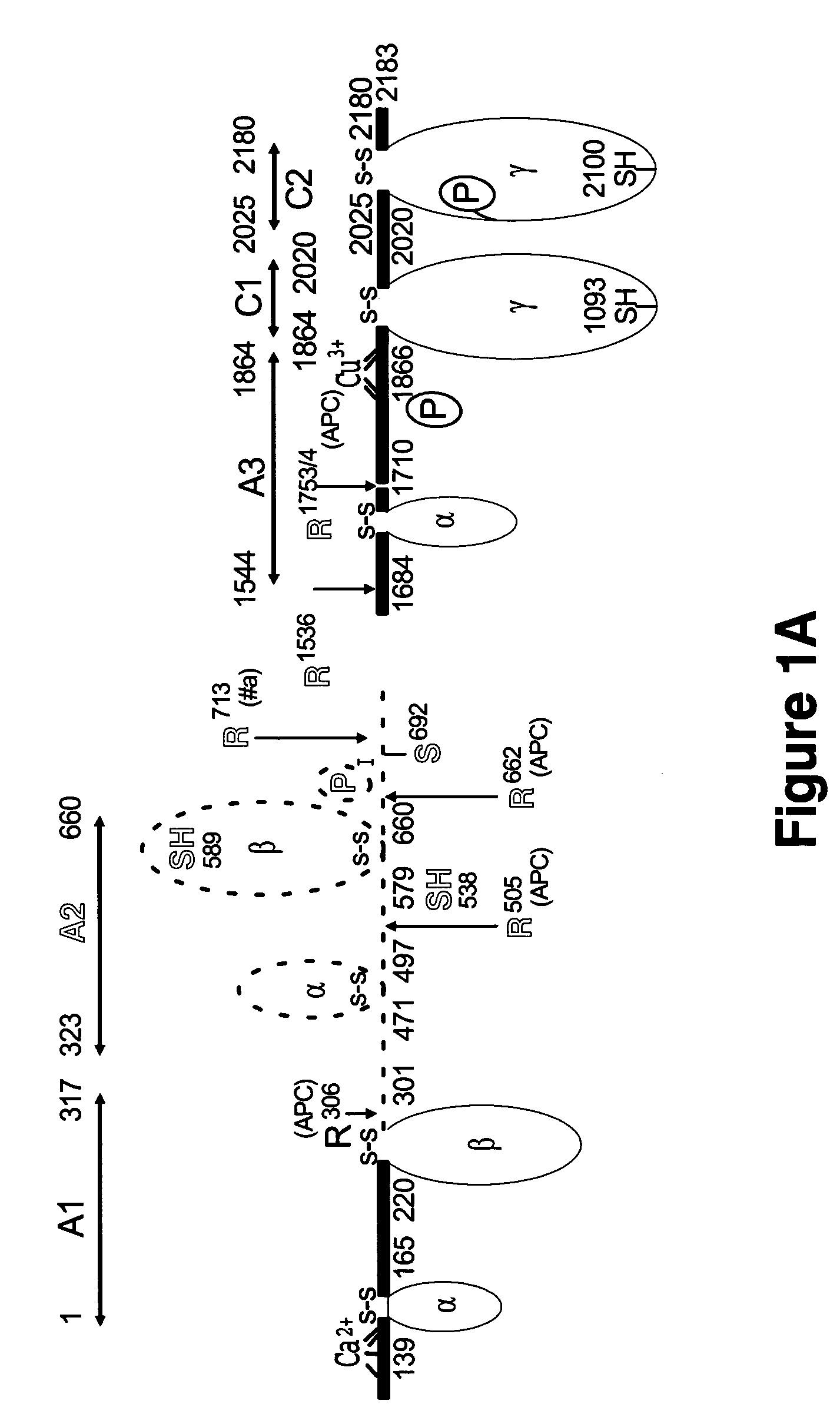
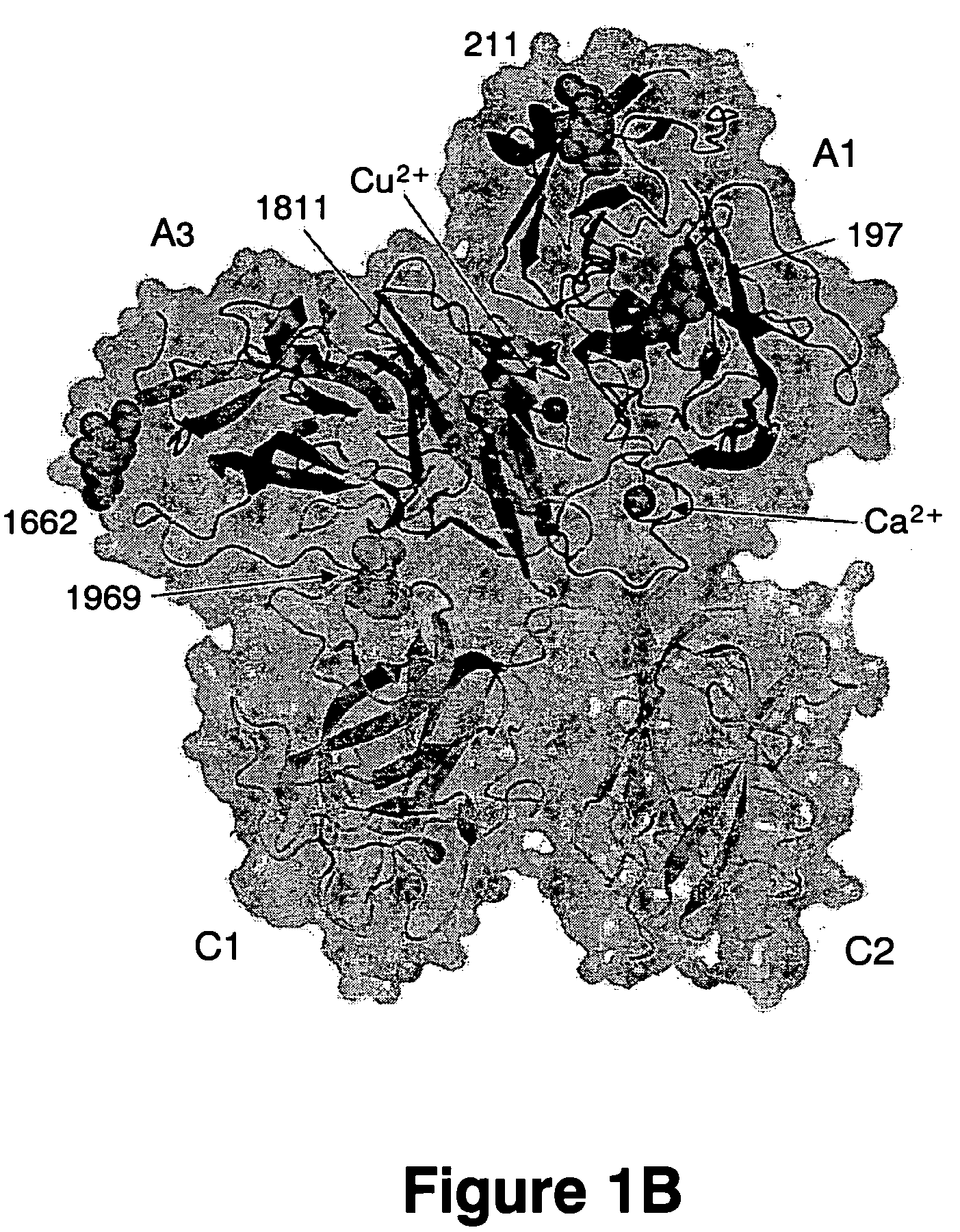
The present invention shows the crystal structure of protein C inactivated factor Va (A1-A3-C1-C2) that depicts a novel domain arrangement. The newly disclosed orientation has implications for binding to membranes essential for function. A high-affinity calcium binding site and a copper binding site have been identified, neither of which show a direct involvement in chain association. This structure represents the largest physiologically relevant fragment of factor Va solved to date and provides a new scaffold for generation of models of coagulation factors.

Description

[0001] This work was supported by grants from the National Institutes of Health, HL64891 and HL34575 Department of Energy Grant ER45828. The Government has certain rights in the invention. This application claims benefit of U.S. Provisional Application Ser. No. 60 / 572,040 filed May 18, 2004. [0002] The atomic coordinates and structure factors have been deposited in the protein databank, www.rcsb.org (PDB ID code 1SDD).FIELD OF THE INVENTION [0003] The present invention relates to crystals of factor Vai, an inactivated form of factor V and more particularly to the high resolution structure of Vai obtained by x-ray diffraction. The invention further relates to methods of using the crystal structure coordinates and models of the Vai crystal structure to screen and design therapeutic drugs for intervention in biological processes associated with blood coagulation. BACKGROUND OF THE INVENTION [0004] Design of new drugs has evolved over the years from laborious synthesis of a few lead com...

Claims


Application Information

Patent Timeline
IPC IPC(8): C07K14/745G01N33/48A61K38/48
CPCA61K38/00C07K14/745C12N2517/00G01N2500/00G01N33/86G01N2333/7456C12Q1/56
Inventor EVERSE, STEPHEN J.ADAMS, TY E.HOCKIN, MATTHEW F.MANN, KENNETH G.
Owner EVERSE STEPHEN J
Learn More