
Endoluminal device with extracellular matrix material and methods

a technology of extracellular matrix and endoluminal device, which is applied in the field of medical devices, can solve the problems of increasing frictional forces, weakening or even tearing of blood vessels and ducts, and functional vessels of human and animal bodies, and achieves the effect of reducing the endoleakage of endoluminal devi

Active Publication Date: 2005-12-08
COOK MEDICAL TECH LLC +1
View PDF61 Cites 152 Cited by
  • Summary
  • Abstract
  • Description
  • Claims
  • Application Information

AI Technical Summary

Benefits of technology

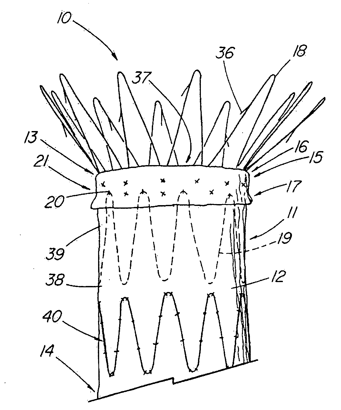
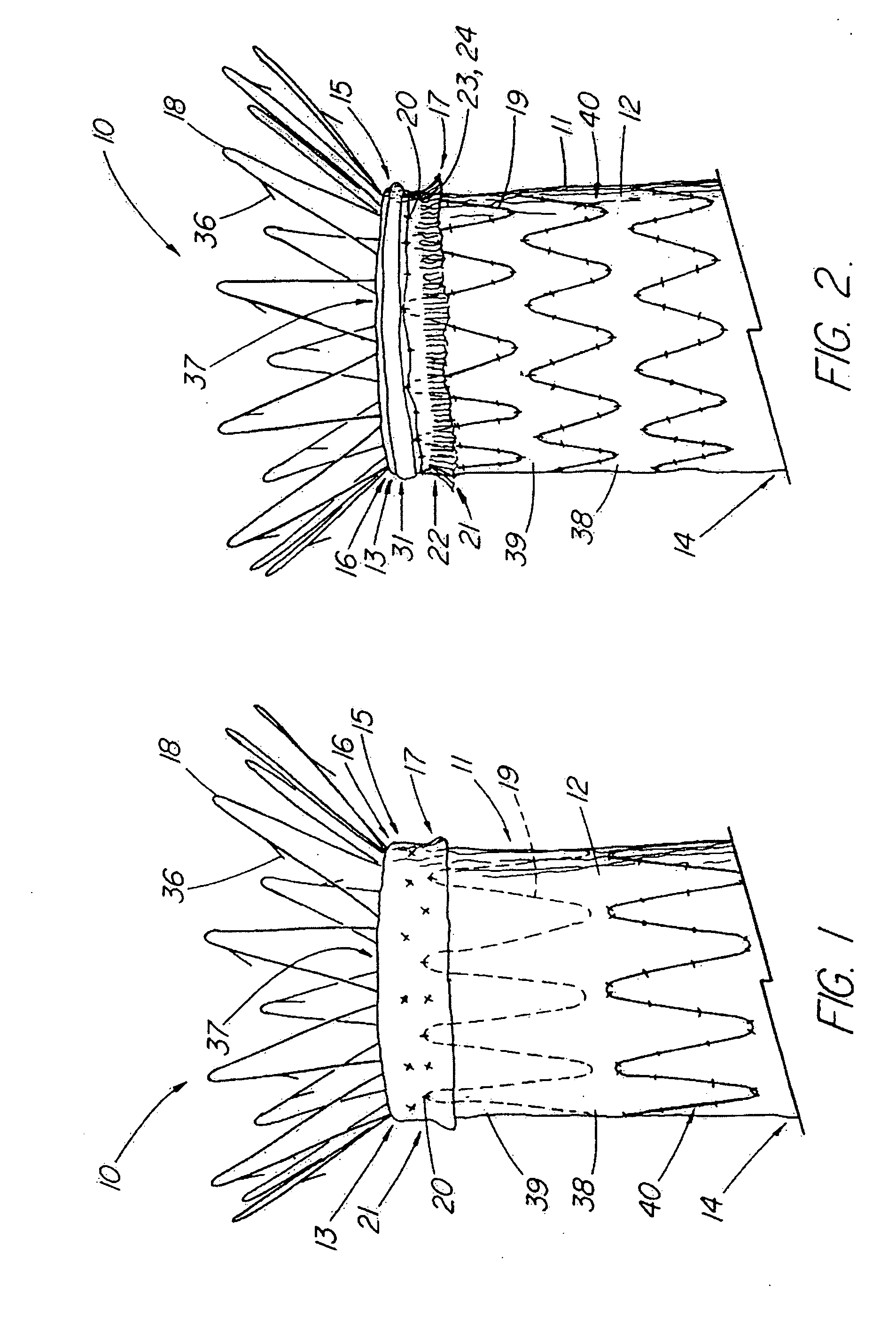
[0018] In yet another aspect of the invention there is a method of reducing endoluminal device endoleaks that comprises providing an endoluminal device comprising a tubular graft supported by a stent. The tubular graft comprises a synthetic material and a bioremodelable material. The bioremodelable material forms an exterior surface in at least one band adjacent at least one of the proximal and distal openings. The method further comprises deploying the endoluminal device into a body lumen.

Problems solved by technology

The functional vessels of human and animal bodies, such as blood vessels and ducts, occasionally weaken or even rupture.
For example, the aortic wall can weaken, resulting in an aneurysm.
Upon further exposure to hemodynamic forces, such an aneurysm can rupture.
The practice of over-sizing a device for the lumen into which it is to be placed may also increase these frictional forces.
Through the physiological process of arterial disease and aneurysm growth, both fixation and sealing may become compromised.
Naturally, endoleaks can be a serious problem, preventing the prosthesis from performing its function.
An endoleak, even a relatively small one, may result in the aneurysm repressurizing, increasing the risk of rupture.
Furthermore, the fixation provided by barbs can be under risk due to the failure of the fixation barbs or local tearing of the aortic wall.
Loss of fixation is highly undesirable and may also contribute to patient mortality and morbidity.
For some stent grafts, loss of fixation and resulting migration may result in the occlusion of one or more blood vessels.

Method used


Image

  • Endoluminal device with extracellular matrix material and methods
  • Endoluminal device with extracellular matrix material and methods
  • Endoluminal device with extracellular matrix material and methods

Examples


example 1

[0225] Thirty feet of whole intestine from a mature adult hog is rinsed with water. This material is then treated in a 0.2% by volume peracetic acid in a 5% by volume aqueous ethanol solution for a period of two hours with agitation. The tela submucosa layer is then delaminated in a disinfected casing machine from the whole intestine. The delaminated tela submucosa is rinsed four (4) times with sterile water and tested for impurities or contaminants such as endotoxins, microbial organisms, and pyrogens. The resultant tissue was found to have essentially zero bioburden level. The tela submucosa layer separated easily and consistently from the whole intestine and was found to have minimal tissue debris on its surface.

example 2

[0226] A ten foot section of porcine whole intestine is washed with water. After rinsing, this section of tela submucosa intestinal source material is treated for about two and a half hours in 0.2% peracetic acid by volume in a 5% by volume aqueous ethanol solution with agitation. Following the treatment with peracetic acid, the tela submucosa layer is delaminated from the whole intestine. The resultant tela submucosa is then rinsed four (4) times with sterile water. The bioburden was found to be essentially zero.

example 3

[0227] A small section of the tela submucosa intestinal material was subcutaneously implanted in a rat. Within 72 hours, significant angiogenesis was observed.


PUM


Abstract

An endoluminal device comprises a stent and a tubular graft supported by the stent. The graft has a proximal and a distal opening and comprises a synthetic material and a bioremodelable material. The bioremodelable material is disposed on an exterior surface in at least one band adjacent at least one of the proximal and distal openings.

Description

RELATED APPLICATIONS [0001] This application is a continuation-in-part of U.S. application Ser. No. 10 / 644,129, filed Aug. 20, 2003 which claims the benefit of U.S. Provisional Application Ser. No. 60 / 404,662, filed August 20, 2002, which are incorporated herein by reference. This application also claims the benefit of U.S. Provisional Application Ser. No. 60 / 572,806, filed May 20, 2004, which is also incorporated by reference.TECHNICAL FIELD [0002] This invention relates to medical devices and more particularly, to endoluminal devices suitable for various medical applications and the methods for making and using such endoluminal devices. BACKGROUND OF THE INVENTION [0003] This invention will be discussed in relation to endoluminally deployed stent grafts but the invention is not so limited and can also be applied to the grafts for the human or animal body and where biological fixation is a desired or necessary function. [0004] Throughout this specification, when discussing the appl...

Claims


Application Information

Patent Timeline
Patent Type & Authority Applications(United States)
IPC IPC(8): A61F2/00A61F2/06
CPCA61F2/0077A61F2/07A61F2/848A61F2002/061A61F2002/065A61F2002/075A61F2002/8483A61F2002/8486A61F2250/0065A61F2/89A61F2/915A61F2230/005A61F2230/0054A61F2230/0067
Inventor BAHLER, CLINTON D.DEBRUYNE, MICHAEL P.FEARNOT, NEAL E.LEEWOOD, ALAN R.MEAD, JASON A.OSBORNE, THOMAS A.SUN, JICHAO
Owner COOK MEDICAL TECH LLC
Learn More