
Robotic surgical tool with ultrasound cauterizing and cutting instrument

a robotic surgical and ultrasound technology, applied in the field of surgical instruments, can solve the problems of traditional minimally invasive surgical instruments, deny the surgeon the flexibility of tool placement in open surgery, and minimally invasive techniques, so as to improve the efficiency of robotic surgery, facilitate the positioning of the probe tip, and reduce the risk of surgical complications.

Inactive Publication Date: 2005-01-27
INTUITIVE SURGICAL
View PDF43 Cites 266 Cited by
  • Summary
  • Abstract
  • Description
  • Claims
  • Application Information

AI Technical Summary

Benefits of technology

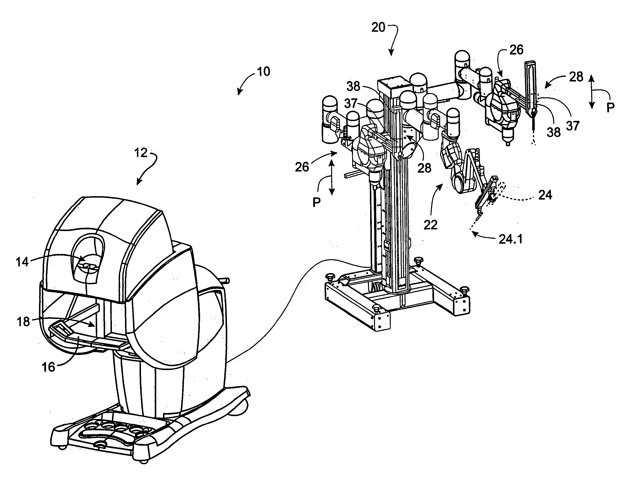
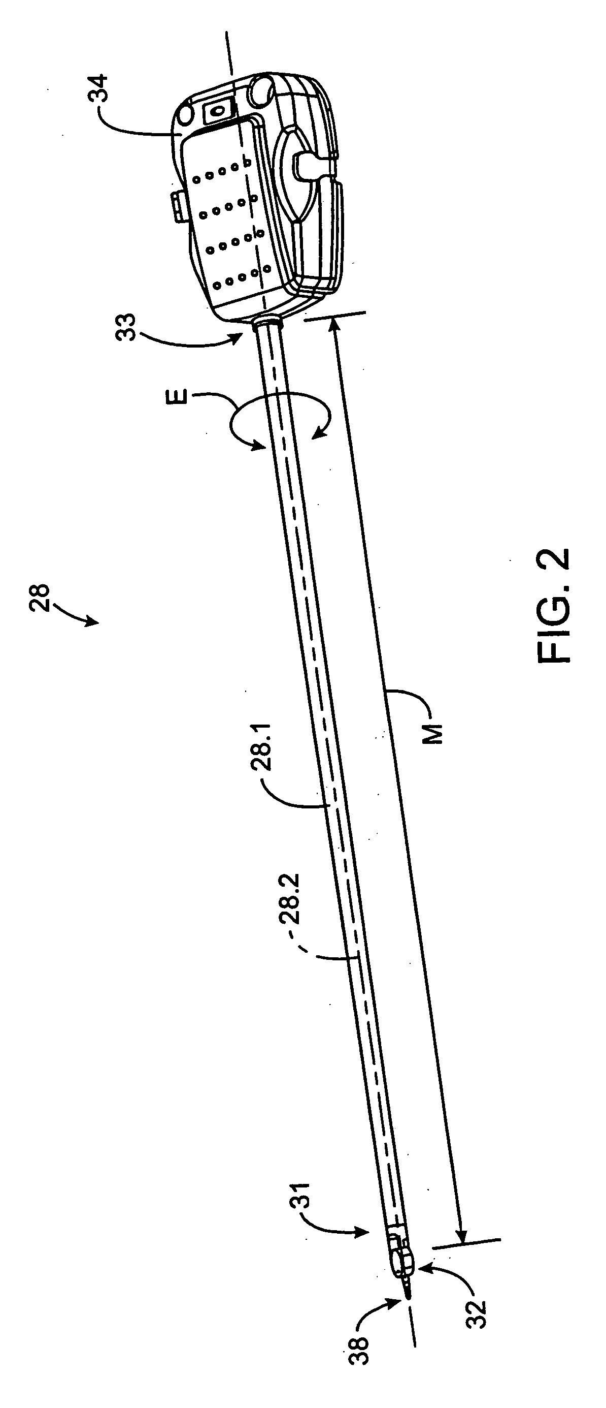
[0015] Surgical apparatus and methods for enhancing robotic surgery generally include a surgical instrument with an elongate shaft having an ultrasound probe, an end effector at the distal end of the shaft, and a base at the proximal end of the shaft. The end effector includes an ultrasound probe tip and the surgical instrument is generally configured for convenient positioning of the probe tip within a surgical site by a robotic surgical system. Ultrasound energy delivered by the probe tip may be used to cut, cauterize, or achieve various other desired effects on tissue at a surgical site. By providing ultrasound energy via a robotic surgical instrument for use with a robotic surgical system, the apparatus and methods of the present invention enable the advantages associated with ultrasound to be combined with the advantages of minimally invasive robotic surgery.
[0031] To reduce costs and for manufacturing convenience, the instrument may include OEM parts. For example, the instrument probe assembly may include parts or components generally similar or identical to parts or components (OEM components) of current or future commercially-available endoscopic instruments for surgical or diagnostic uses (OEM medical systems), including manually operated instruments. The surgical instruments of the invention may perform some or all of the functions of such OEM medical systems. For example, the instrument probe assembly of the surgical instruments of the invention may include OEM components of ultrasound treatment probes, electrocautery probes, ultrasound diagnostic probes, diagnostic imagery probes. In further examples, the instrument probe assembly may include suitable OEM components of biopsy probes, suction probes, substance injection probes, surgical accessory application probes, stapler probes, tissue grasping and cutting probes, and the like. Likewise, the instrument probe assembly may combine more than one of the medical functions of the above described instruments.

Problems solved by technology

Only a small percentage of procedures currently use minimally invasive techniques, however, because certain instruments, systems and methods are not currently available in a form for providing minimally invasive surgery.
For example, traditional minimally invasive surgical instruments often deny the surgeon the flexibility of tool placement found in open surgery.
Difficulty is experienced in approaching the surgical site with the instruments through the small incisions.
Additionally, the added length of typical endoscopic instruments often reduces the surgeon's ability to feel forces exerted by tissues and organs on the end effector.
Furthermore, coordination of the movement of the end effector of the instrument as viewed in the image on the television monitor with actual end effector movement is particularly difficult, since the movement as perceived in the image normally does not correspond intuitively with the actual end effector movement.
Accordingly, lack of intuitive response to surgical instrument movement input is often experienced.
Such a lack of intuitiveness, dexterity and sensitivity of endoscopic tools has been found to be an impediment in the increased the use of minimally invasive surgery.
Currently, however, ultrasound instruments for use with a robotic surgical system are not available.

Method used


Image

  • Robotic surgical tool with ultrasound cauterizing and cutting instrument
  • Robotic surgical tool with ultrasound cauterizing and cutting instrument
  • Robotic surgical tool with ultrasound cauterizing and cutting instrument

Examples


embodiment 300

[0123]FIGS. 20 through 23 illustrate an alternative example of an instrument embodiment 300 including aspects of the invention.

embodiment 80

[0124] It should be noted that much of the description above with respect to the robotic instrument embodiment 80 of FIGS. 10-19, including incorporated references, is also relevant with respect to instrument 300, since in many cases generally similar structures of each instrument serve equivalent functions.

[0125] For convenience and to minimize manufacturing costs, selected OEM components of commercially available instruments may optionally be included in the instrument 300 described herein. FIGS. 24-27 are sheets of reproductions of the FIGS. 26-36 of U.S. Pat. No. 6,280,407, issued Aug. 28, 2001 to Manna, et al., entitled “Ultrasonic Dissection And Coagulation System”, and assigned to United States Surgical Corporation of Norwalk, Conn., the entire contents of which are hereby incorporated by reference. The patent describes, among other things, a hand-held ultrasonic treatment instrument example generally similar to the AutoSonix* Ultra Shears* made by United States Surgical Corp...


PUM


Abstract

A surgical instrument for enhancing robotic surgery generally includes an elongate shaft with an ultrasound probe, an end effector at the distal end of the shaft, and a base at the proximal end of the shaft. The end effector includes an ultrasound probe tip and the surgical instrument is generally configured for convenient positioning of the probe tip within a surgical site by a robotic surgical system. Ultrasound energy delivered by the probe tip may be used to cut, cauterize, or achieve various other desired effects on tissue at a surgical site. In various embodiments, the end effector also includes a gripper, for gripping tissue in cooperation with the ultrasound probe tip. The base is generally configured to removably couple the surgical instrument to a robotic surgical system and to transmit forces from the surgical system to the end effector, through the elongate shaft. A method for enhancing robotic surgery generally includes coupling the surgical instrument to a robotic surgical system, positioning the probe tip in contact with tissue at a surgical site, and delivering ultrasound energy to the tissue.

Description

CROSS-REFERENCES TO RELATED APPLICATIONS [0001] This application is a divisional application of U.S. patent application Ser. No. 10 / 126,499 filed on Apr. 18, 2002, which claims the benefit of prior provisional application No. 60 / 285,485, filed on Apr. 19, 2001, under 37 CFR §1.78(a)(4), the full disclosures of which are incorporated herein by reference.BACKGROUND OF THE INVENTION [0002] The present invention generally relates to surgical apparatus and methods. More specifically, the invention relates to a surgical instrument and method for use with a robotic surgical system, the instrument including an ultrasonic probe. [0003] Minimally invasive surgical techniques generally reduce the amount of extraneous tissue damage during surgical procedures, thereby reducing patient recovery time, discomfort, and deleterious side effects. One effect of minimally invasive surgery, for example, is reduced post-operative hospital recovery times. Because the average hospital stay for a standard su...

Claims


Application Information

Patent Timeline
IPC IPC(8): A61B17/00A61B17/32A61B19/00
CPCA61B17/320068A61B19/22A61B19/2203A61B19/5212A61B17/320092A61B2019/2223A61B2019/2234A61B2019/2242A61B2019/2292A61B2017/00477A61B8/4405A61B34/70A61B34/71A61B90/361A61B34/30A61B34/37A61B2034/305A61B34/76A61B2017/320094A61B2017/320093A61B2017/320071A61B2017/320095A61B2017/320069
Inventor ANDERSON, STEPHEN C.JULIAN, CHRISTOPHER A.
Owner INTUITIVE SURGICAL
Learn More