
Method and system for myocardial infarction repair

a myocardial infarction and repair method technology, applied in the field of myocardial infarction repair, can solve the problems of creating electrical abnormalities, 4-6 times higher risk of heart failure in survivors of ami, and no current or proposed therapy addresses myocardial necrosis

Inactive Publication Date: 2004-05-06
MEDTRONIC INC
View PDF25 Cites 10 Cited by
  • Summary
  • Abstract
  • Description
  • Claims
  • Application Information

AI Technical Summary

Benefits of technology

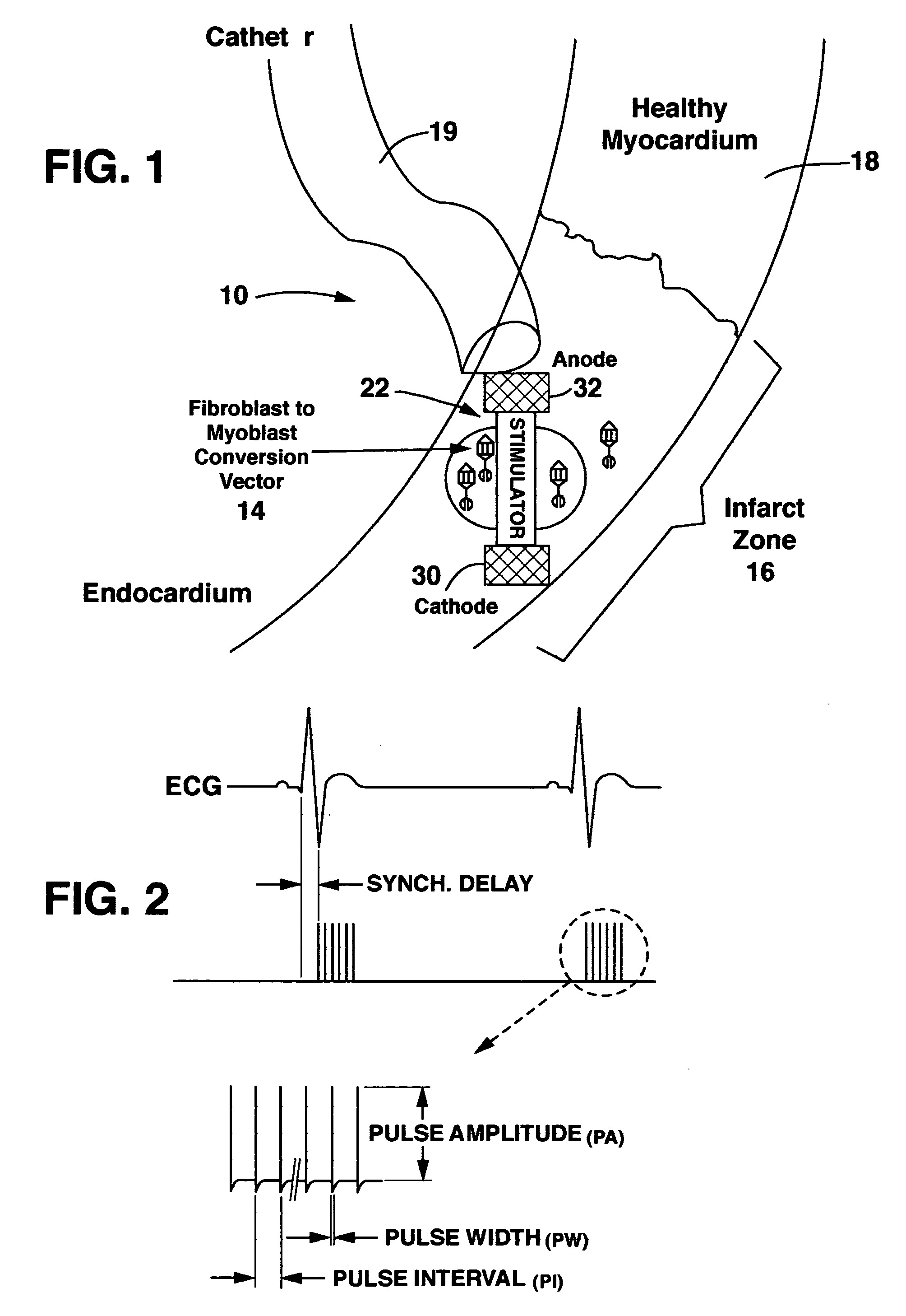
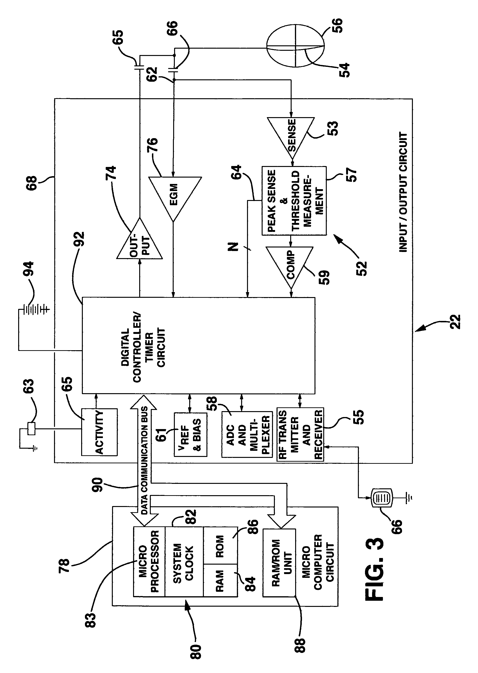
The present invention provides methods and systems for repairing damaged heart tissue after a myocardial infarction or other heart disease. This involves combining a cell repopulation source with an electrical stimulation device to create new contractile tissue that can improve heart function. The cell repopulation source can be implanted into the damaged heart tissue and the electrical stimulation device can be a muscle stimulator or other device that can stimulate the new contractile tissue. The invention has advantages of restoring elasticity and contractility to the heart tissue, increasing heart function, reducing remodeling, and decreasing morbidity and mortality.

Problems solved by technology

This fibrous scar does not significantly contribute to the contraction of the heart and can, in fact, create electrical abnormalities.
Those who survive AMI have a 4-6 times higher risk of developing heart failure.
However, none of the current or proposed therapies address myocardial necrosis (i.e., degradation and death of the cells of the heart muscle).
Although these approaches produced intriguing research studies, cells from established cell lines are typically rejected from the human recipient.
Although their results showed the efficacy of transplanted cell technology, this approach would not likely be effective in clinical medicine since the syngeneic fetal cardiac tissue will not be available for human patients.
This method, however, is highly invasive, which compromises its feasibility on human MI patients.
However, its viability has not been demonstrated in vivo.

Method used


Image

  • Method and system for myocardial infarction repair
  • Method and system for myocardial infarction repair
  • Method and system for myocardial infarction repair

Examples


example 1

[0105] Transformation of Fibroblasts in situ and Electrical Stimulation

[0106] Adenovirus expressing myogenin (Myogen adenovirus / cDNA, which can be produced according to the method described by Murry et al., J. Clin. Invest., 98, 2209-2217 (1196)) was injected directly to the myocardium using a 100 microliter syringe. 10.sup.9 pfu (pfu-plaque forming units-one pfu is approximately 50 adenovirus particles) were diluted with saline to form a 100 microliter solution. This solution was kept on dry ice until the injection, and delivered in four equal amounts to the perimeter of the infarct zone, 90 degrees apart.

[0107] A histopathological assessment of the treated tissue was done to assess the extent of fibroblast transformation. Tissue was processed for histology and stained with H&E and Masson's Trichrome according to standard methods.

[0108] Immunohistochemical staining was also done to determine whether there was myogenin expression in the treated tissue. Eight m frozen sections were c...

example 2

[0111] Injection of Contractile Cells and Electrical Stimulation in Canines

[0112] Growth and Passage Information for Skeletal Myoblast Cells

[0113] 1. Growth Medium Formulation:

[0114] 81.6% M199 (Sigma, M-4530)

[0115] 7.4% MEM (Sigma, M-4655)

[0116] 10% Fetal Bovine Serum (Hyclone, Cat.# A-1115-L)

[0117] 1.times.(1%) Penicillin / Streptomycin (Final Conc. 100,000 U / L Pen. / 10 mg / L Strep., Sigma, P-0781).

[0118] 2. Cell Passage Information:

[0119] A. Seeding densities of 1.times.10.sup.4 cells / cm.sup.2 will yield an 80% confluent monolayer in approximately 96 hours.

[0120] B. Split ratios of 1:4-1:6 will yield a confluent monolayer within 96 hours.

[0121] C. Do not allow the cells to become confluent. Cell to cell contact will cause the cells to differentiate into myotubes.

[0122] 3. Passage Information:

4 Flask Size ml of HBSS ml of Trypsin Solution ml of Media / Flask T-25 3 3 10 T-75 5 5 20-35 T-150 10-15 10-15 40-60 T-225 15-25 15-25 60-125

[0123] A. Remove culture medium from T-flask.

[0124] B. ...


PUM

PropertyMeasurementUnit
timeaaaaaaaaaa
concentrationaaaaaaaaaa
diameteraaaaaaaaaa

Abstract

An implantable system is provided that includes: a cell repopulation source comprising genetic material, undifferentiated and / or differentiated contractile cells, or a combination thereof capable of forming new contractile tissue in and / or near an infarct zone of a patient's myocardium; and an electrical stimulation device for electrically stimulating the new contractile tissue in and / or near the infarct zone of the patient's myocardium or otherwise damaged or diseased myocardial tissue.

Description

FIELD OF THE INVENTION[0001] The present invention relates to methods and implantable systems to reverse damage to heart muscle following myocardial infarction and more generally in and / or near damaged or diseased myocardial tissue. Specifically, this involves the repopulation of the damaged or diseased myocardium with undifferentiated or differentiated contractile cells, which additionally may be formed in situ through the use of genetic engineering techniques, and augmentation with electrical stimulation.BACKGROUND OF THE INVENTION[0002] Coronary Artery Disease (CAD) affects 1.5 million people in the USA annually. About 10% of these patients die within the first year and about 900,000 suffer from acute myocardial infarction. During CAD, formation of plaques under the endothelial tissue narrows the lumen of the coronary artery and increases its resistance to blood flow, thereby reducing the O.sub.2 supply. Injury to the myocardium (i.e., the middle and thickest layer of the heart w...

Claims


Application Information

Patent Timeline
Patent Type & Authority Applications(United States)
IPC IPC(8): A61N1/36A61N1/362A61N1/372A61N1/375C12N5/02C12N5/077
CPCA61N1/327A61N1/36042C12N5/0657A61N1/37205A61N1/3756A61N1/362A61N1/3629
Inventor SOYKAN, ORHANDONOVAN, MAURA G.
Owner MEDTRONIC INC
Learn More