
Early detection of inflammation and infection using infrared thermography

a technology of infrared thermography and inflammation, applied in the field of infrared thermography imaging in animals, can solve the problems of inability to detect inflammation, redness, warmth (altered heat pattern), pus formation, and swelling of normal tissues, and achieves the effects of reducing the number of infrared thermography images, reducing the detection cost, and improving the detection efficiency

Inactive Publication Date: 2004-01-29
SCHAEFER ALLAN L +3
View PDF24 Cites 61 Cited by
  • Summary
  • Abstract
  • Description
  • Claims
  • Application Information

AI Technical Summary

Benefits of technology

0021] The present invention is based, in part, on the surprising discovery that temperature differences less than 1.degree. C. are clinically significant. This discovery was made possible by employing induction models of mastitis and BVD that allowed the Applicants to evaluate inflammation or infection resulting from known etiologies and to compare the infrared charact...

Problems solved by technology

These events result in swelling, redness, warmth (altered heat patterns), and pus formation at the site of injury.
When this delicately balanced interplay is disrupted, the inflammatory response may result in considerable damage to normal tissue of uncontrolled inflammatory responses, clinical intervention is needed to prevent tissue damage and organ dysfunction.
However, non-invasive techniques for the detection of inflammation remain elusive.
Therefore, these techniques are not useful for detecting the early phase in the development of inflammation.
The induction of a biological response by a radiopharmaceutical is a major drawback of using scintigraphic imaging.
However, this method is only useful for detecting the late stages in the development of inflammation.
The inability to diagnose and image inflammation in vivo continues to be a major obstacle to the successful treatment of inflammatory disorders.
This method is invasive and often results in an amount of healthy tissue being removed along with the tissue suspected of being affected by inflammation.
The appearance of localized swelling, discoloration and tissue debris are often apparent and significant tissue damage can result.
However, immunological assays for detecting viral or bacterial infections require a laboratory and someone with technical expertise to perform the assays.
Further, the biological samples required to perform immunological assays are not easily obtained from an animal.
Additionally, the immunological assays are too costly for individual or sporadic infections and are generally not performed until clinical symptoms have manifested.
The disease is of great concern in the dairy industry, where significant economic loss can occur due to the requirement to not use the affected milk for human consumption and due to the shortened milking life of the affected animals.
However, given the expense and labour for the treatment of mastitis, treatment is usually not initiated until the condition is diagnosed clinically.
However, none are particularly effective at early detection (e.g., within the first few hours) of mastitis onset and, as described by Batra and McAllister (1984), these aforementioned procedures often have an unacceptably high percentage of false negatives (i.e., failure to identify an infected cow).
Unfortunately, these types of tests are not particularly effective in detecting the earliest onset or subclinical cases of mastitis.
Furthermore, the need to capture the animal and collect milk samples complicates the use of this method.
Furthermore, some diagnostic tools, such as rectal temperature, while usually efficacious, are often not as sensitive as would be desired or are simply impractical.
BVD is a significant problem in North American cattle populations, causing high morbidity and mortality especially in veal, dairy and beef populations (Cortese et al., 1998, J. Am. Vet. Med.
Further, the ability to obtain a reliable vaccine has remained elusive (Cortese et al., 1998, supra).
The immunological assays are laborious, time consuming and expensive, and require the collection of a biological sample.
The early infrared thermography detection systems were bulky, complex, and required frequent recharging with liquid nitrogen.
Furthermore, the spatial resolution was poor, the exposure time was long, and the minimum resolvable temperature difference was large for the infrared thermography systems.
Reliable detection of inflammation was not achieved.
In addition, many physicians and veterinarians were not adequately trained to interpret the data from the infrared imagery and there was a high false positive rate.
Thus, the infrared thermography was branded as a failure and has not been explored much by the medical or veterinary communities for the past three decades.

Method used


Image

  • Early detection of inflammation and infection using infrared thermography
  • Early detection of inflammation and infection using infrared thermography
  • Early detection of inflammation and infection using infrared thermography

Examples


Embodiment Construction

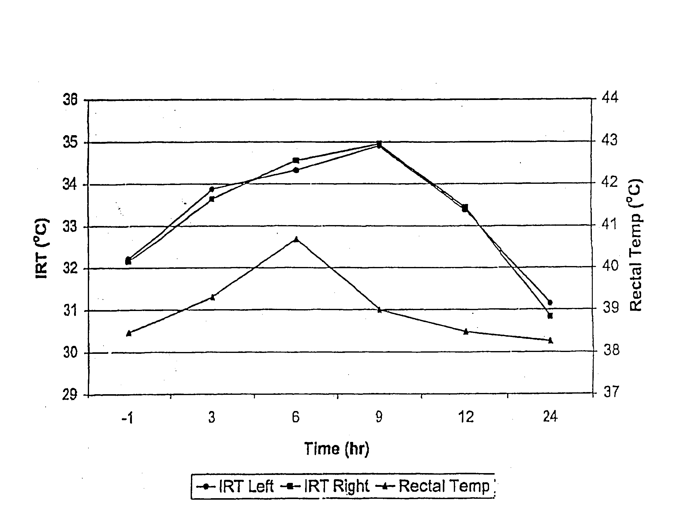
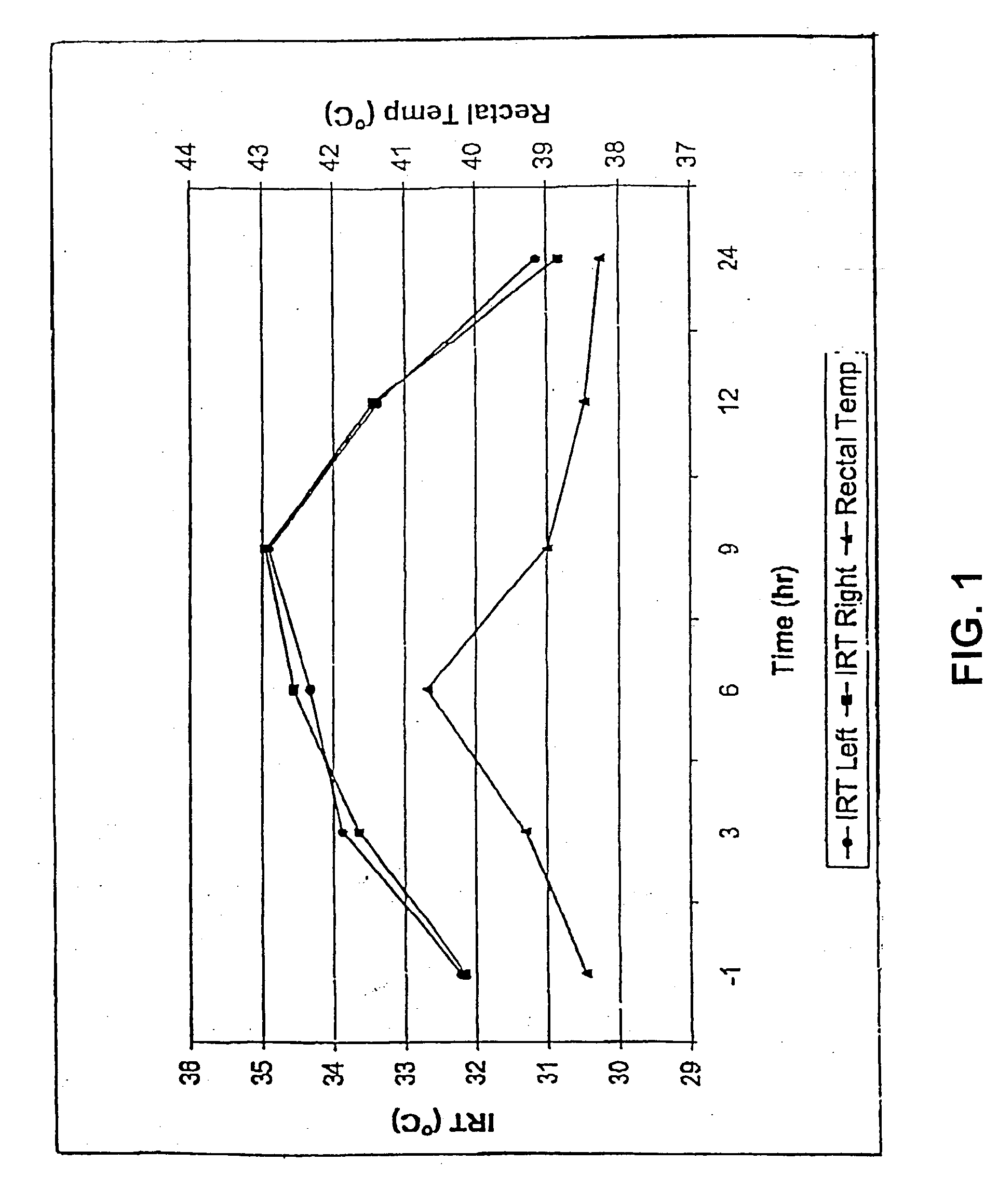
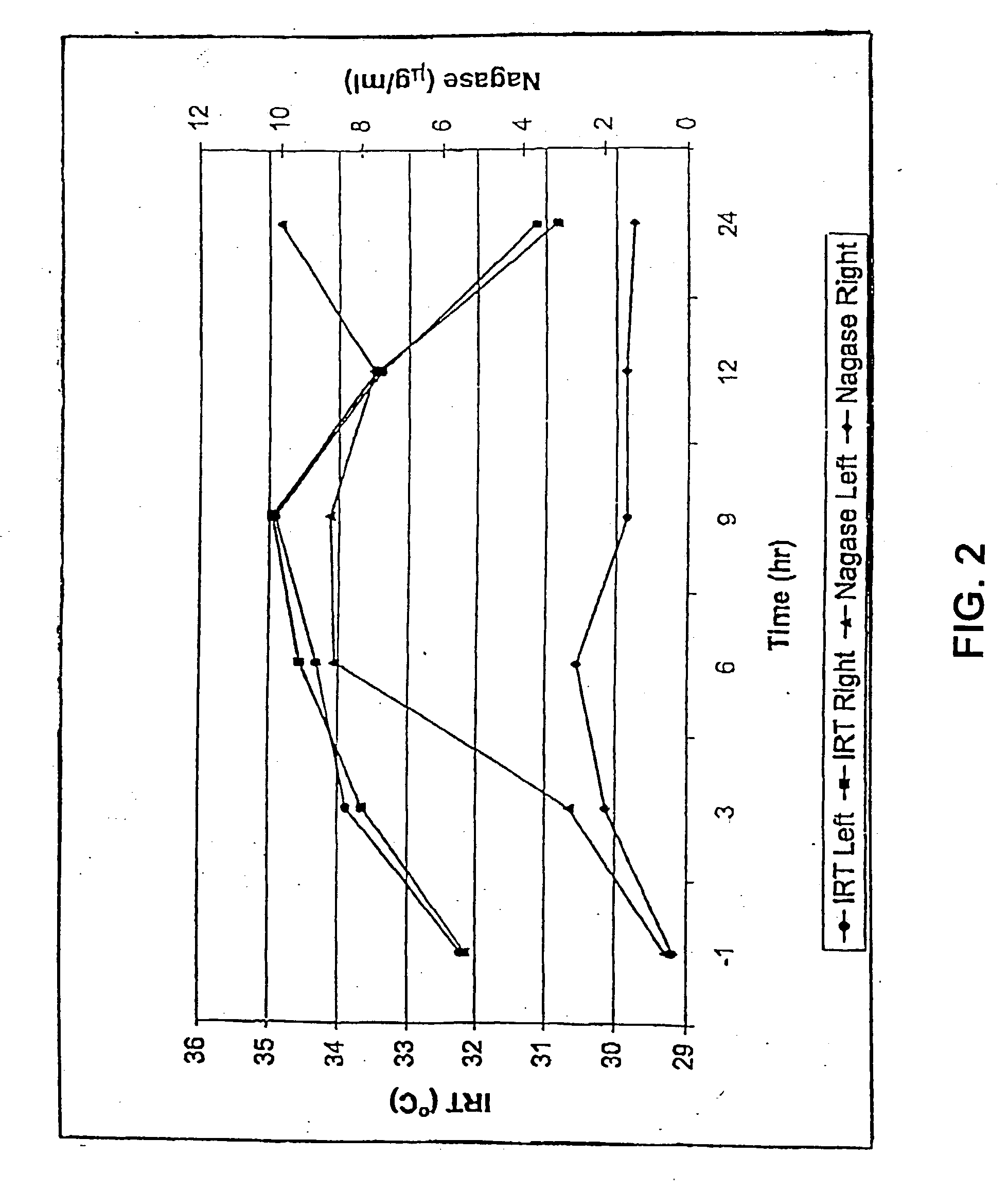
[0031] The present invention relates to the use of infrared thermography for the early or subclinical detection of inflammation in animals. The present invention also relates to the use of infrared thermography in the diagnosis of diseases or disorders that induce inflammation and / or induce the catabolism of tissues. The present invention provides methods for detecting inflammation of an anatomical structure of an animal, preferably a mammal and more preferably a non-human animal. The present invention further provides methods for detecting infection of an anatomical structure of an animal, preferably a mammal. In one embodiment, the present invention provides methods for detecting infection of an anatomical structure in a non-human animal. In yet another embodiment, the present invention provides methods for detecting infection in humans. The term "anatomical structure" used herein refers to any definable area of an animal, preferably a tissue or a joint of an animal, that radiates...


PUM


Abstract

The present invention provides a method for the detection of inflammation in animals using infrared thermography. The invention also provides a method for the detection of diseases or disorders that induce inflammation using infrared thermography. The present invention further provides a method for the detection of infections in animals using infrared thermography.

Description

[0001] This is a continuation of U.S. patent application Ser. No. 09 / 533,400, filed Mar. 22, 2000, which in turn is a continuation-in-part of U.S. patent application Ser. No. 09 / 274,032, filed Mar. 22, 1999, now abandoned, both of which are incorporated herein by reference in their entirety.1. FIELD OF THE INVENTION[0002] The invention relates to the use of infrared thermography imaging in animals for the early detection of inflammation. The invention further relates to the use of infrared thermography in animals for the early detection of infection.2. BACKGROUND OF THE INVENTION[0003] Inflammation plays a fundamental role in host defenses and the progression of immune-mediated diseases. The inflammatory response is initiated in response to tissue injury (e.g., trauma, ischemia, and foreign particles) and infection by a complex cascade of events, including chemical mediators (e.g., cytokines and prostaglandins) and inflammatory cells (e.g., leukocytes). The inflammatory response is ...

Claims


Application Information

Patent Timeline
IPC IPC(8): G01N25/72
CPCG01N25/72A01J5/0138A61B5/01
Inventor SCHAEFER, ALLAN L.SCOTT, SHANNON L.LACASSE, PIERRETONG, ALAN K.W.
Owner SCHAEFER ALLAN L
Learn More