
Co-extruded, multi-lumen medical lead

a medical lead and multi-lumen technology, applied in the field of co-extruded multi-lumen medical lead, can solve the problems of untoward effect on device performance and patient well-being, deformation of the insulation or fracture of the lead conductor, and disadvantageous large lead body,

Inactive Publication Date: 2001-08-30
MEDTRONIC INC
View PDF0 Cites 40 Cited by
  • Summary
  • Abstract
  • Description
  • Claims
  • Application Information

AI Technical Summary

Problems solved by technology

The lead conductors and insulation are subjected to cumulative mechanical stresses, as well as material reactions as described below, that can result in degradation of the insulation or fractures of the lead conductors with untoward effects on device performance and patient well being.
Such relatively large and stiff lead bodies were disadvantageous in a number of respects.
The available bio-compatible conductor material alloy presented an impedance that limited current carrying capacity.
The large diameter body made it difficult to implant more than one lead through the venous system.
The relatively high column strength was often still insufficient to maintain the pace / sense electrodes in the atrial appendage or ventricular apex, and physicians often resorted to leaving the stylets in place, resulting in fracture of the lead conductor and lead body sheath when the stylet wire broke.
Once the lead bodies fibrosed in, they were difficult to retract from the heart if they needed to be replaced.
Finally, the lead conductors tended to fracture at stress sites, in bipolar leads sometimes due to stresses applied unevenly to the side-by-side arrangement of the conductor coils.
The increased number of separate polarity and insulated coiled wire conductors is difficult to accommodate in the conventional coaxial coiled wire conductor winding arrangement having a desired, small, lead body outer diameter.
Moreover, the use of thin polyurethane inner and outer sheathes along with certain lead conductor alloys became problematic as the bio-stability of such lead materials in chronic implantation came into question as described in commonly assigned U.S. Pat. No. 5,419,921.
The loss of support of the lead conductors upon splitting of the lead body sheath webs can also result in excessive bending and eventual fracture of the conductor.
This problem is exacerbated when lead conductors of differing types each having a differing bending stiffness are enclosed in the outer lead conductor lumens leading to a lead body that is more flexible when bent in one direction than when bent in another direction.

Method used


Image

  • Co-extruded, multi-lumen medical lead
  • Co-extruded, multi-lumen medical lead
  • Co-extruded, multi-lumen medical lead

Examples


Embodiment Construction

[0033] The present invention finds particular utility in the fabrication and implantation of cardiac leads, e.g., atrial and / or ventricular pacing leads and / or cardioversion / defibrillation leads having elongated lead bodies and lead conductors that are subject to fracture. Preferred embodiments of such lead conductor fabrications of such endocardial cardiac leads that are implanted transvenously will be described in detail. But, it is to be understood that the present invention is not limited to the same. The present invention can be implemented in the fabrication and use of other epicardial cardiac leads that are implanted subcutaneously and in electrical leads intended to be disposed within the patients body, including nerve, brain, organ, and muscle stimulation leads.

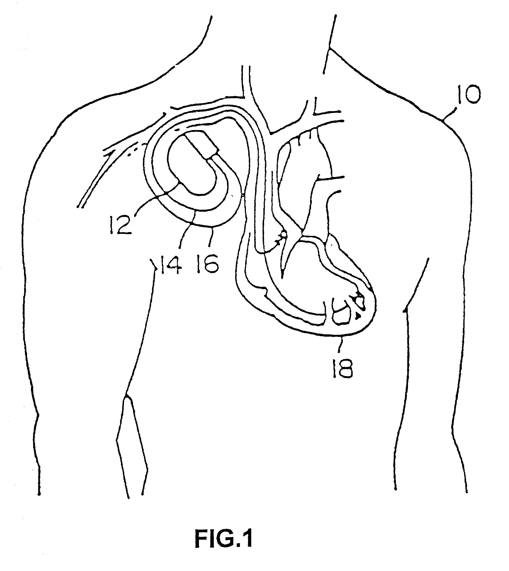
[0034] FIG. 1 depicts a typical arrangement of a pacing or implantable cardioverter / defibrillator (ICD) system implanted in a patient 10, the system comprising a subcutaneously disposed implantable pulse generator (I...


PUM


Abstract

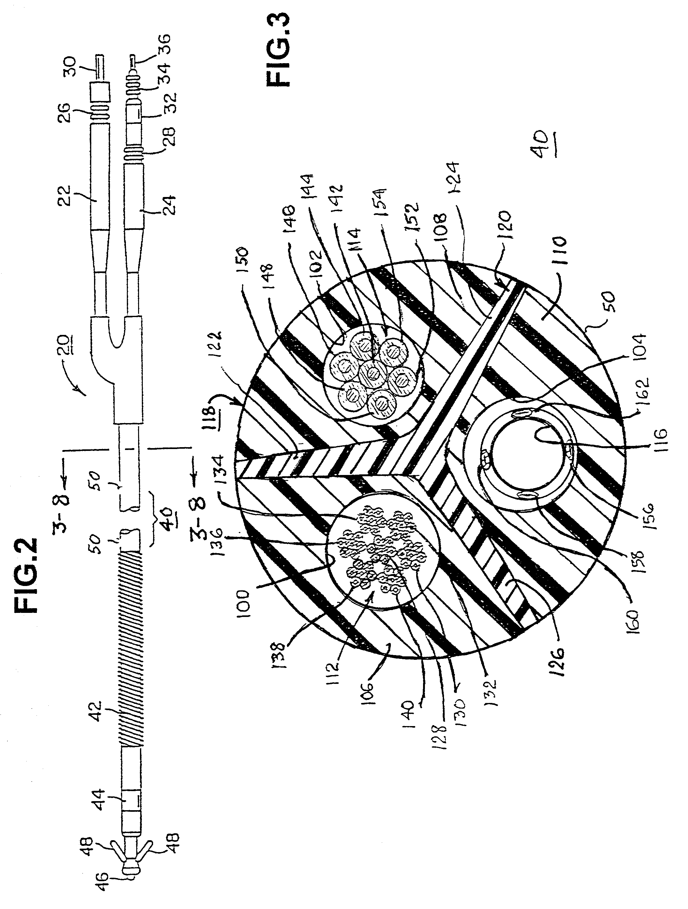
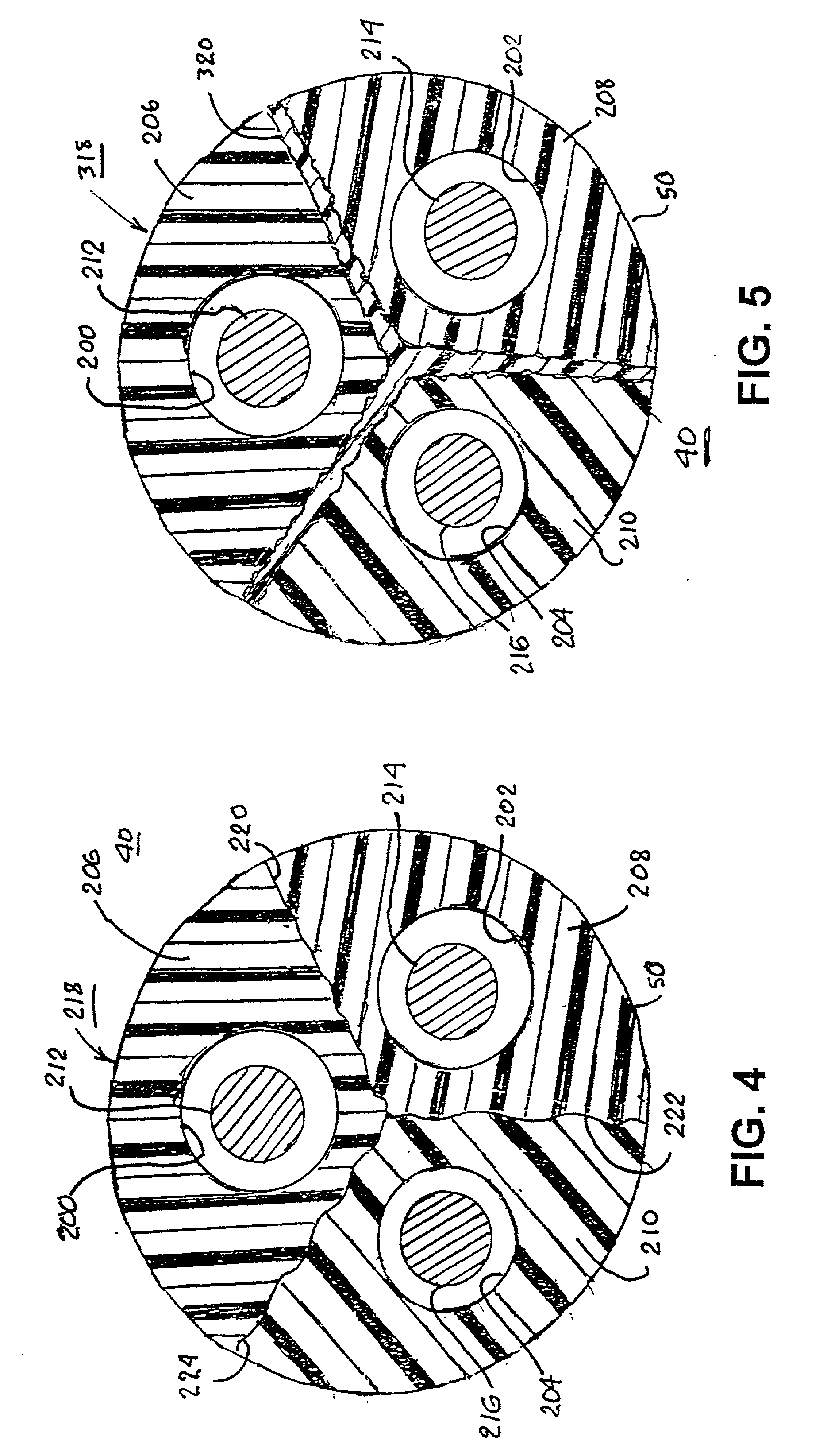
Medical electrical leads for sensing or electrical stimulation of body organs or tissues, particularly implantable cardiac leads for delivering pacing pulses and cardioversion / defibrillation shocks, and / or sensing the cardiac electrogram (EGM) or other physiologic data and their methods of fabrication are disclosed. A lead body sheath is co-extruded in a co-extrusion process using biocompatible, electrically insulating, materials of differing durometers in differing axial sections thereof, resulting in a unitary lead body sheath having differing stiffness sections including axial segments or webs or lumen encircling rings or other structures in its cross-section. The lead body sheath is co-extruded to have an outer surface adapted to be exposed to the environment or to be enclosed within an outer sheath and to have a plurality of lead conductor lumens for receiving and enclosing a like plurality of lead conductors of the same or differing types. The lead body sheath can be co-extruded of a plurality of sheath segments containing a lead conductor lumen and formed of a first durometer material or of differing durometer materials. A web of a further durometer material can be co-extruded extending between the adjoining boundaries of the axial sheath segments and bonding the adjacent segments together. The lead body sheath can be tailored to exhibit differing bending stiffnesses away from the lead body sheath axis in selected polar directions around the 360° circumference of the sheath body.

Description

[0001] Reference is hereby made to commonly assigned, U.S. patent application Ser. No. 08 / 990,647 filed Dec. 15, 1997, for MEDICAL ELECTRICAL LEAD in the name of Alan Rausch et al.[0002] The present invention relates to medical electrical leads for sensing or electrical stimulation of body organs or tissues and their method of fabrication, such leads having multiple electrical conductors encased in a lead body, and particularly to implantable cardiac leads for delivering electrical stimulation to the heart, e.g., pacing pulses and cardioversion / defibrillation shocks, and / or sensing the cardiac electrogram (EGM) or other physiologic data.[0003] Implantable medical electrical stimulation and / or sensing leads are well known in the fields of cardiac stimulation and monitoring, including cardiac pacing and cardioversion / defibrillation, and in other fields of electrical stimulation or monitoring of electrical signals or other physiologic parameters of the body. A pacemaker or cardioverter...

Claims


Application Information

Patent Timeline
Patent Type & Authority Applications(United States)
IPC IPC(8): A61N1/05
CPCA61N1/05A61N1/056
Inventor BORGERSEN, SVENN E.KRAMER, HANS W.
Owner MEDTRONIC INC
Learn More