Methods and systems for synthetic computed tomography (CT) image creation

a synthetic computed tomography and image creation technology, applied in the field of biological imaging methods and systems, can solve the problems of inaccuracy of the method assignment of the hounsfield unit to each point in the synthetic ct, the patient is expensive and time-consuming to undergo both mr and ct scanning, and the radiation therapy planning process is inaccuracy, so as to achieve more accurate transfer of normal tissue segmentation, improve similarity, and accurate assignment of the hounsfield uni

Active Publication Date: 2021-05-25
MEMORIAL SLOAN KETTERING CANCER CENT
View PDF8 Cites 0 Cited by
  • Summary
  • Abstract
  • Description
  • Claims
  • Application Information

AI Technical Summary

Benefits of technology

[0008]Presented herein are systems and methods for synthetic CT image creation that allow MR-only radiotherapy of cancer patients, e.g., head and neck (H&N) cancer patients, prostate cancer patients, patients with cancer of the pelvis, abdomen cancer patients, patients with cancer of the extremities, brain cancer patients, or thorax cancer patients. Also presented are methods of automatic segmentation of normal tissue in CT or MR images of cancer patients. Furthermore, the methods and systems described herein also feature image processing techniques that improve the similarity between CT and MR images prior to CT-MR image registration, as well as standardization of the MR intensity histograms prior to MR-MR registration. Application of the techniques result in more accurate assignment of the Hounsfield unit to each point in the synthetic CT compared to other atlas-based methods, providing for more accurate dosing in MR-only radiotherapy simulation and planning. Furthermore, application of the same techniques can result in more accurate transfer of normal tissue segmentations between MR or CT image pairs.
[0013]In certain embodiments, the one or more co-registered atlas CT images are each registered to a standardized atlas MR image from the representative population using a rigid+deformable registration to construct an atlas of CT-MR, then used to generate the synthetic CT image for the subject. In certain embodiments, bone intensity in each of the atlas CT images are suppressed in creating the CT-MR atlas, thereby improving registration of the CT-MR atlas.

Problems solved by technology

It can be expensive and time consuming for a patient to undergo both MR and CT scanning.
In addition, the act of registration of the MR and CT scans can introduce inaccuracies into the radiation therapy planning process.
However, previous methods lack accurate assignment of the Hounsfield unit to each point in the synthetic CT.
Visualization and manual segmentation of the normal tissues of a patient's CT or MR images is a time consuming process.
However, previous methods may lack accurate definition of the normal tissues in the patient's images.

Method used


Image

  • Methods and systems for synthetic computed tomography (CT) image creation
  • Methods and systems for synthetic computed tomography (CT) image creation
  • Methods and systems for synthetic computed tomography (CT) image creation

Examples


experimental example

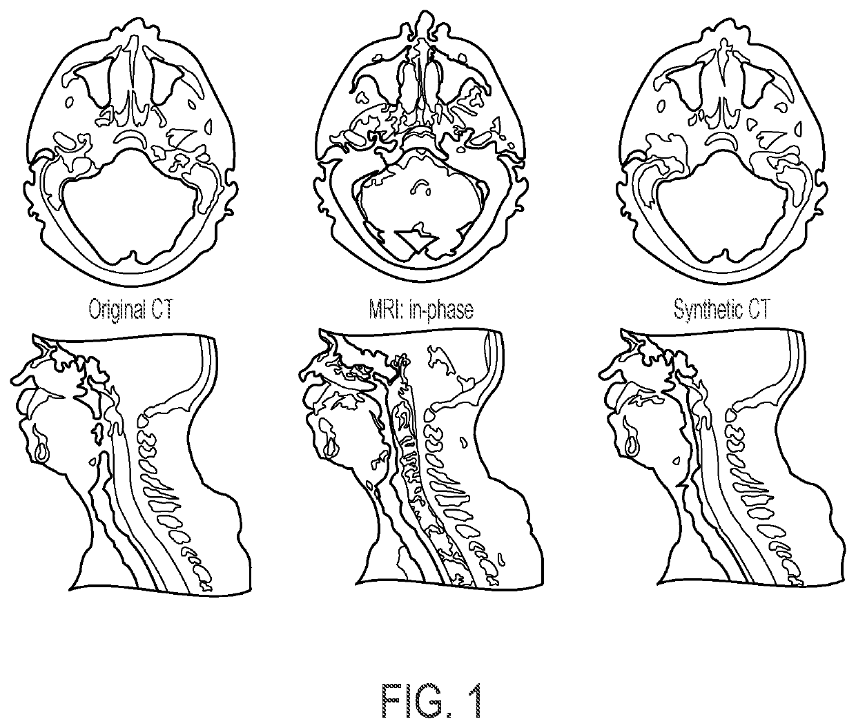
[0040]Eleven sets of CT and MM (in-phase, Philips mDixon sequence) scans were randomly selected from a pool of H&N cancer patients. A bias field correction algorithm was primarily applied to each MRI scan to eliminate the intensity variation due to B0 and B1 field inhomogeneity and tissue susceptibility effect. A landmark-based MM standardization technique was then used to standardize the MR intensity histograms wherein each landmark, a total of four, corresponds to a different histogram extremum. Using a rigid+deformable registration, a CT scan from each patient was registered to the standardized MRI to construct an atlas of CT-MRI. To improve the performance of the registration, bone intensity in the CT image was suppressed to increase the image intensity similarity between CT and MRI scans. CT image was initially clustered into classes of air, bone and soft tissue. The cluster center of the bone class was then transformed to the air class to suppress the bone signal. To synthesiz...


PUM


Abstract

Described herein are systems and methods for synthetic CT image creation that allow MR-only radiotherapy of cancer patients, e.g., head and neck (H&N) cancer patients, prostate cancer patients, patients with cancer of the pelvis, abdomen cancer patients, patients with cancer of the extremities, brain cancer patients, or thorax cancer patients. The methods and systems described herein feature image processing techniques that improve the similarity between CT and MR images prior to CT-MR image registration, as well as standardization of the MR intensity histograms prior to MR-MR registration. Application of the techniques result in more accurate assignment of the Hounsfield unit to each point in the synthetic CT compared to other atlas-based methods, providing for more accurate dosing in MR-only radiotherapy simulation and planning.

Description

CROSS REFERENCE TO RELATED APPLICATION[0001]The application is a 371 National Stage entry of International Application No. PCT / US2017 / 061038, which claims the benefit of U.S. Application Ser. No. 62 / 469,140 filed on Mar. 9, 2017, the disclosures of each of which are hereby incorporated by reference in their entireties. This application also claims the benefit of U.S. Application Ser. No. 62 / 420,499 filed on Nov. 10, 2016 and U.S. Application Ser. No. 62 / 445,937 filed on Jan. 13, 2017.GOVERNMENT SUPPORT[0002]This invention was made with government support under CA008748 awarded by the National Institutes of Health. The government has certain rights in the invention.TECHNICAL FIELD[0003]This invention relates generally to methods and systems of in vivo biological imaging (e.g. clinical and / or research). More particularly, in certain embodiments, the invention relates to systems and methods for generating synthetic computed tomography (CT) images from magnetic resonance images (MRI).BA...

Claims


Application Information

Patent Timeline
Patent Type & Authority Patents(United States)
IPC IPC(8): G06K9/00A61B6/03G06T7/30A61B6/00A61N5/10G01R33/56G06T5/50
CPCA61B6/032A61B6/5247A61N5/103G01R33/5608G06T7/30G06T5/50G06T2207/10081G06T2207/10088G06T3/0081
Inventor HUNT, MARGIE A.DEASY, JOSEPH OWENFARJAM, REZAAPTE, ADITYATYAGI, NEELAMVEERARAGHAVAN, HARINIZAKIAN, KRISTEN L.
Owner MEMORIAL SLOAN KETTERING CANCER CENT
Why Eureka
Try Eureka