Ultrasound transducer holder
a transducer and ultrasonic technology, applied in ultrasonic/sonic/infrasonic image/data processing, ultrasonic/sonic image/data processing, applications, etc., can solve the problems of musculoskeletal disorders, manual process can be tiring for the technician, and the ultrasonic procedure is not always reproducible between different technicians or scans, etc., to achieve a higher elastic modulus
- Summary
- Abstract
- Description
- Claims
- Application Information
AI Technical Summary
Benefits of technology
Problems solved by technology
Method used
Image
Examples
example 1
of a Soft Robotic Ultrasound Transducer Holder
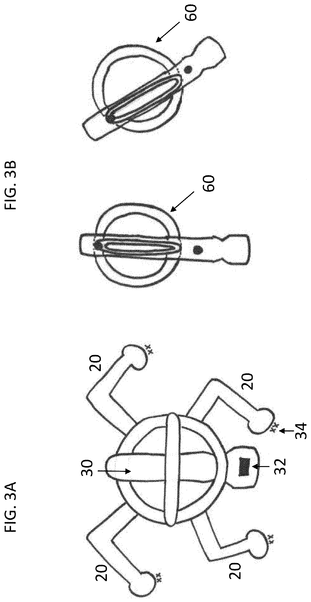
[0074]The soft robotic actuator (20, 22) has an air inlet (36) and two semicircular air channels (38) and can move in different directions based on air flow. If one chamber (38) is pressurized, then the soft actuator (20, 22) bends. If both air chambers (38) are pressurized, then the soft actuator (20, 22) elongates and gives linear actuation. If various actuators are controlled with an on / off pulse, the actuators can walk.
[0075]Linear actuators (22) are placed in the holder base horizontally. The holder has a center recess of 50.8 mm on the octagon and the probe has a diameter of 25.4 mm, so there is a clearance gap of 25.4 mm to which probe (30) can be tilted. The angle relationship is given in Tables 2 and 3.
[0076]
TABLE 2Tilt angle with linear actuator movementActuator Movement (mm)6.3512.7019.0525.4Tilt Angle, degrees4°8°12°16°
[0077]
TABLE 3Vertical height adjustment with tilt angleTilt Angle, degrees4°8°12°16°Height change (mm)0.341....
example 2
g an Echocardiogram
[0079]The devices described herein may be used to perform an echocardiogram to obtain different ultrasound image views of the heart. A technician or operator will mount ultrasound transducer (30) in base (10) and / or top frame (16). Ring (18) in the center of top frame (16) will snap around probe (30) to keep it in place. Top frame (16) is adjustable in height so probe (30) can be adjusted up or down in the assembly during mounting. Once transducer probe (30) is mounted in assembly (70), the assembly can be placed on the chest of a subject above the heart. A strap or belt can be used if needed to hold maintain general position. The air source is then turned on and probe (30) can be directed to move to a specific location by interacting with the microprocessor. The assembly can be manipulated to tilt transducer (30) to obtain ultrasound images of the heart at different angles. Three views of the heart that can be imaged include the parasternal long axis view, which ...
example 3
g Musculoskeletal Conditions in Motion
[0080]An ultrasound technician attaches the transducer holder to a patient with musculoskeletal pain. The patient reports significant pain and spasms in his shoulder when raising his arm. The straps on the transducer holder keep the transducer probe in close contact with the patient's shoulder, such as when the patient raises their arm. The transducer holder positions the ultrasound transducer in close proximity to a joint, ligaments, muscles, and / or nerves of the shoulder of the patient, e.g., while the patient is in motion, thereby assisting the technician in analyzing the patient's condition while freeing the hands of the technician to perform other tasks during the procedure. A series of imaging tests can be performed that assist the technician with diagnosing the patient's condition. The results of the imaging tests can be used, e.g., in combination with artificial intelligence and data mining from the captured images, to predict positions ...
PUM
 Abstract
Description
Claims
Application Information
 - R&D
- Intellectual Property
- Life Sciences
- Materials
- Tech Scout
- Unparalleled Data Quality
- Higher Quality Content
- 60% Fewer Hallucinations
Browse by: Latest US Patents, China's latest patents, Technical Efficacy Thesaurus, Application Domain, Technology Topic, Popular Technical Reports.
© 2025 PatSnap. All rights reserved.Legal|Privacy policy|Modern Slavery Act Transparency Statement|Sitemap|About US| Contact US: help@patsnap.com


