Photonic probe apparatus with integrated tissue marking facility

a probe apparatus and tissue marking technology, applied in the field of photonic probe apparatus and a method, can solve the problems of large observer-to-observer variation, large differences in the appearance of (pre)cancerous tissue, and the physician cannot directly see the lesion, so as to simplify the excising procedure, improve the classification of tissue, and eliminate one.

Active Publication Date: 2020-07-21
KONINKLJIJKE PHILIPS NV
View PDF20 Cites 1 Cited by
  • Summary
  • Abstract
  • Description
  • Claims
  • Application Information

AI Technical Summary

Benefits of technology

[0013]It would be advantageous to achieve a methodology and an apparatus for detecting and marking cancerous tissue which can improve the classification of the tissue. It would also be advantageous to achieve a methodology and an apparatus for detecting and marking cancerous tissue which can simplify the excising procedure. In general, the invention preferably seeks to mitigate, alleviate or eliminate one or more of the above mentioned disadvantages singly or in any combination.

Problems solved by technology

The appearance of (pre)cancerous tissue can show very large differences depending on the body site, as can the difficulties and consequences related to excision of malignant tissue.
In practice however, the physician cannot directly see the lesion.
Both colposcopy images and acetowhitening are notoriously hard to interpret with large observer-to-observer variations.
Also these methods provide only information about tissue which can be assessed visually.
For suspected cervical cancer, acetowhitened tissue regions are notoriously hard to interpret and only the Ectocervix is marked up.
Acetowhitening thereby involves the main disadvantages that it is very difficult to apply any objective threshold in the classification of the malignancy of the tissue and that the marking is not permanent leading to practical difficulties when performing excision procedures.
Additionally, the endocervix is difficult to mark up, and especially it will be difficult to detect marked tissue in the endocervix.

Method used


Image

  • Photonic probe apparatus with integrated tissue marking facility
  • Photonic probe apparatus with integrated tissue marking facility
  • Photonic probe apparatus with integrated tissue marking facility

Examples


Embodiment Construction

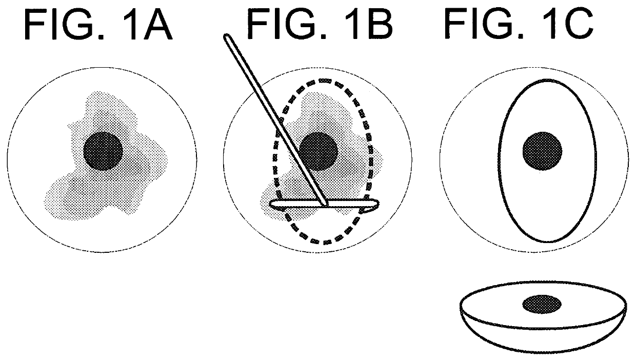
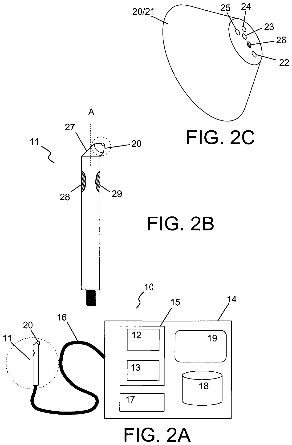
[0042]An embodiment of the invention is illustrated in FIG. 2A, showing a photonic probe apparatus 10 comprising a probe 11, a light source 12 connected to the probe for illuminating tissue, and a light sensor 13 connected to the probe to receive and detect light collected from the tissue region through the probe 11, preferably through a tip 20 of the probe. An analyzing unit 14 is connected to the light sensor 13 for determining whether a threshold measure of probability of a cancerous or precancerous lesion in the probed tissue region in contact with the probe is exceeded based on an output signal from the light sensor 13. The apparatus also comprises an integrated tissue marking facility (not shown in FIG. 2A) which can be activated to mark the probed tissue region through the probe when the threshold measure is exceeded.

[0043]The light source 12 may involve more light sources such as a combination of monochromatic and broad spectral light sources. The light source 12 may be inte...


PUM


Abstract

The invention relates to a photonic probe apparatus and a method for probing tissue to detect and mark biological tissue with cancerous or precancerous states. The apparatus involves a probe for illuminating tissue and collecting light from an illuminated tissue region through the probe, a unit for analyzing collected light to determine whether a threshold measure of probability of a cancerous or precancerous lesion in the probed tissue region in contact with the probe is exceeded, and an integrated tissue marking facility which can be activated to mark the probed tissue region through the probe when the threshold measure is exceeded. The photonic probe apparatus and the method are especially suitable for probing regions in squamous and columnar epithelia to detect and mark regions with cervical cancer or cervical intraepithelial neoplasia (CIN).

Description

CROSS-REFERENCE TO PRIOR APPLICATIONS[0001]This application is the U.S. National Phase application under 35 U.S.C. § 371 of International Application Serial No. PCT / IB2013 / 051063, filed on Feb. 8, 2013, which claims the benefit of U.S. application Ser. No. 61 / 597,872, Filed on Feb. 13, 2012. These applications are hereby incorporated by reference herein.FIELD OF THE INVENTION[0002]The invention relates to a photonic probe apparatus and a method for probing tissue to detect and mark tissue with cancerous or precancerous states. In particular, the invention relates to such photonic probe apparatus having an integrated tissue marking facility.BACKGROUND OF THE INVENTION[0003]Cancer that originates in squamous epithelia can develop in a large number of organs or tissues. The appearance of (pre)cancerous tissue can show very large differences depending on the body site, as can the difficulties and consequences related to excision of malignant tissue. This is especially relevant for cervi...

Claims


Application Information

Patent Timeline
Patent Type & Authority Patents(United States)
IPC IPC(8): A61B18/14A61B5/00A61B90/00A61B18/00A61B17/00A61B1/00A61B1/07A61B1/06A61B1/04
CPCA61B5/742A61B5/0075A61B5/7475A61B5/0062A61B1/043A61B5/7405A61B18/14A61B5/7275A61B5/7282A61B5/0084A61B5/0071A61B2017/00473A61B2018/00589A61B1/00165A61B2576/00A61B2090/3908A61B1/0623A61B2090/3937A61B2560/0443A61B2562/0238A61B1/00179A61B2018/00982A61B1/07
Inventor MUELLER, MANFREDKESWARPU, PAYALGUPTA, VIPINFIRTION, CELINE
Owner KONINKLJIJKE PHILIPS NV
Why Eureka
Try Eureka