
Non-invasive skin collection system

a skin collection and non-invasive technology, applied in the field of non-invasive skin collection systems, can solve the problems of increased risk of metastasis, difficult visible observation and pathologic assessment of these lesions, and significant burden on the healthcare system of melanoma, so as to reduce the risk of metastasis, the effect of reducing the survival rate and high cos

Active Publication Date: 2020-07-14
DERMTECH INC
View PDF13 Cites 5 Cited by
  • Summary
  • Abstract
  • Description
  • Claims
  • Application Information

AI Technical Summary

Benefits of technology

[0005]Lesions positive for melanoma are subsequently staged to determine if the tumor remains in situ or if it has undergone invasion. For early melanoma, this staging can be difficult and is also dependent on the area selected for examination. It is important to identify the stage accurately, because invasive melanoma has a lower survival rate, requires more extensive medical treatment, surveillance, work up and has a higher cost. The medical work up for invasive melanoma may include a sentinel lymph node biopsy surgical procedure to determine if the melanoma has metastasized. This is a significant surgical procedure with associated high cost and morbidity with results that can be ambiguous.

Problems solved by technology

Because approximately 62% of melanomas and 45% of melanoma deaths occur prior to age 65, melanoma places significant burdens on the healthcare system.
However, once the cancer advances into the deeper layers of skin, the risk of metastasis increases.
Visible observation and pathologic assessment of these lesions is challenging, making melanoma one of the top five misdiagnosed cancers.
Similar to visual evaluation of pigmented lesions, histopathologic assessment of pigmented lesions is also subjective and challenging.
Because patients can have many atypical lesions, the decision of which lesions to biopsy can be challenging and it is not practical to biopsy all lesions.
Of the millions of surgical biopsies performed each year on selected pigmented skin lesions, over 95% are negative for melanoma and represent an unnecessary surgical procedure.
For early melanoma, this staging can be difficult and is also dependent on the area selected for examination.
This is a significant surgical procedure with associated high cost and morbidity with results that can be ambiguous.

Method used


Image

  • Non-invasive skin collection system
  • Non-invasive skin collection system
  • Non-invasive skin collection system

Examples


example 1

Care Skin Sample Collection

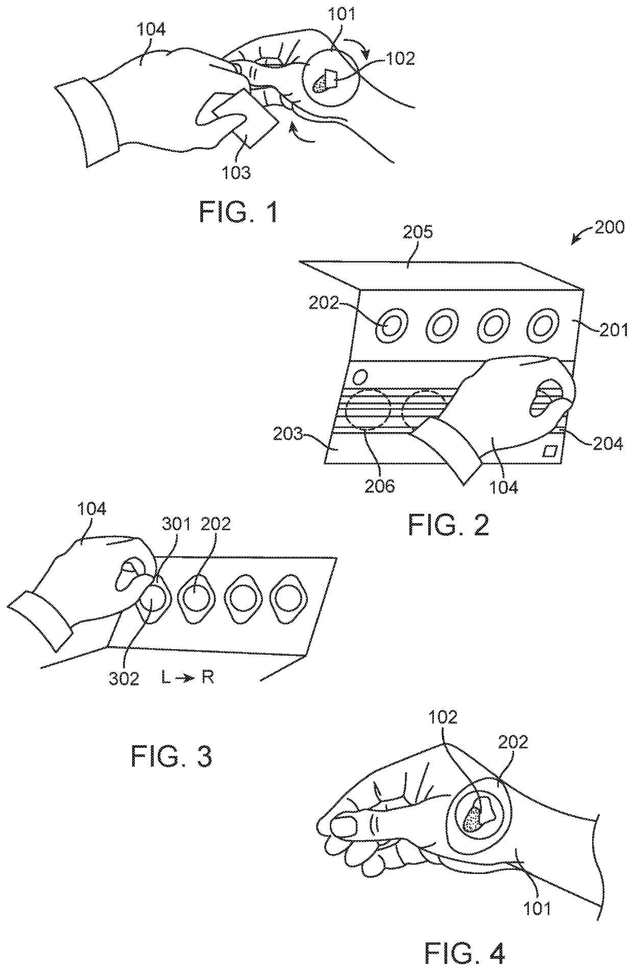
[0095]A pigmented lesion located on the hand of a subject is selected for skin sampling. The skin sampling area contains a minimal amount of hair, is not irritated and has not been previously biopsied. The lesion is about 8 mm in size. As exemplified in FIG. 1, the skin sampling area (101) comprising the skin lesion (102) is cleansed with an alcohol pad (103) by a practitioner (104) wearing gloves, and the skin is allowed to air dry for 5 minutes.

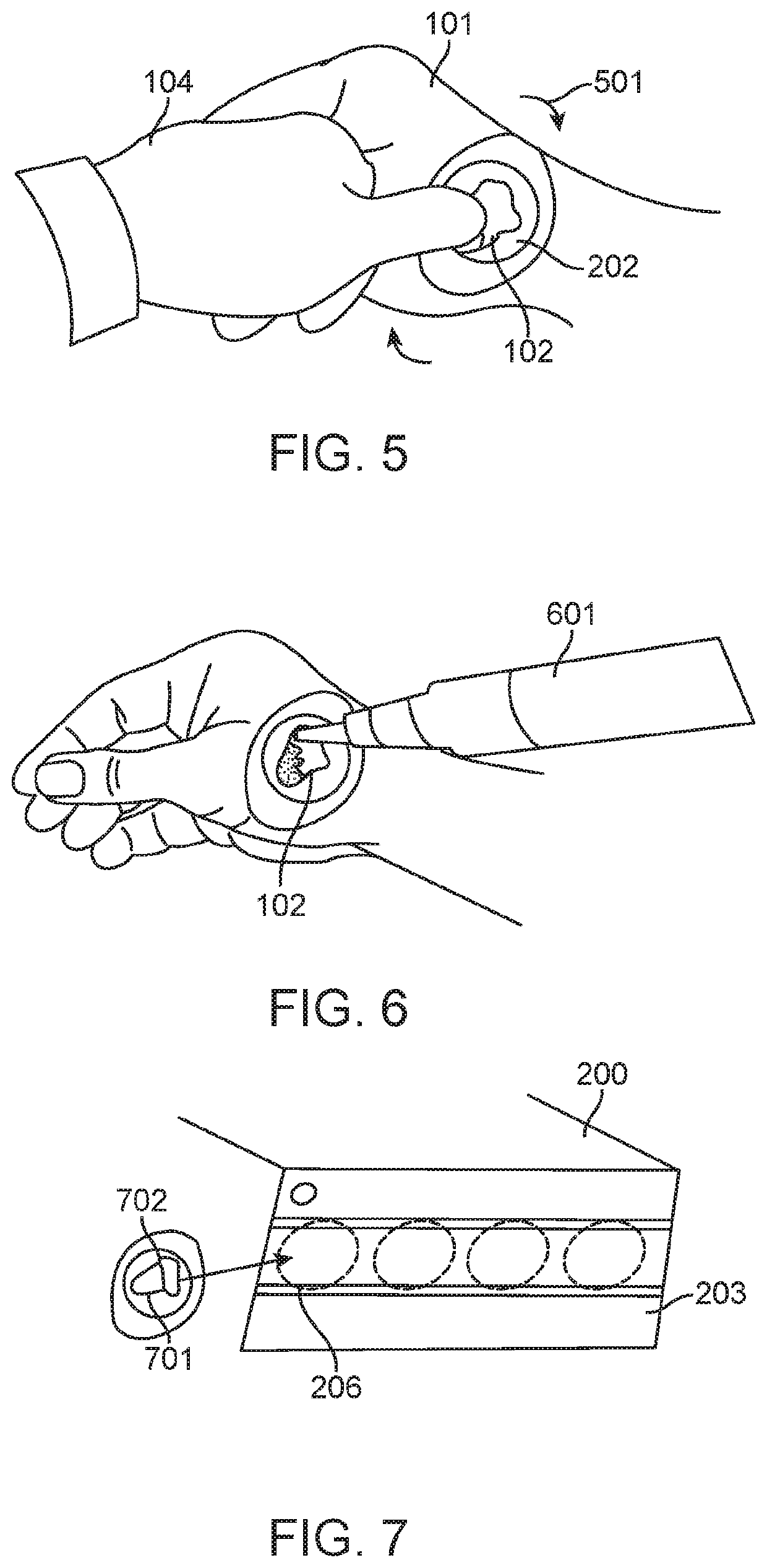
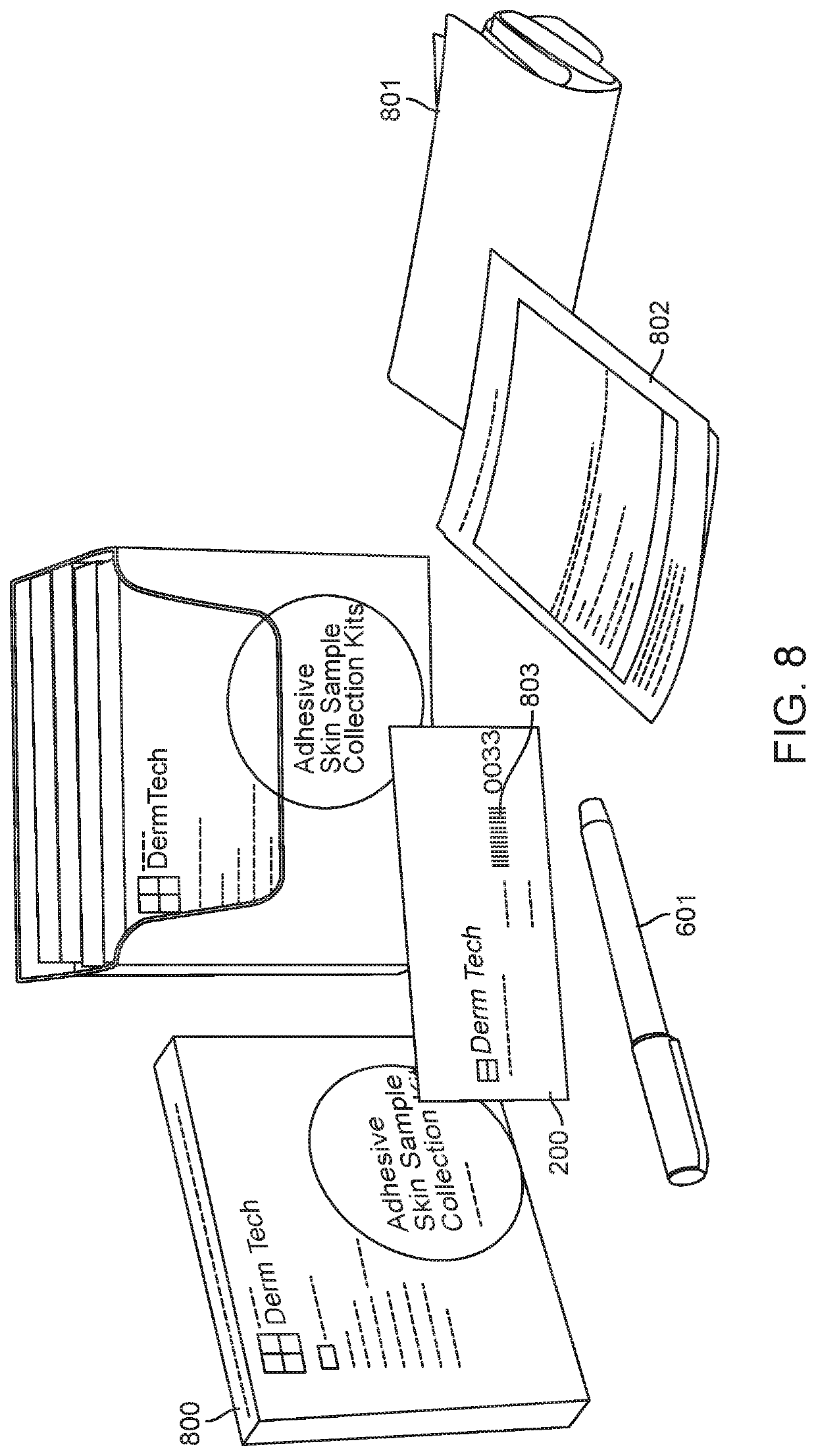
[0096]A tri-fold skin sample collector is removed from an adhesive skin sample collection kit exemplified by FIG. 8. FIG. 2 exemplifies the tri-fold skin sample collector (200) comprising a peelable release panel (201) comprising four adhesive tapes (202), a placement area panel (203) comprising a removable liner (204), and a clear panel (205). The tri-fold skin sample collector has a barcode specific for the subject. The removable liner is removed from the placement area panel (203), exposing four regions (206) d...

example 2

le Collection

[0099]A pigmented lesion located on the upper back of a subject is selected for skin sampling. The skin sampling area contains a minimal amount of hair, is not irritated and has not been previously biopsied. The lesion is about 15 mm in size. The lesion is sampled utilizing an adhesive skin sample collection kit. The skin sample collection kit includes an instructions for use sheet (or an instruction manual). The lesion is sampled by a capable person who has read and understood the skin sample collection kit instructions for use sheet.

[0100]A pair of gloves is removed from the skin sample collection kit and the fitted onto the person performing the skin sampling procedure. The skin sampling area comprising the pigmented lesion is cleansed with an alcohol pad provided in the adhesive skin sample collection kit and the skin is allowed to air dry.

[0101]A tri-fold skin sample collector is removed from the adhesive skin sample collection kit. The tri-fold skin sample collect...

example 3

n System

[0104]The adhesive skin sample collection kit components are stored in a cardboard box (800) as exemplified in FIG. 8. The kit contains a tri-fold skin sample collector (200) comprising four adhesive tapes, instructions for use sheet, a marking pen, a pre-paid, self-addressed shipping package (801), and a shipping label (802). The tri-fold skin sample collector comprises three panels including a peelable release panel comprising the four adhesive tapes, a placement area panel comprising a removable liner and a clear panel. The tri-fold skin sample collector further comprises a unique barcode (803) configured to identify a subject. The adhesive tapes stored on the peelable release panel have an expiry date of 2 years from the date of manufacture. The skin sample collection kit is stored between 10® C. and 30® C. The instructions for use sheet (or instruction manual) include all information necessary to enable a person to understand and perform the method. The instructions for...


PUM


Abstract

The subject matter described herein provides non-invasive tape stripping methods for the collection of a skin sample. The tape stripping method includes applying and removing at least one adhesive tape, provided that a skin sample is adhered to the adhesive tape after removal. The at least one adhesive tape is supplied in an adhesive skin sample collection kit. The adhesive skin sample collection kit further comprises a sample collector and instructions for use sheet.

Description

CROSS REFERENCE[0001]This application is a U.S. National Phase of International Application No. PCT / US2016 / 030287, filed Apr. 29, 2016, which claims the benefit of U.S. Provisional Application No. 62 / 156,091, filed May 1, 2015, which is incorporated herein by reference in its entirety.BACKGROUND OF THE INVENTION[0002]Skin diseases are some of the most common human illnesses and represent an important global burden in healthcare. Three skin diseases are in the top ten most prevalent diseases worldwide, and eight fall into the top 50. When considered collectively, skin conditions range from being the second to the 11th leading causes of years lived with disability.SUMMARY OF THE INVENTION[0003]Skin diseases include eczema, psoriasis, acne vulgaris, pruritus, alopecia areata, decubitus ulcer, urticaria, scabies, fungal skin diseases, impetigo, abscess, bacterial skin diseases, cellulitis, lupus, viral warts, molluscum contagiosum and cancers, such as melanomas. Indeed, the deadliest sk...

Claims


Application Information

Patent Timeline
Patent Type & Authority Patents(United States)
IPC IPC(8): A61B10/02A61B10/00A61B50/30A61B17/00A61B50/00
CPCA61B10/0096A61B10/00A61B50/30A61B10/02A61B2050/0065A61B2017/00951A61B2050/3015A61B2017/00761A61B2010/0225A61B2017/00774
Inventor PALMER, TARA J.ALSOBROOK, II, JOHN P.DOBAK, JOHN
Owner DERMTECH INC
Learn More