
Methods and systems for adaptive scan control

a technology of adaptive scanning and control method, applied in the field of non-invasive diagnostic imaging, can solve the problem of taking five minutes alone, and achieve the effect of substantially reducing the time of ischemic stroke assessment, radiotherapy dose, and contrast dos

Active Publication Date: 2019-08-20
GENERAL ELECTRIC CO
View PDF85 Cites 4 Cited by
  • Summary
  • Abstract
  • Description
  • Claims
  • Application Information

AI Technical Summary

Benefits of technology

This approach significantly reduces the time and radiation / contrast dose required for ischemic stroke assessment, enabling faster and more accurate diagnosis by seamlessly combining CT angiography and perfusion scans while avoiding venous contamination.

Problems solved by technology

However, the timing bolus scan alone takes five minutes, and performing CTA and CTP studies separately requires five to seven minutes between acquisitions to allow contrast washout.

Method used


Image

  • Methods and systems for adaptive scan control
  • Methods and systems for adaptive scan control
  • Methods and systems for adaptive scan control

Examples


Embodiment Construction

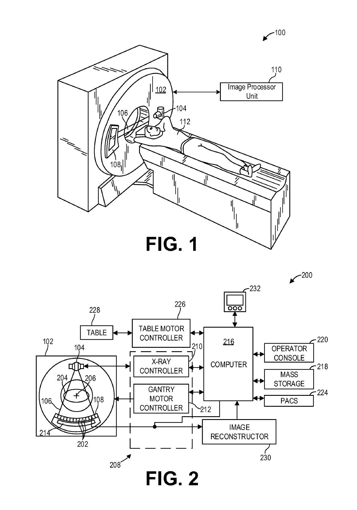
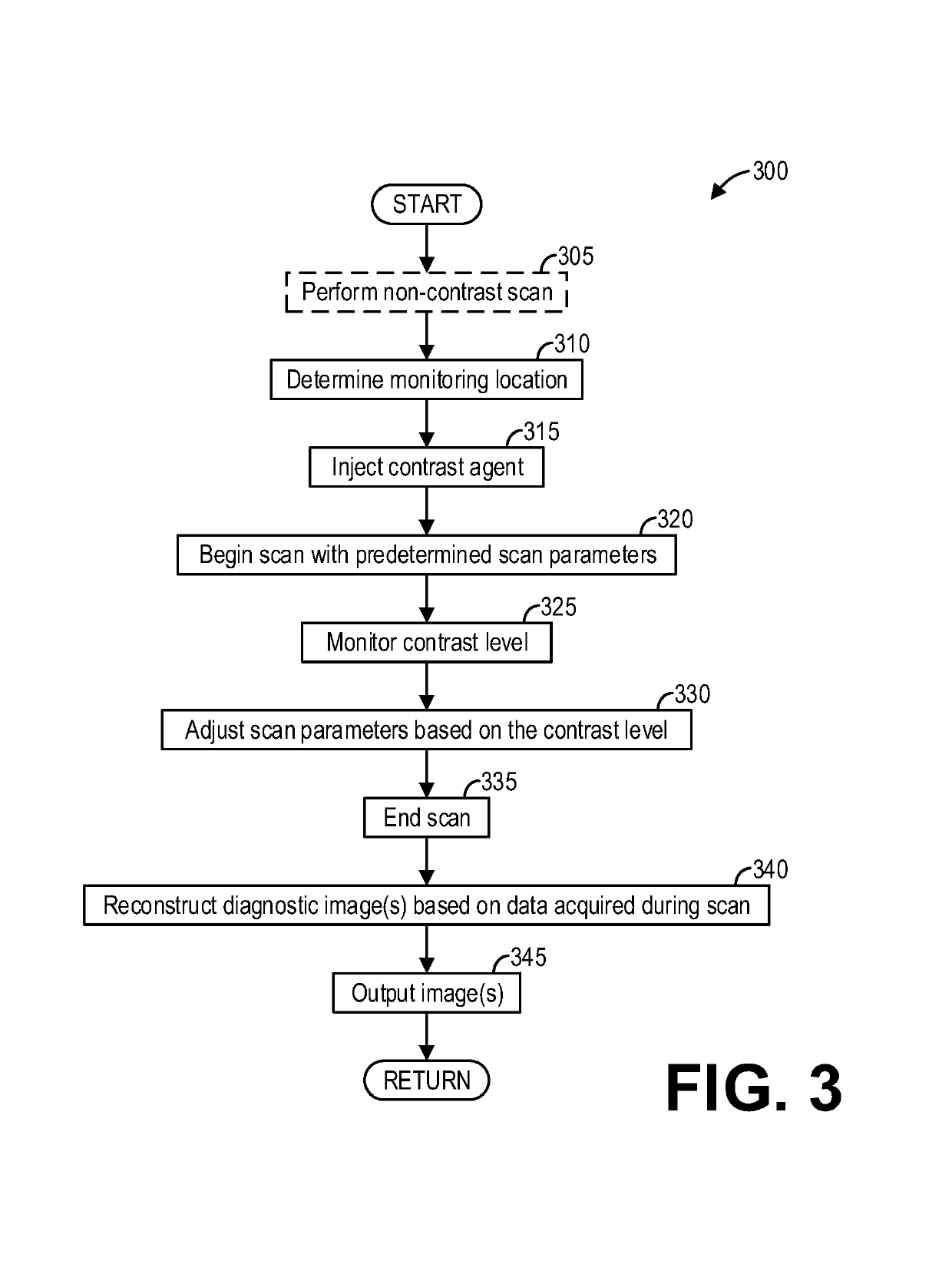
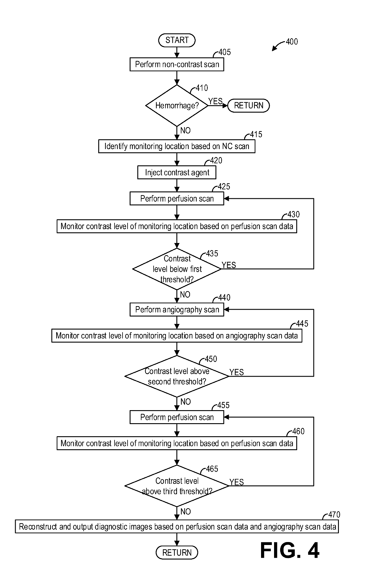
[0014]The following description relates to various embodiments of medical imaging systems. In particular, methods and systems are provided for adaptively controlling a diagnostic scan by monitoring contrast enhancement. An example of a computed tomography (CT) imaging system that may be used to acquire images processed in accordance with the present techniques is provided in FIGS. 1 and 2. A method for adaptive scan control, such as the method shown in FIG. 3, may include monitoring contrast levels during a scan and adjusting scan parameters responsive thereto. Such a method enables personalization of scan protocols on a patient-by-patient basis. Furthermore, by adapting scans based on monitored contrast levels in real-time, multiple scan protocols may be combined into a single scan. As an example, a method, such as the method depicted in FIGS. 4 and 5, includes interleaving CT angiography (CTA) and CT perfusion (CTP) scans into a single scan by switching scan protocols responsive t...


PUM


Abstract

Methods and systems are provided for adaptive scan control. In one embodiment, a method comprises: while performing a scan of a scan subject, processing acquired projection data to measure a contrast level; responsive to the contrast level increasing above a first threshold, automatically switching the scan from a first scan protocol to a second scan protocol; responsive to the contrast level decreasing below a second threshold, automatically switching the scan from the second scan protocol to the first scan protocol; and responsive to the contrast level decreasing below a third threshold, automatically ending the scan. In this way, multiple scan protocols, such as angiography and perfusion scan protocols, can be interleaved within a single scan without the use of a separate timing bolus scan.

Description

FIELD[0001]Embodiments of the subject matter disclosed herein relate to non-invasive diagnostic imaging, and more particularly, to real-time adaptive scanning.BACKGROUND[0002]Non-invasive imaging technologies allow images of the internal structures of a patient or object to be obtained without performing an invasive procedure on the patient or object. In particular, technologies such as computed tomography (CT) use various physical principals, such as the differential transmission of x-rays through the target volume, to acquire image data and to construct tomographic images (e.g., three-dimensional representations of the interior of the human body or of other imaged structures).[0003]For emergency room (ER) stroke management, time is critical to determine a proper course of treatment. For every minute a large vessel ischemic stroke is untreated, the average patient loses 1.9 million neurons. For each hour in which a treatment fails, the patient loses as many neurons as it does in al...

Claims


Application Information

Patent Timeline
Patent Type & Authority Patents(United States)
IPC IPC(8): A61B6/03A61B6/00
CPCA61B6/504A61B6/032A61B6/481A61B6/486A61B6/542A61B6/507A61B6/5217A61B6/54A61B6/501G16H50/30
Inventor VAZ, MICHAEL SARJUNETT, ELIZABETH JANUSPITTERLE, DAVID JOSEPHCHEVALIER, DAVID ERIKHAMMOND, CHRISTINE CAROLLEWIS, CHELSEY
Owner GENERAL ELECTRIC CO
Learn More