
Automatic left ventricular function evaluation

a left ventricular and function evaluation technology, applied in the field of automatic left ventricular function evaluation, can solve the problems of difficult task, inability to accurately detect the patient, and rare use in everyday practice, and achieve the effect of reducing the noise of the echocardiogram

Active Publication Date: 2018-09-18
DIACARDIO
View PDF109 Cites 2 Cited by
  • Summary
  • Abstract
  • Description
  • Claims
  • Application Information

AI Technical Summary

Benefits of technology

[0027]In some embodiments, the method further comprises, prior to performing the image mapping, reducing noise in the echocardiogram.
[0047]In some embodiments, said processing unit is further configured to, prior to performing the image mapping, reduce noise in the echocardiogram.

Problems solved by technology

As this technique is operator-dependent and time consuming, it is rarely used in everyday practice.
Automatic LV contour detection can be described as a segmentation problem—the problem of separating muscle from blood, which is often considered to be a difficult task in ultrasound images.
As with all Doppler techniques, TDI measures motions parallel to the direction of the ultrasound beam; further, the Doppler-based methods obtain tissue velocity information from fixed beam position, which may not be accurate since the heart is constantly moving.
Therefore, TDI is not conventionally used for regional wall motion evaluation; however, it was found to be helpful in evaluating diastolic function by measuring the overall longitudinal left ventricular (LV) relaxation at the mitral annulus.
Several difficulties may arise when using such methods.

Method used


Image

  • Automatic left ventricular function evaluation
  • Automatic left ventricular function evaluation
  • Automatic left ventricular function evaluation

Examples


experiment 1

[0153]Automatic EF evaluation of a patient with normal global LV systolic function was performed: EF=75%. EDV, ESV and EF results are summarized in Table 2, which compares the present automatic method with manual tracing done by an expert:

[0154]

TABLE 2EDV (ml)ESV (ml)EF (%)Expert962475Algorithm992574

experiment 2

[0155]Automatic EF evaluation of a patient previously diagnosed with severely reduced global LV systolic function and dilated LV: EF=25%. EDV, ESV and EF results are summarized in Table 3, which compares the present automatic method with manual tracing done by an expert:

[0156]

TABLE 3EDV (ml)ESV (ml)EF (%)Expert25318925Algorithm25117530

experiment 3

[0157]Automatic, global diastolic function evaluation of a patient previously determined to have normal global LV diastolic function was performed: A filling rate curve 1600 (FIG. 16) exhibited a normal E / A ratio.


PUM


Abstract

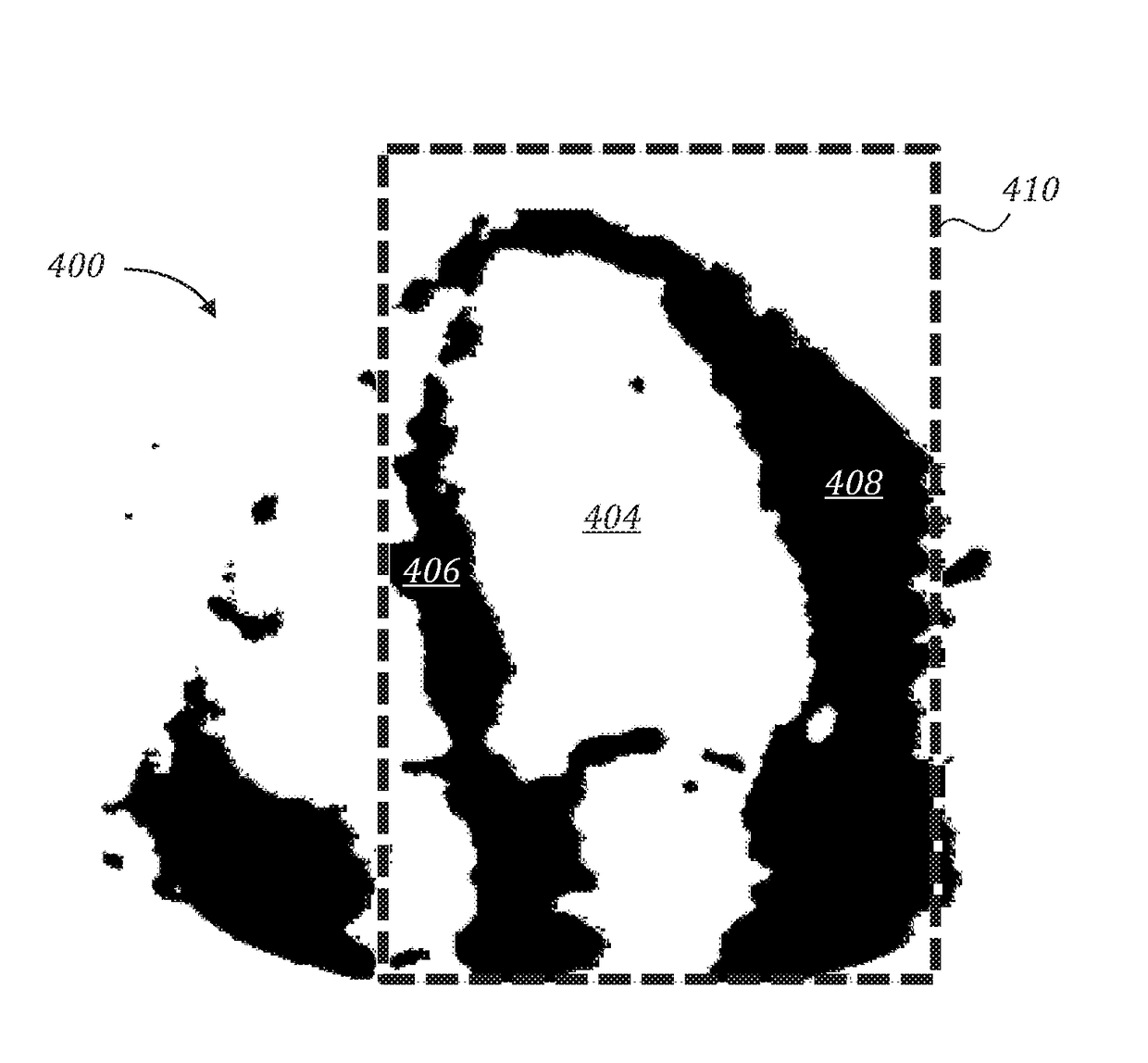
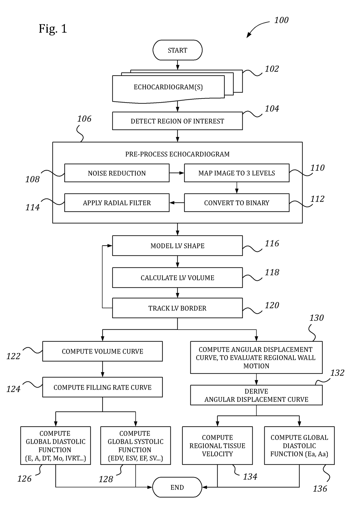
A method for automatic left ventricular (LV) inner border detection, the method comprising: performing image mapping on an echocardiogram, to produce a multi-level image map; converting the image map into a binary image, by attributing pixels of one or more darker levels of the image map to the LV cavity and pixels of one or more lighter levels of the image map to the myocardium; applying a radial filter to contours of the myocardium in the binary image, to extract an approximate inner border of the LV; and performing shape modeling on the approximate inner border, to determine the LV inner border.

Description

RELATED APPLICATION DATA[0001]This application is a U.S. National Stage of International Application No. PCT / IL2011 / 050084, filed Dec. 29, 2011, which claims the benefit of U.S. Provisional Application No. 61 / 427,830, filed Dec. 29, 2010, the contents of each of which are herein expressly incorporated by reference for all purposes.FIELD OF THE INVENTION[0002]The invention relates to automatic left ventricular (LV) function evaluation.BACKGROUND OF THE INVENTION[0003]Echocardiography is an important, widely-used imaging technique for clinical diagnosis of heart diseases. Due to its low cost and non-invasive nature, ultrasonic imaging is an integral part of modern diagnostic-imaging facilities, allowing qualitative and quantitative performance evaluation of the cardiovascular system, including LV function.[0004]Since many of the cardiac diseases in adults result in abnormalities of LV function, the LV function evaluation is a very important part of almost every echocardiographic exami...

Claims


Application Information

Patent Timeline
Patent Type & Authority Patents(United States)
IPC IPC(8): G01S15/89A61B8/00A61B8/06A61B8/08G06T7/00G06T7/12G06T7/136G06T7/174G06T7/181
CPCA61B8/065A61B8/0883A61B8/5223A61B8/543G06T7/0016G06T2207/10016G06T2207/10132G06T2207/20032G06T2207/30048G06T7/12G06T7/174G06T7/181G06T7/136G16H50/30G06T7/0012
Inventor GUTERMAN, HUGOLIEL, NOAHYAACOBI, MARINA
Owner DIACARDIO
Learn More