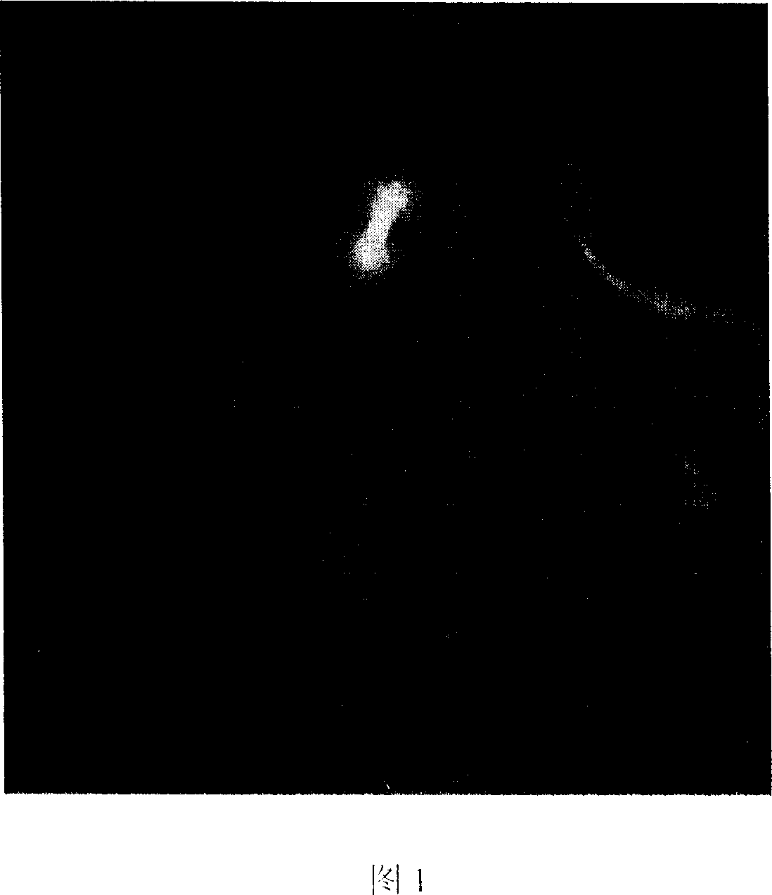
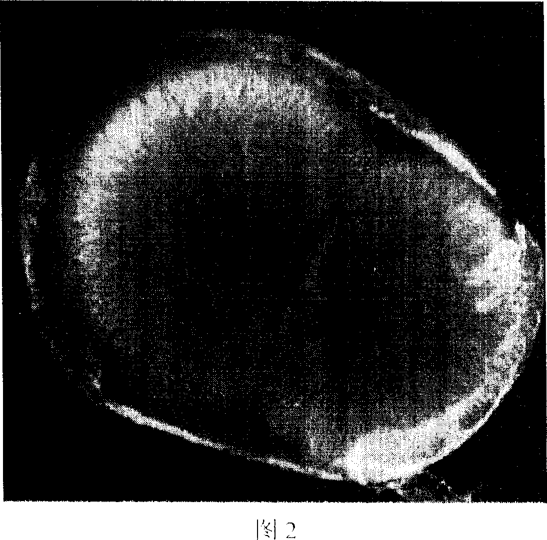
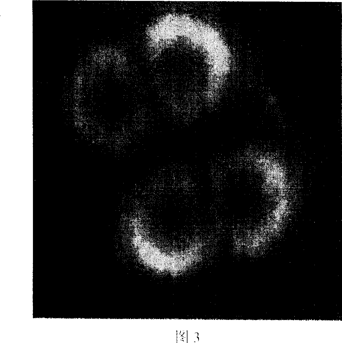
Immunofluorescence dyeing viewing method for left-eyed flounder zygote microtubule skeleton
A technique for immunofluorescence staining and fertilized eggs, which is applied in the direction of biochemical equipment and methods, microorganisms, microorganisms, etc., to achieve the effects of complete blastodisc structure, sufficient antibody binding, and reduced quenching speed
- Summary
- Abstract
- Description
- Claims
- Application Information
AI Technical Summary
Problems solved by technology
Method used
Image
Examples
Embodiment 1
[0028] 1) When the flounder fertilized eggs develop to the germinal disc, prepare the FGP-fix fixative solution prepared by the improved PIPES. The method is: first prepare the microtubule polymerization solution-80mM K-PIPES, 5mM EGTA, 100mM MgCl 2 , 400mM NaCl, pH 6.8, stored at 4°C; before fixation, add formaldehyde, glutaraldehyde, and Triton X-100 to the microtubule polymerization solution, and the addition amounts account for 3.7% and 0.25% of the concentration of the microtubule polymerization solution, respectively , 0.5% prepared as a fixative.
[0029] 2) The samples were fixed, and the fertilized eggs were incubated in the fixative at room temperature for 3 hours, then washed twice with PBST buffer, wherein the PBST buffer contained 128mM NaCl, 2mM KCl, 8mMNaH 2 PO 4 , 2mMKH 2 PO 4 , and Tween-20, pH 7.2, the concentration percentage of Tween-20 added is 0.1% of the Tween phosphate buffer solution, and finally stored in PBST at 4°C.
[0030] 3) Under a dissectin...
Embodiment 2
[0036] 1) When turbot fertilized eggs develop to 2-cells, prepare FGP-fix fixative solution prepared by improved PIPES, the method is: first prepare microtubule polymerization solution-90mM K-PIPES, 7mM EGTA, 150mMMgCl 2 , 450mM NaCl, pH 6.9, stored at 4°C; before fixation, add formaldehyde, glutaraldehyde, and Triton X-100 to the microtubule polymerization solution in amounts of 3.8%, 0.4% and 0.75%, prepared as a fixative.
[0037] 2) Fix the sample. After the fertilized eggs are incubated in the fixative at room temperature for 6 hours, wash twice with PBST buffer, wherein the PBST buffer contains 128mM NaCl, 2mM KCl, 8mMNaH 2 PO 4 , 2mMKH 2 PO 4 , and Tween-20, pH 7.3, the concentration percentage of Tween-20 added is 0.2% of the Tween phosphate buffer solution, and finally stored in PBST at 4°C.
[0038] 3) Under a dissecting microscope, the fertilized eggs with a relatively normal development state are selected, and the blastodisc is separated from the whole fertiliz...
Embodiment 3
[0044] 1) Fix the fertilized eggs of turbot at the 4-cell stage, and prepare the FGP-fix fixative solution prepared by improved PIPES. The method is: first prepare the microtubule polymerization solution 100mM K-PIPES, 10mM GTA, 200mM MgCl 2 , 500mM NaCl, pH 7.0, stored at 4°C; before fixation, add formaldehyde, glutaraldehyde, and Triton X-100 to the microtubule polymerization solution in amounts of 4.0%, 0.5% and 1%, prepared as a fixative.
[0045] 2) Fix the fertilized eggs of turbot at the 4-cell stage. After the fertilized eggs are incubated in the fixative at room temperature for 4 hours, they are washed twice with PBST buffer, wherein the PBST buffer contains 128mM NaCl, 2mM KCl, 8mM NaH 2 PO 4 , 2mM KH 2 PO 4 , and Tween-20, pH 7.4, the concentration percentage of Tween-20 added is 0.3% of the Tween phosphate buffer solution, and finally stored in PBST at 4°C.
[0046] 3) Under a dissecting microscope, the fertilized eggs with a relatively normal development state...
PUM
 Abstract
Description
Claims
Application Information
 - Generate Ideas
- Intellectual Property
- Life Sciences
- Materials
- Tech Scout
- Unparalleled Data Quality
- Higher Quality Content
- 60% Fewer Hallucinations
Browse by: Latest US Patents, China's latest patents, Technical Efficacy Thesaurus, Application Domain, Technology Topic, Popular Technical Reports.
© 2025 PatSnap. All rights reserved.Legal|Privacy policy|Modern Slavery Act Transparency Statement|Sitemap|About US| Contact US: help@patsnap.com


