
Positioning method and apparatus of ultrasonic diagnosis device for surgery supervision

A positioning equipment, ultrasonic diagnosis technology, applied in the directions of sonic diagnosis, ultrasonic/sonic/infrasound diagnosis, infrasound diagnosis, etc. Real-time surgical monitoring and other problems, to achieve the effect of proper fixation and convenient adjustment

Inactive Publication Date: 2007-03-14
王敏岐
View PDF5 Cites 6 Cited by
  • Summary
  • Abstract
  • Description
  • Claims
  • Application Information

AI Technical Summary

Problems solved by technology

[0009] But the fixing device of B-mode ultrasonic monitoring of prior art still has limitations: the fixing structure of real-time monitoring B-ultrasonic detection diagnosis is more complicated, or the fixed effect needs to be improved, or does not consider B-type ultrasonic monitoring instrument from screen or probe Considering the comprehensive considerations, there is still no reasonable and reliable solution for setting the screen or probe position, or the fixed clamping device needs a more reasonable design, or the probe position of the B-type ultrasonic monitor is difficult to reach an ideal place and cannot adapt Monitoring of various real-time operations

Method used


Image

  • Positioning method and apparatus of ultrasonic diagnosis device for surgery supervision
  • Positioning method and apparatus of ultrasonic diagnosis device for surgery supervision
  • Positioning method and apparatus of ultrasonic diagnosis device for surgery supervision

Examples


Embodiment Construction

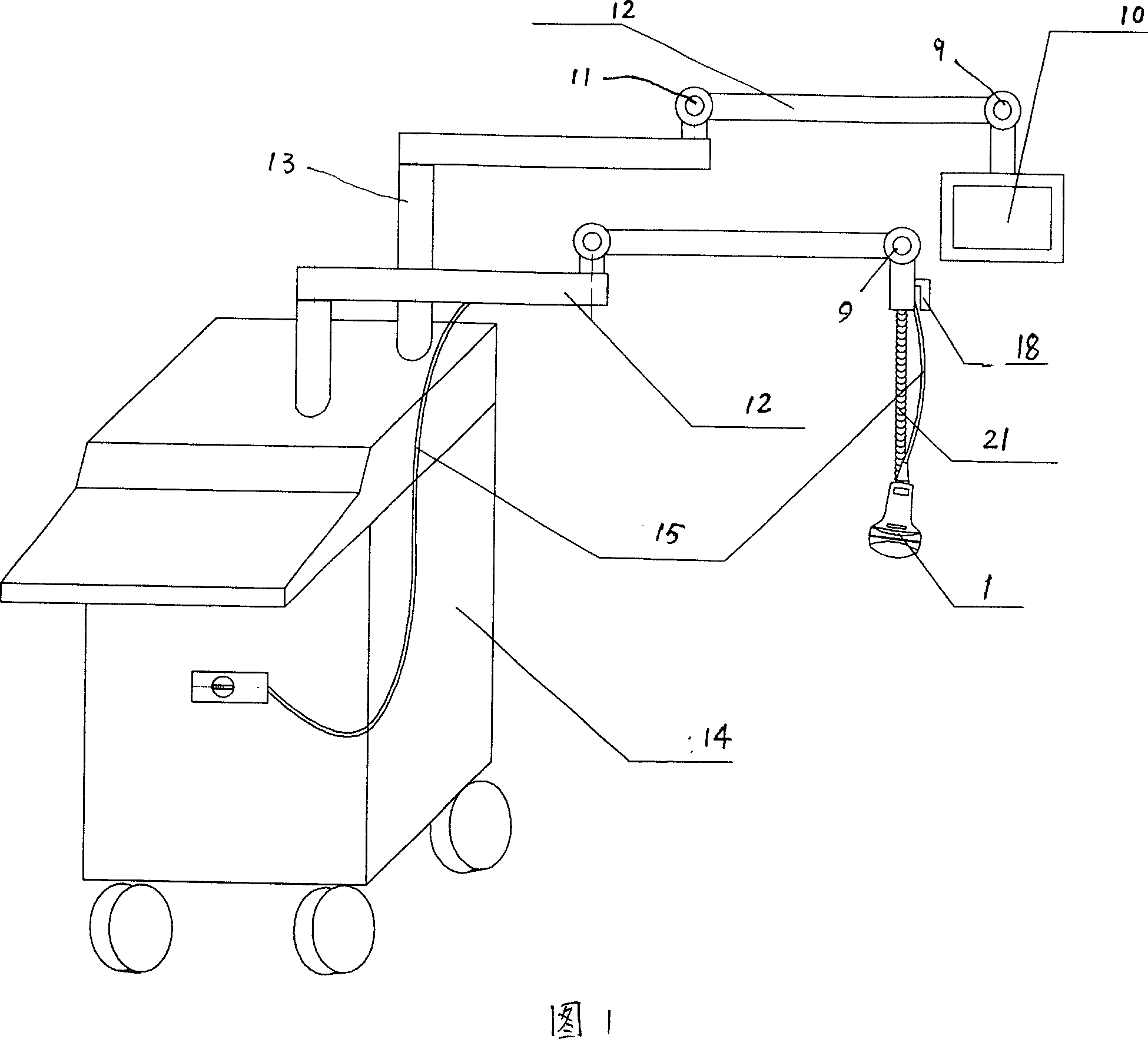
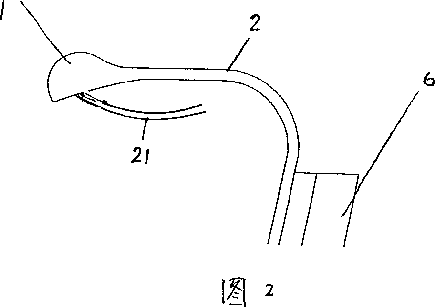
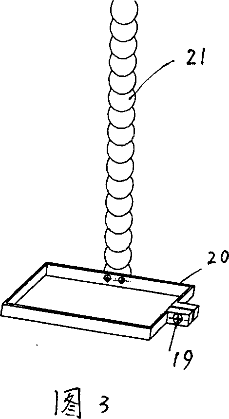
[0025] Examples are described below. The device of each of the embodiments can make it easy for the surgeon to align the B-ultrasound screen and the probe (also known as the transmitter) used. The embodiment of the positioning equipment of the ultrasonic diagnostic apparatus used for surgical monitoring of the present invention comprises a fixed B-ultrasound screen (monitor) 10 or a probe 1 or a combined B-type ultrasonic diagnostic instrument host 14 for positioning, and adopts a serpentine soft arm 21 ( Bead rope) structure to fix the probe, or adopt universal joint structure 9. A separate curved cantilever type extension bracket 8 can also be used. The universal joint structure generally includes a universal joint ball and a ball bowl. The universal joint ball extends out of the supporting shaft. The two structures are multi-dimensionally fixed in any direction: the B-ultrasound probe or screen is positioned and the universal joint ball is extended out of the supporting sh...


PUM


Abstract

A locating method of ultrasonic equipment for medical diagnosis for monitoring operation, equipped with the screen location device of multidimensional fixed B model ultrasonic monitoring instrument, or the locating device for instant position of B model ultrasonic monitoring instrument. The position of the probe or the screen of monitoring instrument is positioned by the bend cantilever three-dimensional extend bracket with serpentine flexible beam structure or combined with gimbal joint. The locating device has a positioned instrument to fix the screen or the probe of B model ultrasonic monitoring instrument, which adopts the bend cantilever extend bracket, serpentine flexible beam structure or gimbal joint to fix the screen or the probe. The invention which overcomes the disadvantages of the present locating device, is convenient to adjust, and makes the probe position of B model ultrasonic monitoring instrument easy for operation.

Description

technical field [0001] The invention relates to a positioning method and equipment for an ultrasonic diagnostic device used for surgical monitoring (especially real-time monitoring). Background technique [0002] It is very important to carry out the operation under the condition of direct "field of view". In many cases, the operation is performed under the condition of "field of view" provided by X-ray, endoscope, and B-ultrasound. Otherwise, surgical operations are carried out blindly, such as interventional treatment using X-ray monitoring; such as gynecological abortion operations only rely on the "feeling" transmitted by manual instruments and the experience of surgeons to perform operations. However, if the doctor is not sure about the position and size of the uterus, it is possible to puncture the uterus. Especially in the case of cervical stenosis or uterine tumors (endometrioma or sarcoma) the risk of piercing the uterus is greater. ...

Claims


Application Information

Patent Timeline
Patent Type & Authority Applications(China)
IPC IPC(8): A61B8/00A61B19/00
Inventor 王敏岐
Owner 王敏岐
Learn More